




Abstract
The diacylglycerol kinases (DGKs) are a family of proteins that catalyze the phosphorylation of the cell membrane lipid diacylglycerol (DG), a cellular component that is important in lipid biochemistry and signal transduction, into phosphatidic acid. DG-mediated signal transduction downstream of the T cell receptor has previously been reported to be terminated in most cases by one of 10 DGK isoforms, DGKζ. In this study, we performed immunohistochemical analysis using a rabbit anti-DGKζ monoclonal antibody (mAb) (clone EPR22040-80) against tissues from the tonsils of a patient with oropharyngeal squamous cell carcinoma. We demonstrated that many DGKζ-expressing T cells are localized in the tonsils. We further characterized the binding epitope using an enzyme-linked immunosorbent assay and found that Pro790, Gln791, Gly792, and Leu795 residues of DGKζ are important for facilitating anti-DGKζ mAb binding to DGKζ. This anti-DGKζ mAb could be valuable in immunohistochemical analyses in determining the distribution of DGKζ-expressing T cells in pathophysiological tissues.
Introduction
Diacylglycerol kinases (DGKs) are a family of proteins that phosphorylate the cell membrane lipid diacylglycerol (DG) into phosphatidic acid.(1–3) DG functions as an important second messenger in T cells.(4) DG-mediated signal transduction downstream of T cell receptors (TCRs) is terminated by DGKα and DGKζ, 2 of the 10 DGK isoforms.(5) DGKζ has been shown to be the dominant isoform.(6) T cells deficient in either DGKα or DGKζ are hyper-responsive, leading to enhanced proliferation and secretion of cytokines in response to TCR activation.(7–9) Riese et al. demonstrated that CD8+ T cells deficient in DGKs exhibit enhanced activity against xenografts after adoptive transfer of T cells when expressing TCRs or chimeric antigen receptors specific for tumor antigens.(10) Jing et al. reported that targeting DGKζ may increase the efficacy of adoptive T cell and immune checkpoint therapies in the treatment of leukemia.(11)
In the present study, we selected a commercially available rabbit anti-DGKζ monoclonal antibody (mAb) that is advantageous for immunohistochemical analysis and further characterized its binding epitope using enzyme-linked immunosorbent assay (ELISA).
Materials and Methods
Immunohistochemical analyses
We used tissues from a patient with oropharyngeal squamous cell carcinoma who had undergone surgery at the Sendai Medical Center. Informed consent for sample procurement and subsequent data analyses was obtained from the patient or the patient's guardian. The tissue samples were processed to produce 4-μm paraffin-embedded tissue sections that were autoclaved in EnVision FLEX Target Retrieval Solution High pH (Agilent Technologies, Inc., Santa Clara, CA) for 20 minutes and then blocked using the SuperBlock T20 (PBS) blocking buffer (Thermo Fisher Scientific, Inc., Waltham, MA). Samples were incubated with rabbit anti-DGK
Enzyme-linked immunosorbent assay
The DGKζ peptides synthesized using PEPScreen (Sigma-Aldrich Corp., St. Louis, MO) were immobilized on Nunc MaxiSorp 96-well immunoplates (Thermo Fisher Scientific, Inc.) at concentrations of 10 μg/mL for 30 minutes at 37°C. Following blocking with SuperBlock T20 (PBS) blocking buffer, the plates were incubated with 1 μg/mL of rabbit anti-DGKζ mAb, followed by 1:1000 dilution of peroxidase-conjugated anti-rabbit IgG (Agilent Technologies, Inc.). The enzymatic reaction was performed using 1-Step Ultra TMB-ELISA (Thermo Fisher Scientific, Inc.). Optical density was measured at 655 nm using an iMark microplate reader (Bio-Rad Laboratories, Inc., Berkeley, CA). These reactions were performed at 37°C using a total sample volume of 50–100 μL.
Inhibition assay
The 4-μm paraffin-embedded tissue sections were directly autoclaved in EnVision FLEX Target Retrieval Solution High pH (Agilent Technologies, Inc.) for 20 minutes and blocked using the SuperBlock T20 (PBS) blocking buffer (Thermo Fisher Scientific, Inc.), incubated with a rabbit anti-DGK
Results
For immunohistochemical analysis, we used formalin-fixed and paraffin-embedded (FFPE) sections, including the tonsils of a patient with oropharyngeal squamous cell carcinoma, because DGKζ is known to be expressed in activated T cells.(7–9) As shown in Figure 1, the rabbit anti-DGK
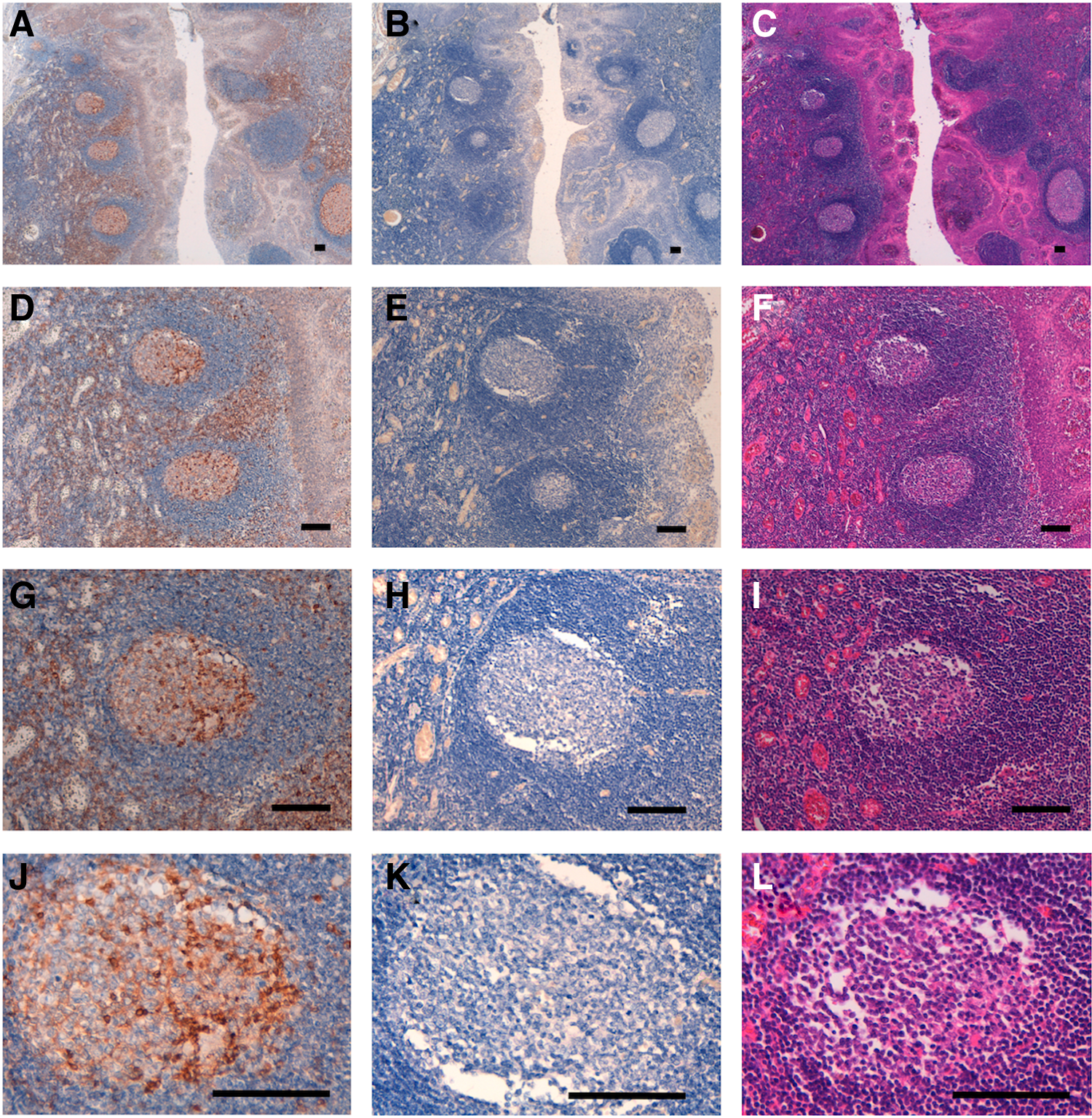
Immunohistochemical analysis of DGKζ against oropharyngeal squamous cell carcinomas. Tissue sections were incubated with EPR22040-80
Next, we examined the binding epitope of EPR22040-80. We first produced 92 synthetic peptides of DGKζ, in which all cysteine residues were converted into serine residues to avoid self-aggregation (Table 1). Using ELISA, we demonstrated that the 781–800 position peptide was detected by EPR22040-80. We also synthesized 20 peptides, including a number of point mutations of the peptide at positions 781–800 (Table 2). Almost all the point mutations were detected by EPR22040-80, except for P790A, Q791A, G792A, and L795A, indicating Pro790, Gln791, Gly792, and Leu795 are included in the critical epitope of EPR22040-80. The epitope of EPR22040-80 is summarized in Figure 2.
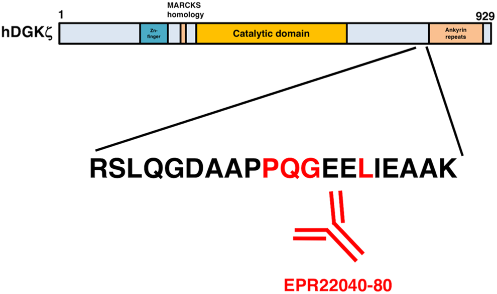
Schematic illustration of EPR22040-80 epitope. Red amino acids, strong reaction with EPR22040-80.
Determination of EPR22040-80 Epitope by Enzyme-Linked Immunosorbent Assay
+++, OD655 ≧ 0.6; —, OD655 < 0.2.
Determination of EPR22040-80 Epitope by Enzyme-Linked Immunosorbent Assay Using Point Mutants
Mutated amino acids (Ala or Gly) are shown in bold letters.
+++, OD655 ≧ 0.6; ++, 0.4 ≦ OD655 < 0.6; +, 0.2 ≦ OD655 < 0.4; —, OD655 < 0.2.
We further performed an inhibition assay using immunohistochemistry. Although EPR22040-80 reacted with the T cells of oropharyngeal squamous cell carcinoma, these reactions were completely neutralized by R781A. L795A did not block the reactions of EPR22040-80 (Fig. 3). These data supported the contention that Leu795 of DGKζ is critical for EPR22040-80 detection.
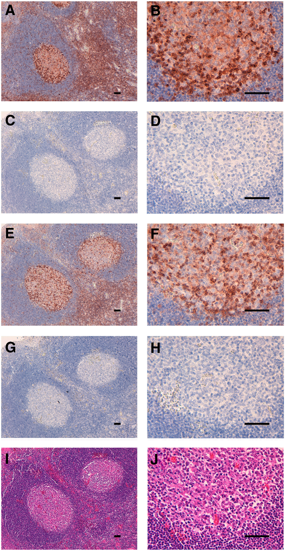
Inhibition assay. Tissue sections were incubated with EPR22040-80
Discussion
We have developed anti-DGKα (clone: DaMab-2)(12) and anti-DGKγ (clone: DgMab-6)(13) mAbs, both of which are extremely useful for immunocytochemical analysis. We characterized the binding epitope of DaMab-2 using Western blots and revealed that the Cys246, Lys249, Pro252, and Cys253 residues of DGKα are important for binding of DaMab-2 to DGKα.(14) These findings could be applied for the production of more functional anti-DGKα mAbs. We have not developed anti-DGKζ mAbs, which are useful for immunocytochemical and immunohistochemical analyses. In particular, it is difficult to develop mAbs for immunohistochemical analyses against FFPE tissue sections. Therefore, we first characterized commercially available anti-DGKζ mAbs. Among several anti-DGKζ mAbs, a rabbit anti-DGKζ mAb (clone: EPR22040-80 from Abcam) stained the T cells of tonsils very strongly. According to the Abcam product datasheet, EPR22040-80 is produced by immunizing recombinant fragments within human DGKζ (700 aa–1000 aa). However, the critical epitope of EPR22040-80 has not been determined. In this study, ELISA demonstrated that Pro790, Gln791, Gly792, and Leu795 of DGK
Footnotes
Acknowledgments
We thank Takuro Nakamura, Miyuki Yanaka, Kayo Hisamatsu, Saori Handa, Yoshimi Nakamura, and Maki Takahashi for their excellent technical assistance. This research was supported, in part, by AMED under Grant Nos. JP18am0101te078 (Y.K.), JP18am0301010 (Y.K.), and JP18ae0101028 (Y.K.), and by JSPS KAKENHI Grant no. 17K07299 (M.K.K.) and Grant no. 16K10748 (Y.K.).
Author Disclosure Statement
Y.K. received research funding from Ono Pharmaceutical Co., Ltd. The other authors have no conflict of interest.