
Abstract
Podoplanin (PDPN)/T1 alpha is known as a specific marker of lymphatic endothelial cells and type I alveolar cells. Sensitive and specific monoclonal antibodies (mAbs) for PDPN are needed for immunohistochemical analyses. Recently, we developed an anticetacean PDPN mAb, PMab-237. Herein, immunohistochemical analyses showed that PMab-237 strongly detected pulmonary type I alveolar cells, renal podocytes, and lymphatic endothelial cells of the harbor porpoise. These findings suggest that PMab-237 may be useful for immunohistochemical analyses for cetacean tissues.
Introduction
Podoplanin (PDPN)/T1
Materials and Methods
Immunohistochemical analyses
Cetacean tissues were collected from two harbor porpoises, and were fixed in 15% neutral buffered formalin at Obihiro University of Agriculture and Veterinary Medicine (Hokkaido, Japan). Histological sections (4-μm thick) were directly autoclaved in EnVision FLEX Target Retrieval Solution High pH (Agilent Technologies, Inc., Santa Clara, CA) for 20 minutes. Then, sections were blocked using the SuperBlock T20 (PBS) Blocking Buffer (Thermo Fisher Scientific, Inc., Waltham, MA), incubated with PMab-237 (5 μg/mL) for 1 hour at room temperature, and treated with the Envision+Kit for mouse (Agilent Technologies, Inc.) for 30 minutes. Color was developed using 3,3′-diaminobenzidine tetrahydrochloride (Agilent Technologies, Inc.) for 2 minutes, and counterstaining was performed using hematoxylin (FUJIFILM Wako Pure Chemical Corporation, Osaka, Japan).
Results and Discussion
Although we have established many anti-PDPN mAbs, such as antihuman,(11) antimouse,(12) antirat,(13) antirabbit,(14) antibovine,(15) antidog,(16) anticat,(17) antipig,(18) and antihorse(19) PDPNs, cetacean PDPN was not recognized by those anti-PDPN mAbs (data not shown). Then, two mice were recently immunized with cetacean PDPN-expressing CHO-K1 (CHO/wPDPN) cells using CBIS method.(20) Wells positive for CHO/wPDPN and negative for CHO-K1 were selected by flow cytometry. PMab-237 (IgG1, kappa) was finally selected. Western blot analysis performed using PMab-237 demonstrated that PMab-237 detects wPDPN as a 40-kDa band in CHO/wPDPN cells. Furthermore, the immunohistochemical analyses revealed that PMab-237 strongly stained CHO/wPDPN cells, whereas it did not react with CHO-K1 cells, suggesting that PMab-237 might be applicable for immunohistochemical analyses against cetacean tissues.
In this study, we performed immunohistochemical analyses against the harbor porpoise. PDPN has been reported to be expressed in several tissues, including lung, kidney, and colon(1); we selected those tissues for immunohistochemical analyses. Expectedly, PMab-237 stained type I alveolar cells of lung (Fig. 1A, B), podocytes of kidney (Fig. 2A, B), and lymphatic endothelial cells of colon (Fig. 3A, B). These staining patterns are consistent with our previous immunohistochemical studies using several anti-PDPN mAbs.(18,22) No staining was observed without primary antibodies against type I alveolar cells of lung (Fig. 1C, D), podocytes of kidney (Fig. 2C, D), and lymphatic endothelial cells of colon (Fig. 3C, D).

Immunohistochemical analyses for cetacean lung. Cetacean lung sections were incubated with 5 μg/mL of PMab-237
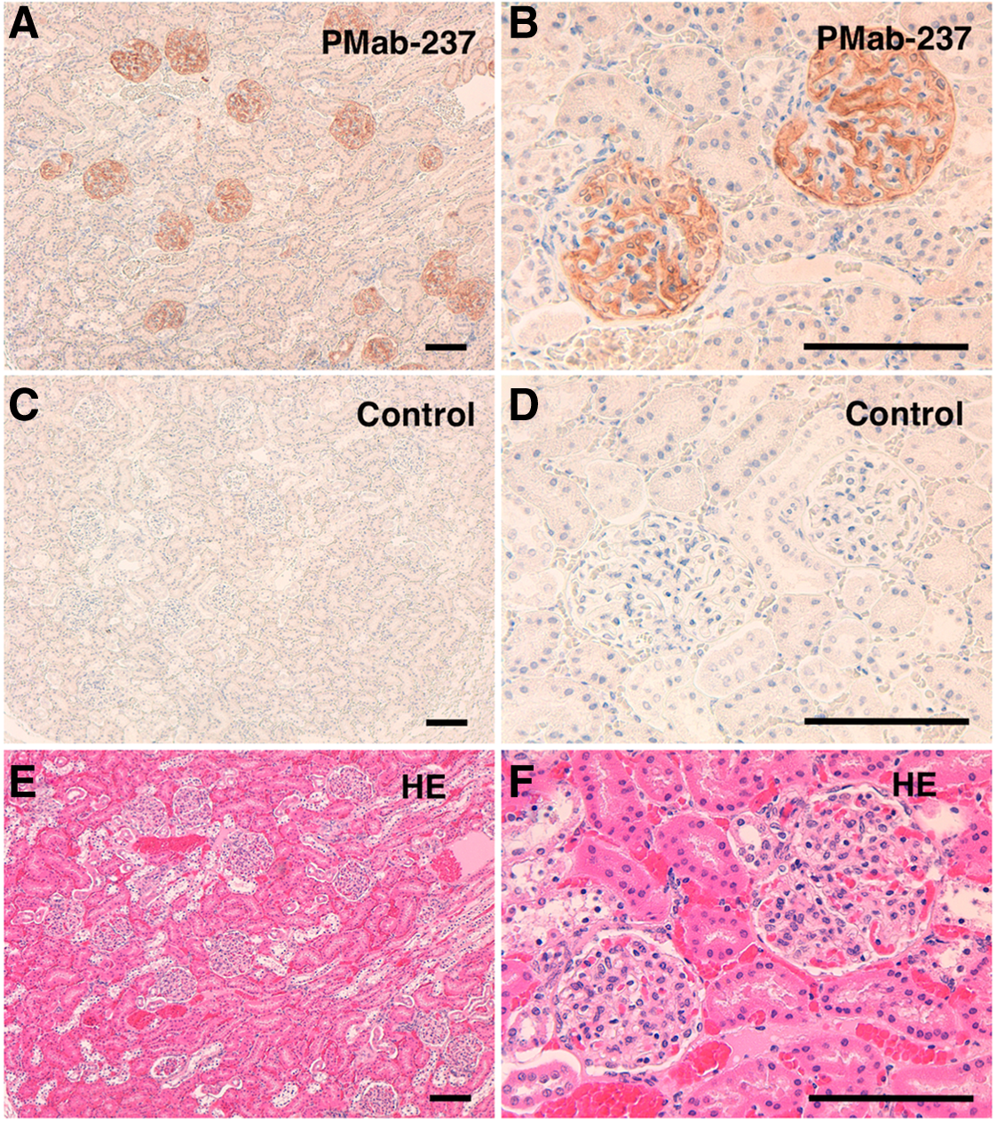
Immunohistochemical analyses for cetacean kidney. Cetacean kidney sections were incubated with 5 μg/mL of PMab-237

Immunohistochemical analyses for cetacean colon. Cetacean colon sections were incubated with 5 μg/mL of PMab-237
Our previous studies showed that PMab-237 cross-reacted with human, bovine, goat, sheep, alpaca, and tiger PDPNs, which were overexpressed in CHO-K1 cells.(20) PMab-237 also reacted with hPDPN, which was overexpressed in a glioblastoma cell line, LN229 (data not shown). Then, we investigated the reactivity of PMab-237 with hPDPN-expressing cancer cell lines, such as a glioblastoma cell (LN319), a lung squamous cell carcinoma cell line (PC-10), and a malignant mesothelioma cell line (NCI-H226) or hPDPN-expressing normal cells, such as lymphatic endothelial cells, a kidney epithelial cell (HEK-293T), and a mesothelial cell line (Met-5A) by flow cytometry. However, PMab-237 did not react any endogenous PDPN by flow cytometry (data not shown), indicating that only overexpressed hPDPN might be detected by PMab-237. Those results demonstrated that PMab-237 could be specific for cetacean PDPN.
In conclusion, PMab-237 is useful for immunohistochemical analyses against the harbor porpoise. The epitope of PMab-237 needs further investigation to clarify the sensitivity and specificity of PMab-237 against cetacean PDPN. PMab-237 could be useful in elucidating the pathophysiological functions of cetacean PDPN in future studies.
Footnotes
Acknowledgments
This research was supported, in part, by AMED under Grant Nos. JP18am0101078 (Y.K.), JP18am0301010 (Y.K.), and JP18ae0101028 (Y.K.), and by JSPS KAKENHI Grant No. 17K07299 (M.K.K.) and Grant No. 16K10748 (Y.K.).
Author Disclosure Statement
No competing financial interests exist.