
Abstract
Podoplanin (PDPN)/T1alpha is expressed on lymphatic endothelial cells, type I alveolar cells of the lungs, and podocytes of the kidney. PDPN possesses three platelet aggregation-stimulating (PLAG) domains (PLAG1, PLAG2, and PLAG3) of the N-terminus and the PLAG-like domains (PLDs). We previously reported an anti-goat PDPN (gPDPN) monoclonal antibody (mAb), PMab-235, which was developed using the Cell-Based Immunization and Screening (CBIS) method. PMab-235 is very useful in flow cytometry, Western blotting, and immunohistochemical analyses; however, the binding epitope of PMab-235 remains to be elucidated. In this study, we investigated the epitopes of PMab-235 using enzyme-linked immunosorbent assay and immunohistochemistry. The results revealed that the critical epitope of PMab-235 produced by CBIS method is Arg75, Leu78, and Pro79 of gPDPN, which is included in PLD. The findings of our study can be applied to the production of more functional anti-gPDPN mAbs.
Introduction
Podoplanin (PDPN)/T1
Because pathophysiological studies of alveolar cells of the lungs, podocytes of the kidneys, or lymphatic endothelial cells of the colon using anti-goat PDPN (gPDPN) mAbs are important in many disorders, we used the Cell-Based Immunization and Screening (CBIS) method and developed specific and sensitive monoclonal antibodies (mAbs) against the gPDPN of 168 amino acids to facilitate the immunohistochemical analysis of paraffin-embedded tissue sections.(20) PMab-235 recognized CHO/gPDPN cells, but showed no reaction with CHO-K1 cells, as assessed by flow cytometry. PMab-235 cross-reacts with bovine PDPN. In contrast, PMab-235 did not react with human, mouse, rat, rabbit, dog, cat, pig, horse, Tasmanian devil, sheep, alpaca, tiger, whale, or bear PDPNs. KD of PMab-235 for CHO/gPDPN cells was determined to be 1.5 × 10⁻⁸, indicating a moderate affinity of PMab-235 for CHO/gPDPN cells. The immunohistochemical analyses revealed that PMab-235 strongly stained type I alveolar cells of lung, podocytes of kidney, and lymphatic endothelial cells of colon. This study aimed to determine the binding epitope of PMab-235 to gPDPN using enzyme-linked immunosorbent assay (ELISA) and immunohistochemistry.
Materials and Methods
Enzyme-linked immunosorbent assay
Synthesized gPDPN peptides using PEPScreen (Sigma-Aldrich Corp., St. Louis, MO) were immobilized on Nunc Maxisorp 96-well immunoplates (Thermo Fisher Scientific, Inc., Waltham, MA) at 10 μg/mL for 30 minutes at 37°C. After blocking with SuperBlock T20 (PBS) Blocking Buffer (Thermo Fisher Scientific, Inc.), the plates were incubated with purified PMab-235 (1 μg/mL), followed by a peroxidase-conjugated anti-mouse IgG (Agilent Technologies, Inc., Santa Clara, CA) dilution of 1:2000. Enzymatic reactions were performed using 1-Step Ultra TMB-ELISA (Thermo Fisher Scientific, Inc.). Optical density was measured at 655 nm using an iMark microplate reader (Bio-Rad Laboratories, Inc., Berkeley, CA). These reactions were performed at 37°C using a total sample volume of 50–100 μL.
Immunohistochemical analyses
Goat tissues (lungs and colons) were previously collected in NIPPON ZENYAKU KOGYO CO., LTD. (Fukushima, Japan), and fixed in 4% paraformaldehyde phosphate buffer solution (Nacalai Tesque, Inc., Kyoto, Japan).(21) The tissues were processed to produce 4-μm paraffin-embedded tissue sections, which were directly autoclaved in citrate buffer (pH 6.0; Nichirei Biosciences, Inc., Tokyo, Japan) for 20 minutes. These tissue sections were then blocked with the SuperBlock T20 (PBS) Blocking Buffer (Thermo Fisher Scientific, Inc.), incubated with PMab-235 (0.01–1 μg/mL) for 1 hour at room temperature, and treated using an Envision + Kit (Agilent Technologies, Inc.) for 30 minutes. Color was developed with 3,3′-diaminobenzidine tetrahydrochloride (DAB; Agilent Technologies, Inc.) for 2 minutes, and counterstaining was performed using hematoxylin (FUJIFILM Wako Pure Chemical Corporation, Osaka, Japan).
Blocking assay using immunohistochemical analyses
Goat tissue sections were blocked with the SuperBlock T20 (PBS) Blocking Buffer (Thermo Fisher Scientific, Inc.), incubated with PMab-235 (0.2 or 0.02 μg/mL) or PMab-235 (0.2 or 0.02 μg/mL) + peptides (1 μg/mL) for 1 hour at room temperature, and treated using an Envision + Kit (Agilent Technologies, Inc.) for 30 minutes. Color was developed with DAB (Agilent Technologies, Inc.) for 2 minutes, and counterstaining was performed using hematoxylin (FUJIFILM Wako Pure Chemical Corporation).
Results
We first synthesized a series of gPDPN peptides, which are summarized in Table 1. Using ELISA, PMab-235 detected a 61–80 peptide (61-TPMPTGTENVTHGHREDLPT-80) of gPDPN, whereas it did not react with the other peptides: 21–40, 31–50, 41–60, 51–70, 71–90, 81–100, 91–110, 101–120, 111–130, and 121–135 peptides (Fig. 1 and Table 1).

Epitope mapping using synthetic peptides of gPDPN. Synthesized gPDPN peptides were immobilized at 10 μg/mL, and were incubated with PMab-235, followed by secondary antibodies. Black bar: positive reaction by PMab-235. gPDPN, goat podoplanin.
Determination of PMab-235 Epitope by Enzyme-Linked Immunosorbent Assay
+++, OD655≧0.6; −, OD655<0.1
We next moved on to the blocking assay to confirm the binding epitope of PMab-235. We first performed immunohistochemical analyses for goat lung tissues. PMab-235 showed very high reaction with type I alveolar cells of goat lungs in a dose-dependent manner (0.01–0.1 μg/mL; Fig. 2). Then, we performed a blocking assay using immunohistochemistry against goat lungs at a concentration of 0.02 μg/mL. PMab-235 reacted with type I alveolar cells of lung (Figs. 3A, B). These reactions were not neutralized by 21–40 peptide in lung (Figs. 3C, D). In contrast, these reactions were partially neutralized by 61–80 peptide in lung (Figs. 3E, F), indicating that 61–80 peptide is an epitope of PMab-235.

Immunohistochemical analyses against goat lung tissues using PMab-235. Histological sections of the goat lungs were directly autoclaved in citrate buffer for 20 minutes. After blocking with SuperBlock T20 (PBS) Blocking Buffer, the sections were incubated with PMab-235 at a concentration of 0.1 μg/mL

Immunohistochemical analyses against goat lung tissues using PMab-235 and peptides of gPDPN. Histological sections of the goat lungs were directly autoclaved in citrate buffer for 20 minutes. After blocking with SuperBlock T20 (PBS) Blocking Buffer, the sections were incubated with
PMab-235 also showed very high reaction with lymphatic endothelial cells of goat colons in a dose-dependent manner (0.1–1 μg/mL; Fig. 4), even at a low concentration of 0.1 μg/mL (Fig. 4G, H). We performed a blocking assay using immunohistochemistry against goat colons at a concentration of 0.2 μg/mL. PMab-235 reacted with lymphatic endothelial cells of colons (Figs. 5A, B). These reactions were not neutralized by 21–40 peptide (Figs. 5C, D). In contrast, these reactions were completely neutralized by 61–80 peptide (Figs. 5E, F), also confirming that 61–80 peptide is an epitope of PMab-235.

Immunohistochemical analyses against goat colon tissues using PMab-235. Histological sections of the goat colons were directly autoclaved in citrate buffer for 20 minutes. After blocking with SuperBlock T20 (PBS) Blocking Buffer, the sections were incubated with PMab-235 at a concentration of 1 μg/mL

Immunohistochemical analyses against goat colon tissues using PMab-235 and peptides of gPDPN. Histological sections of the goat colons were directly autoclaved in citrate buffer for 20 minutes. After blocking with SuperBlock T20 (PBS) Blocking Buffer, the sections were incubated with
We then synthesized point mutants of 61–80 peptides (Table 2). Using ELISA, PMab-235 detected T61A, P62A, M63A, P64A, T65A, G66A, T67A, E68A, N69A, V70A, T71A, H72A, G73A, H74A, E76A, D77A, and T80A. Conversely, it did not detect R75A, L78A, and P79A, indicating that R75A, L78A, and P79A are critical for PMab-235 to detect gPDPN.
Determination of PMab-235 Epitope by Enzyme-Linked Immunosorbent Assay
+++, OD655≧0.6; −, OD655<0.1
Discussion
We previously developed mAbs against human,(22) mouse,(22) rat,(23) rabbit,(24) dog,(25) cat,(26) bovine,(27) horse,(28–30) and pig PDPNs.(31,32) PDPN comprises three tandem-repeat of the “EDxxVTPG” sequences, which were defined to be platelet aggregation-stimulating (PLAG) domains (PLAG1, PLAG2, and PLAG3) of the N-terminus of the PDPN protein.(4) There are also several PLAG-like domains (PLDs) of the “E(D/E)xx(T/S)xx” sequence. PLDs, one of which is also named as PLAG4, are reportedly important for PDPN–CLEC-2 interactions.(33) Almost all mAbs against PDPNs reportedly react with PLAG domains or PLDs.(33,34) As summarized in Figure 6, PMab-235 detected PLD of gPDPN.
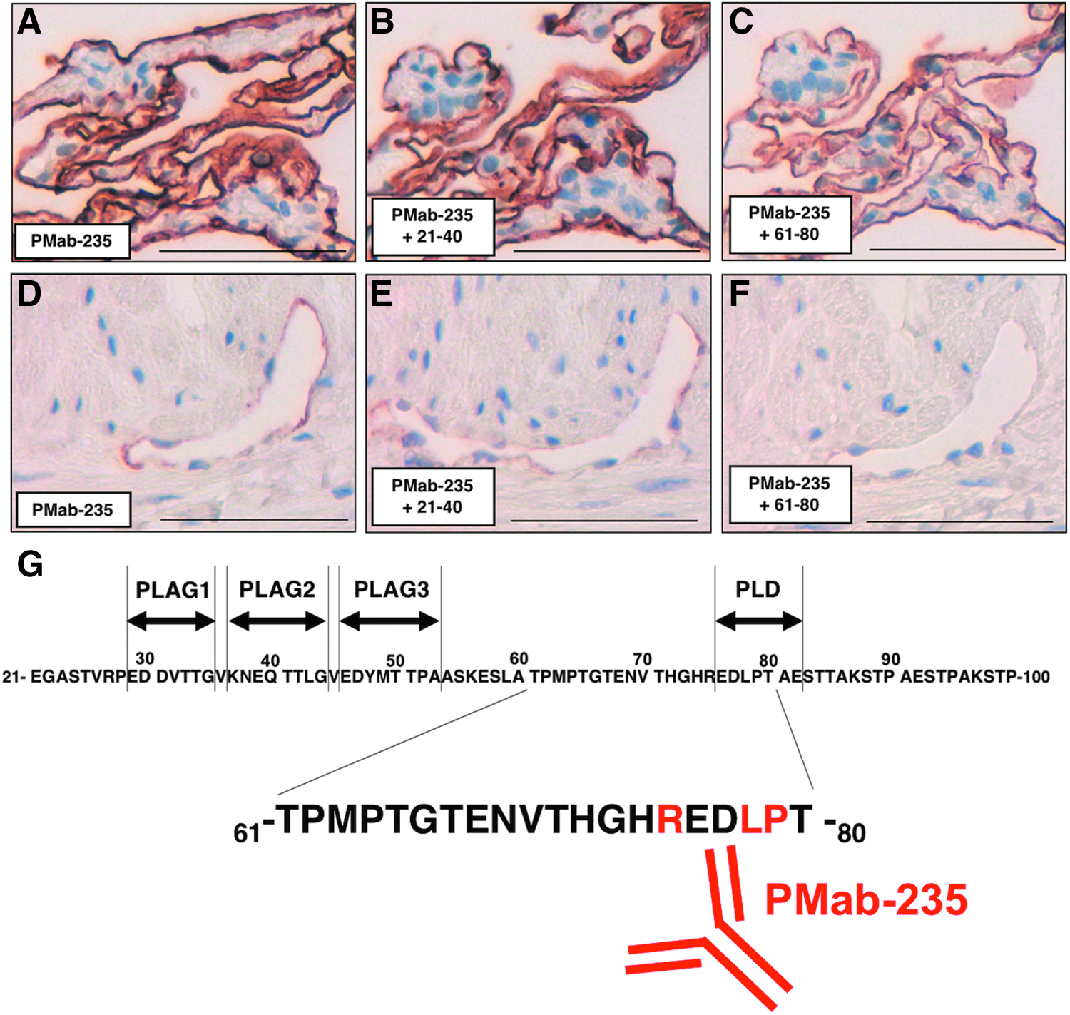
Summary of PMab-235 epitope.
PMab-235 cross-reacted with bovine PDPN (bovPDPN).(20) The corresponding sequence of PMab-235 epitope “REDLPT” is also “REDLPT” about bovPDPN, which indicates 100% identity between gPDPN and bovPDPN. In the blocking assay using immunohistochemistry against goat lungs, the 61–80 peptide of goat PDPN did not completely block the PMab-235 binding to type I alveolar cells (Figs. 3E, F) because PMab-235 was developed using CBIS method, not by immunizing synthetic peptides, then the binding affinity of PMab-235 against type I alveolar cells might be higher than that for the 61–80 peptide.
Taken together, PLD of gPDPN was clarified to be advantageous epitopes for several applications, such as flow cytometry, Western blotting, and immunohistochemical analyses. These findings can be applied to the production of more functional anti-gPDPN mAbs.
Footnotes
Acknowledgments
This research was supported in part by AMED under Grant Nos. JP18am0101078 (Y.K.), JP18am0301010 (Y.K.), and JP18ae0101028 (Y.K.), and by JSPS KAKENHI Grant Nos. 17K07299 (M.K.K.) and 19K07705 (Y.K.).
Author Disclosure Statement
Y.K. received research funding from ZENOAQ RESOURCE CO., LTD. The other authors have no conflict of interest.