
Abstract
Podoplanin (PDPN), which is a mucin-type membrane glycoprotein, is expressed on lymphatic endothelial cells and epithelial cells of many organs. PDPN is also overexpressed in several malignant cancers, and its expression is associated with cancer progression and poor prognosis. Human PDPN possesses three platelet aggregation-stimulating (PLAG) domains and the PLAG-like domain (PLD), which binds to C-type lectin-like receptor-2 (CLEC-2). Previously, we reported a novel antihorse PDPN (horPDPN) monoclonal antibody (mAb), PMab-219, using Cell-Based Immunization and Screening (CBIS) method. PMab-219 specifically detected horPDPN-overexpressed Chinese hamster ovary (CHO)-K1 (CHO/horPDPN) cells and FHK-TcL3.1, a horse kidney cell line, using flow cytometry. In addition, PMab-219 strongly stained horse tissues such as renal podocytes or lymphatic endothelial cells by immunohistochemistry. However, the specific binding epitope of PMab-219 for horPDPN remains to be clarified. In this study, a series of deletion mutants or point mutants of horPDPN were produced for analyzing the PMab-219 epitope using flow cytometry. The analysis of deletion mutants showed that N-terminus of PMab-219 epitope exists between 55th amino acid (aa) and 60th aa of horPDPN. Furthermore, the analysis of point mutants demonstrated that the critical epitope of PMab-219, which was developed by CBIS method, could include Val59, Arg61, Ser62, and Thr63 of horPDPN, indicating that PMab-219 epitope is independent of PLAG domain or PLD of horPDPN.
Introduction
Podoplanin (PDPN/T1alpha/Aggrus) is a type I transmembrane glycoprotein, and is expressed in normal tissues, such as lymphatic endothelial cells, type I lung alveolar cells, and renal podocytes.(1–3) PDPN is known as a platelet aggregation-inducing factor in highly metastatic tumor cells.(1,4) Human PDPN (hPDPN) is frequently overexpressed in various malignant tumors, including lung cancer, mesothelioma, glioblastoma, and osteosarcoma.(5–7) hPDPN possesses three platelet aggregation-stimulating (PLAG) domains (PLAG1, PLAG2, and PLAG3), which exist at the N-terminus of hPDPN.(8) In addition, one PLAG-like domain (PLD) has been reported to be present in the middle of PDPN.(9) PLAG and PLD are highly conserved among mammalian PDPNs.(8) The interaction between PDPN on lymphatic endothelial cells and C-type lectin-like receptor-2 (CLEC-2) on platelets was shown to facilitate embryonic blood/lymphatic vessel separation.(10)
In our previous study, we developed monoclonal antibodies (mAbs) against human,(11) mouse,(11) rat,(12) rabbit,(13) dog,(14) cat,(15) bovine,(16) pig,(17) Tasmanian devil,(18) alpaca,(19) bear,(20) tiger,(21) whale,(22) goat,(23) and horse PDPNs.(24) Although PMab-202, an antihorse PDPN (horPDPN) mAb, was established by immunizing mice with synthetic peptides of horPDPN, it was not applicable for immunohistochemical analysis.(25) Highly sensitive and specific mAbs against horPDPN were needed to analyze horPDPN for pathological investigation using horse tissues.
Recently, we developed a novel anti-horPDPN mAb, PMab-219, using Cell-Based Immunization and Screening (CBIS) method. PMab-219 specifically detected horPDPN by flow cytometry, Western blotting, and immunohistochemical analysis.(24,26) However, the binding epitope of PMab-219 for horPDPN remains to be clarified. Thus, this study aimed to identify the epitope of PMab-219 using deletion mutants and point mutants of horPDPN using flow cytometry.
Materials and Methods
Cell lines
Chinese hamster ovary (CHO)-K1 was obtained from the American Type Culture Collection (ATCC, Manassas, VA). The horPDPN mutation plasmids containing PA16 tag were transfected into CHO-K1 cells using Lipofectamine LTX (Thermo Fisher Scientific, Inc., Waltham, MA). The PA16 tag comprises 16 amino acids (aas) (GLEGGVAMPGAEDDVV). Transiently transfected cells with deletion mutants or point mutants were cultured in Roswell Park Memorial Institute (RPMI) 1640 medium (Nacalai Tesque, Inc., Kyoto, Japan), supplemented with 10% heat-inactivated fetal bovine serum (FBS; Thermo Fisher Scientific, Inc.), 100 U/mL of penicillin, 100 μg/mL of streptomycin, and 25 μg/mL of amphotericin B (Nacalai Tesque, Inc.) at 37°C in a humidified atmosphere of 5% CO2 and 95% air.
Production of horPDPN mutants
Synthesized DNA of horPDPN was subcloned into the pCAG vector (FUJIFILM Wako Pure Chemical Corporation, Osaka, Japan), and PA16 tag was added at the N-terminus. Deletion mutants of horPDPN sequence were produced using a HotStar HiFidelity PCR (Qiagen, Inc., Hilden, Germany) with oligonucleotides. Substitutions of aas to alanine or glycine in horPDPN sequence were conducted by QuikChange lightning site-directed mutagenesis kit (Agilent Technologies, Inc., Santa Clara, CA). PCR fragments bearing the desired mutations were inserted into the pCAG vector using In-Fusion PCR cloning kit (Takara Bio, Inc., Shiga, Japan).
Flow cytometry
Transiently transfected CHO-K1 cells were detached by 0.25% trypsin/1 mM ethylenediaminetetraacetic acid (EDTA; Nacalai Tesque, Inc.) and collected using 10% FBS in RPMI 1640 medium. After washing with 0.1% bovine serum albumin/phosphate-buffered saline, the cells were incubated with anti-horPDPN antibody (PMab-219; 1 μg/mL) or control anti-PA16 tag antibody (NZ-1; 1 μg/mL) for 30 minutes at 4°C. Alexa Fluor 488-conjugated antimouse IgG (1:1000; Cell Signaling Technology, Inc., Danvers, MA) for detection of PMab-219 or Oregon Green antirat IgG (1:2000; Thermo Fisher Scientific, Inc.) for detection of NZ-1 was added to each cell, and was incubated for 30 min at 4°C. Fluorescence data were collected and analyzed using a Cell Analyzer EC800 (Sony Corp., Tokyo, Japan).
Results
Seven deletion mutants of horPDPN, such as dN30 (corresponding to 30–152 aas), dN40 (corresponding to 40–152 aas), dN50 (corresponding to 50–152 aas), dN55 (corresponding to 55–152 aas), dN60 (corresponding to 60–152 aas), dN70 (corresponding to 70–152 aas), and dN80 (corresponding to 80–152 aas) or wild type (WT) of horPDPN (corresponding to 25–152 aas) were generated using CHO-K1 cells (Fig. 1A). All deletion mutants and WT of horPDPN containing N-terminus PA16 tag were detected by NZ-1 (anti-PA16 tag mAb), indicating that the expression level of each construct is very high (Fig. 1B). In contrast, PMab-219 did not react with dN60, dN70, and dN80 (Fig. 1C), suggesting that N-terminus of PMab-219 epitope might exist between 55th aa and 60th aa of horPDPN.
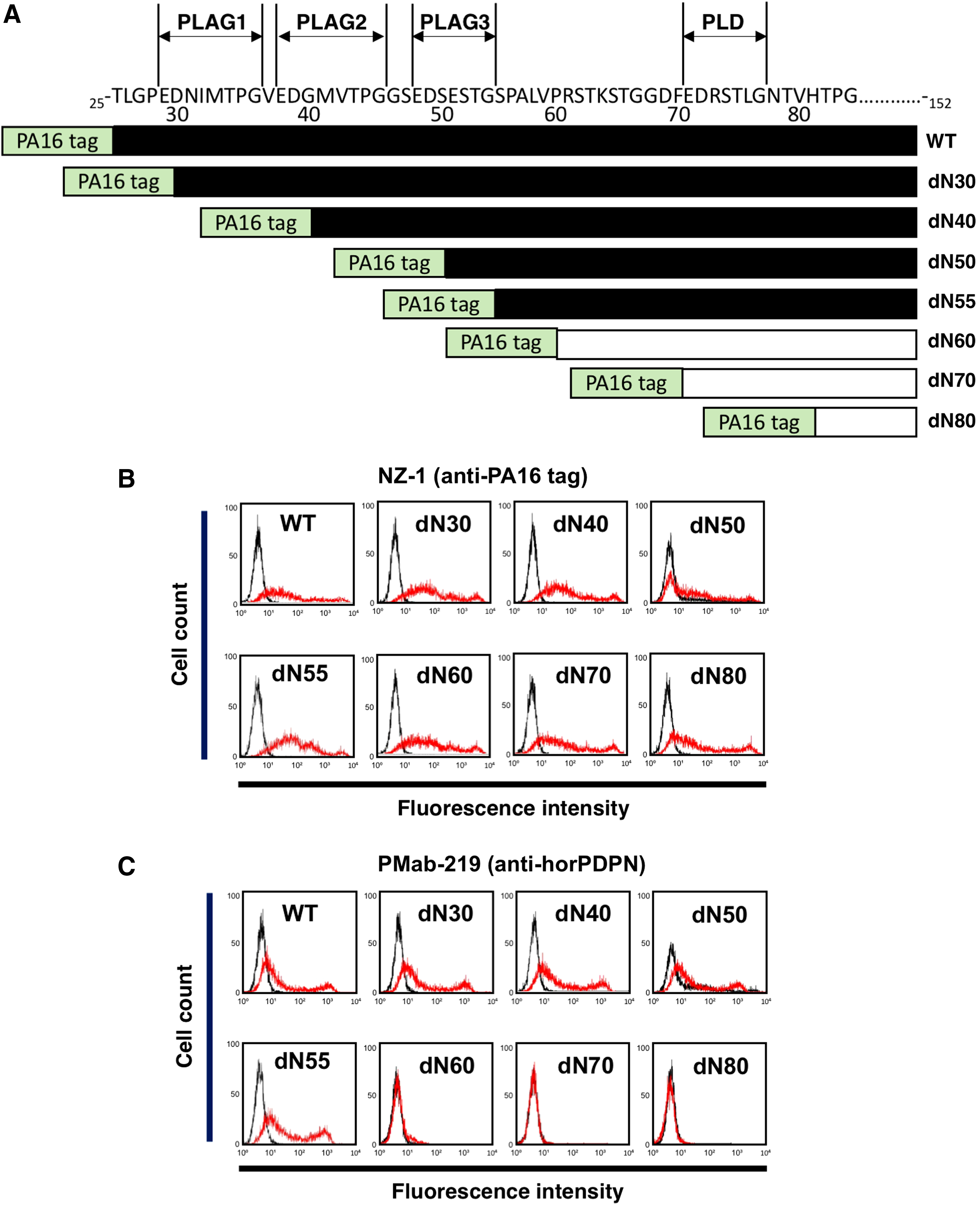
Epitope mapping of PMab-219 using deletion mutants of horPDPN.
To identify the binding epitope of PMab-219, we next produced a series of point mutants of horPDPN in CHO-K1 cells, including S55A, P56A, A57G, L58A, V59A, P60A, R61A, S62A, T63A, K64A, S65A, and T66A. NZ-1 reacted with all point mutants (Fig. 2A). In contrast, PMab-219 did not react with V59A, R61A, and S62A, and weakly recognized T63A (Fig. 2B), indicating Val59, Arg61, Ser62, and Thr63 of horPDPN could be included in the critical epitope of PMab-219 (Fig. 2C).
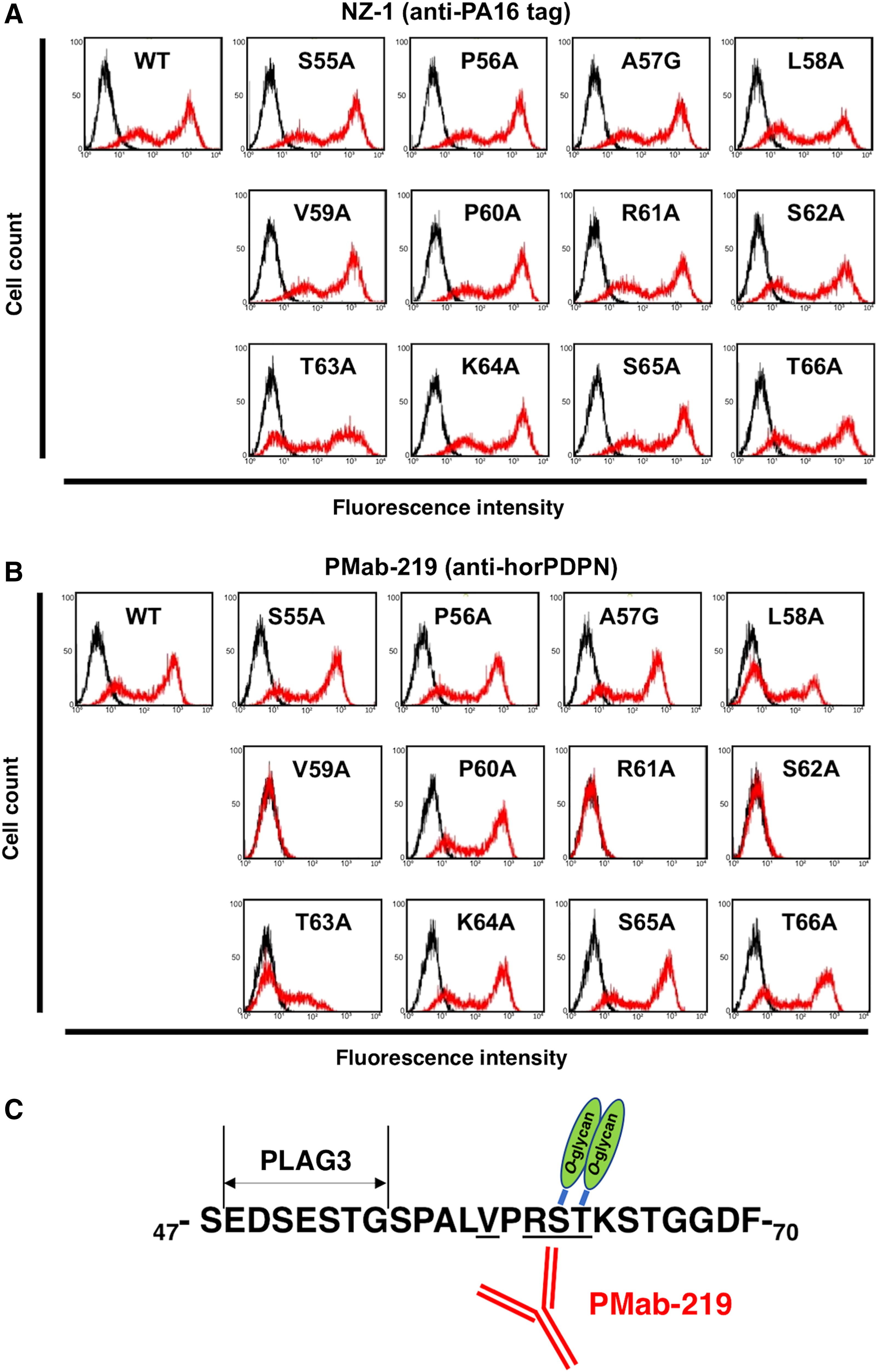
Epitope mapping of PMab-219 using point mutants of horPDPN.
Discussion
We have demonstrated that critical epitope of PMab-219 could include Val59, Arg61, Ser62, and Thr63 of horPDPN using deletion mutants and point mutants of horPDPN. In our previous study, the epitope of PMab-202 (the other anti-horPDPN mAb) was identified as Lys64, Thr66, and Phe70 of horPDPN.(27) Although the binding epitope of both anti-horPDPN mAbs was shown to exist in a similar location of horPDPN, PMab-202 could not stain horse tissues by immunohistochemistry. These results suggested that CBIS method is more advantageous to obtain immunohistochemistry-applicable mAbs. Likewise, we have successfully established immunohistochemistry-applicable anti-PDPN mAbs for pig,(17) Tasmanian devil,(18) alpaca,(19) bear,(20) tiger,(21) whale,(22) and goat PDPNs(23) using CBIS method, but not by immunizing synthetic peptides.
Using glycan-deficient CHO cell lines, such as Lec1 (N-glycan-deficient), Lec2 (sialic acid-deficient), or Lec8 (galactose-deficient),(28) we investigated whether the epitope of PMab-219 could include not only aas but also glycans. Flow cytometric analysis demonstrated that PMab-219 strongly reacts with CHO-K1/horPDPN, Lec1/horPDPN, and Lec2/horPDPN, but weakly reacts with Lec8/horPDPN (data not shown), indicating that galactose might also be included in the PMab-219 epitope. Among Val59, Arg61, Ser62, and Thr63 of horPDPN, Ser62 and Thr63 could be O-glycosylated (Fig. 2C). In a future study, we should determine which O-glycosylation of Ser62 and/or Thr63 is essential for PMab-219 binding to horPDPN.
In conclusion, Val59, Arg61, Ser62, and Thr63 of horPDPN, which are independent of PLAG domain or PLD, are critical for PMab-219-specific binding to horPDPN. PMab-219 could be a useful tool for elucidating the pathophysiological function of horPDPN.
Footnotes
Author Disclosure Statement
No competing financial interests exist.
Funding Information
This research was supported, in part, by AMED under Grant Nos. JP19am0101078 (Y.K.), JP19am0401013 (Y.K.), and JP19ae0101028 (Y.K.), and by JSPS KAKENHI under Grant No. 17K07299 (M.K.K.) and Grant No. 19K07705 (Y.K.).