
Abstract
Podoplanin (PDPN)/T1alpha/Aggrus, a small mucin-type transmembrane glycoprotein, has been shown to be expressed on lymphatic endothelial cells and epithelial cells of many organs. PDPN is also upregulated in many cancers, and is involved in cancer metastasis and malignant progression. Human PDPN possesses three platelet aggregation-stimulating (PLAG) domains and the PLAG-like domain, which bind to C-type lectin-like receptor-2 (CLEC-2). Previously, we reported a novel antipig PDPN (pPDPN) monoclonal antibody (PMab-210) using Cell-Based Immunization and Screening (CBIS) method. PMab-210 specifically detected pPDPN-overexpressed Chinese hamster ovary (CHO)-K1 cells by flow cytometry and Western blot analysis. Immunohistochemical analyses demonstrated that PMab-210 stained pulmonary type I alveolar cells strongly and renal corpuscles weakly in pig or microminipig. However, the specific binding epitope of PMab-210 for pPDPN could not be determined by enzyme-linked immunosorbent assay using a series of pPDPN peptides. In this study, deletion mutants or point mutants of pPDPN were produced for analyzing the PMab-210 epitope using flow cytometry. The analysis of deletion mutants showed that N-terminus of PMab-210 epitope exists between 45th amino acid (aa) and 50th aa of pPDPN. In addition, the analysis of point mutants demonstrated that the critical epitope of PMab-210 could include Glu47, Asp48, Tyr49, Thr50, and Val51 of pPDPN, indicating that PMab-210 epitope is located in PLAG3 domain of pPDPN.
Introduction
Podoplanin (PDPN)/T1alpha/Aggrus is a type I transmembrane sialoglycoprotein consisting of a heavily glycosylated extracellular domain, a single transmembrane, and a short nine amino acid (aa) cytoplasmic tail.(1–3) PDPN has been shown to be expressed on lymphatic endothelial cells, epithelial cells, and fibroblasts of many organs.(2) PDPN-positive cells with the immune cells after myocardial infarction positively affect immune cells recruitment.(4) The PDPN-positive stromal cells play a critical role in a network of immunofibroblasts, which are able to support the earliest phases of tertiary lymphoid structure establishment.(5) PDPN is also upregulated in many cancers, and is involved in cancer metastasis and malignant progression,(6–9) indicating that PDPN possesses many pathophysiological functions in both normal and malignant tissues.
Human PDPN (hPDPN) possesses the EDxxVTPG sequences at the N-terminus of hPDPN, which were defined to be the platelet aggregation-stimulating (PLAG) domains (PLAG1, PLAG2, and PLAG3).(10) In addition, one PLAG-like domain (PLD) of the E(D/E)xx(T/S)xx sequence has been reported to be present in the middle of PDPN.(11) PLD was also called as PLAG4,(12) which is independent of N-terminal PLAG domains. PLAG and PLD are highly conserved among mammalian PDPNs.(10) The interaction between PDPN on lymphatic endothelial cells and C-type lectin-like receptor-2 on platelets was shown to facilitate embryonic blood/lymphatic vessel separation.(13)
Recently, we developed a novel antipig PDPN (pPDPN) monoclonal antibody (mAb), PMab-210, using Cell-Based Immunization and Screening (CBIS) method.(14) PMab-210 specifically detected pPDPN by flow cytometry, Western blotting, and immunohistochemical analysis.(14) However, the binding epitope of PMab-210 for pPDPN remains to be clarified. This study aimed to identify the epitope of PMab-210 using deletion mutants and point mutants of pPDPN using flow cytometry.
Materials and Methods
Cell lines
Chinese hamster ovary (CHO)-K1 was obtained from the American Type Culture Collection (ATCC, Manassas, VA). The pPDPN mutation plasmids containing PA16 tag were transfected into CHO-K1 cells using Lipofectamine LTX (Thermo Fisher Scientific, Inc., Waltham, MA). The PA16 tag comprises 16 aas (GLEGGVAMPGAEDDVV). Transiently transfected cells with deletion mutants or point mutants were cultured in Roswell Park Memorial Institute (RPMI) 1640 medium (Nacalai Tesque, Inc., Kyoto, Japan), supplemented with 10% heat-inactivated fetal bovine serum (FBS; Thermo Fisher Scientific, Inc.), 100 U/mL of penicillin, 100 μg/mL of streptomycin, and 25 μg/mL of amphotericin B (Nacalai Tesque, Inc.) at 37°C in a humidified atmosphere of 5% CO2 and 95% air.
Production of pPDPN mutants
Synthesized DNA of pPDPN was subcloned into the pCAG vector (FUJIFILM Wako Pure Chemical Corporation, Osaka, Japan), and PA16 tag was added at the N-terminus. Deletion mutants of pPDPN sequence were produced using a HotStar HiFidelity Polymerase Kit (Qiagen, Inc., Hilden, Germany) with oligonucleotides. Substitutions of aas to alanine in pPDPN sequence were conducted by QuikChange Lightning Site-Directed Mutagenesis Kits (Agilent Technologies, Inc., Santa Clara, CA). PCR fragments bearing the desired mutations were inserted into the pCAG vector using In-Fusion HD Cloning Kit (Takara Bio, Inc., Shiga, Japan).
Flow cytometry
Transiently transfected CHO-K1 cells were detached by 0.25% trypsin/1 mM ethylenediaminetetraacetic acid (Nacalai Tesque, Inc.) and were collected using 10% FBS in RPMI 1640 medium. After washing with 0.1% bovine serum albumin/phosphate buffered saline, the cells were incubated with an anti-pPDPN antibody (PMab-210; 1 μg/mL) or an anti-PA16 tag antibody (NZ-1; 1 μg/mL) for 30 minutes at 4°C. Alexa Fluor 488-conjugated antimouse IgG (1:1000; Cell Signaling Technology, Inc., Danvers, MA) for detection of PMab-210 or Oregon Green antirat IgG (1:2000; Thermo Fisher Scientific, Inc.) for detection of NZ-1 was added to each cell, and was incubated for 30 minutes at 4°C. Fluorescence data were collected and analyzed using a Cell Analyzer EC800 (Sony Corp., Tokyo, Japan).
Results
Seven deletion mutants of pPDPN, such as dN30 (corresponding to 30–159 aas), dN40 (corresponding to 40–159 aas), dN45 (corresponding to 45–159 aas), dN50 (corresponding to 50–159 aas), dN60 (corresponding to 60–159 aas), dN70 (corresponding to 70–159 aas), and dN80 (corresponding to 80–159 aas), or wild type (WT) of pPDPN (corresponding to 27–159 aas), were generated using CHO-K1 cells (Fig. 1). All deletion mutants and WT of pPDPN containing N-terminal PA16 tag were detected by NZ-1 (an anti-PA16 tag mAb), indicating that the expression level of each construct is very high (Fig. 2A). In contrast, PMab-210 did not react with dN50, dN60, dN70, and dN80 (Fig. 2B), suggesting that N-terminus of PMab-210 epitope might exist between 45th aa and 50th aa of pPDPN.
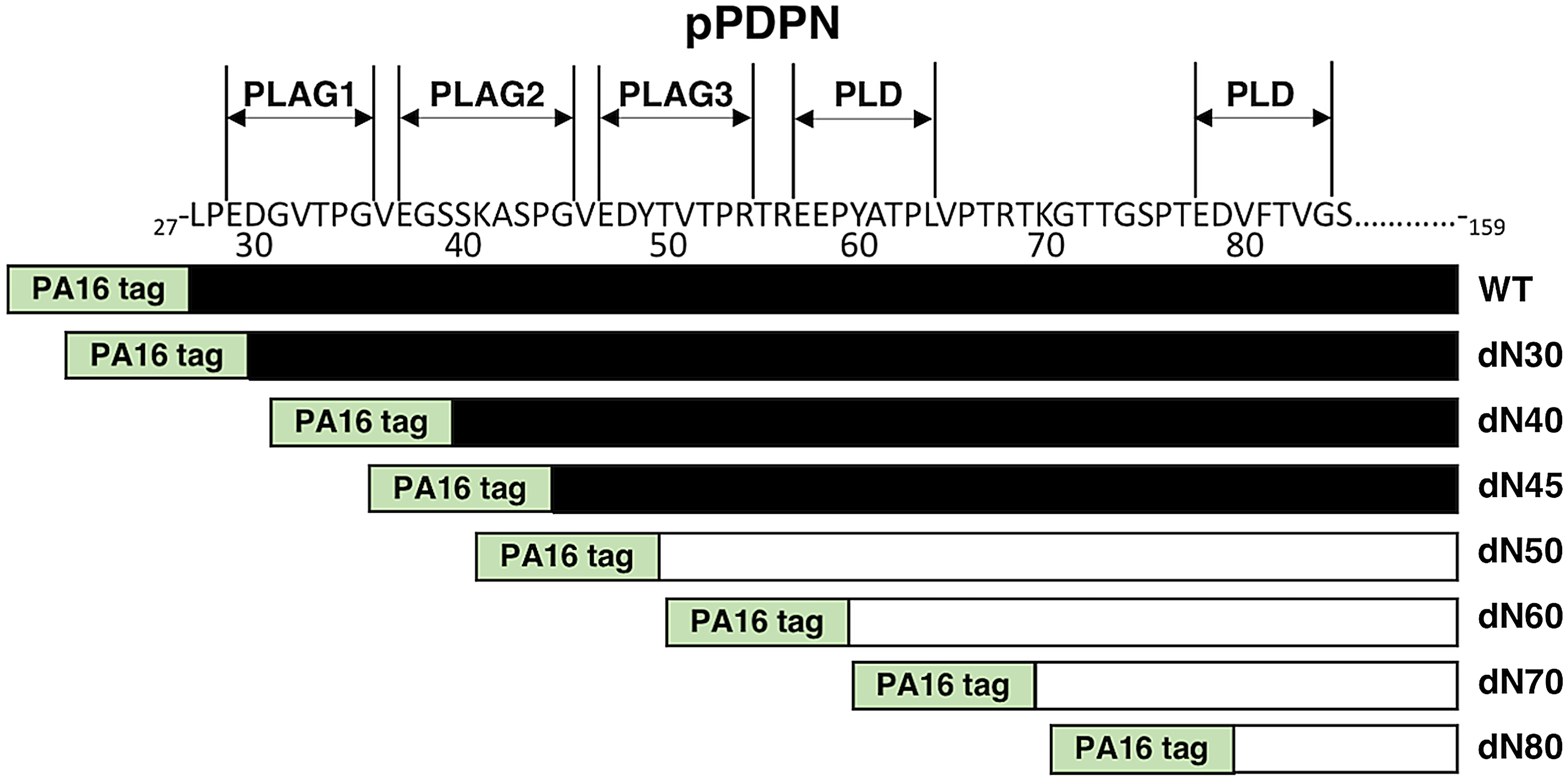
Illustration of WT and seven deletion mutants of pPDPN. Deletion mutants of pPDPN: dN30, dN40, dN45, dN50, dN60, dN70, and dN80. Black bars: the positive reaction of PMab-210. White bars: the negative reaction of PMab-210. PLAG domain, platelet aggregation-stimulating domain; PLD, PLAG-like domain; pPDPN, pig podoplanin; WT, wild type.

Epitope mapping of PMab-210 using deletion mutants of pPDPN.
To identify the binding epitope of PMab-210, we next produced a series of point mutants of pPDPN in CHO-K1 cells, including G45A, V46A, E47A, D48A, Y49A, T50A, V51A, T52A, P53A, R54A, T55A, and R56A. NZ-1 reacted with all point mutants (Fig. 3A). In contrast, PMab-210 did not react with E47A, Y49A, T50A, and V51A, and weakly recognized D48A (Fig. 3B), indicating E47A, D48A, Y49A, T50A, and V51A of pPDPN could be included in the critical epitope of PMab-210 (Fig. 4).

Epitope mapping of PMab-210 using point mutants of pPDPN.
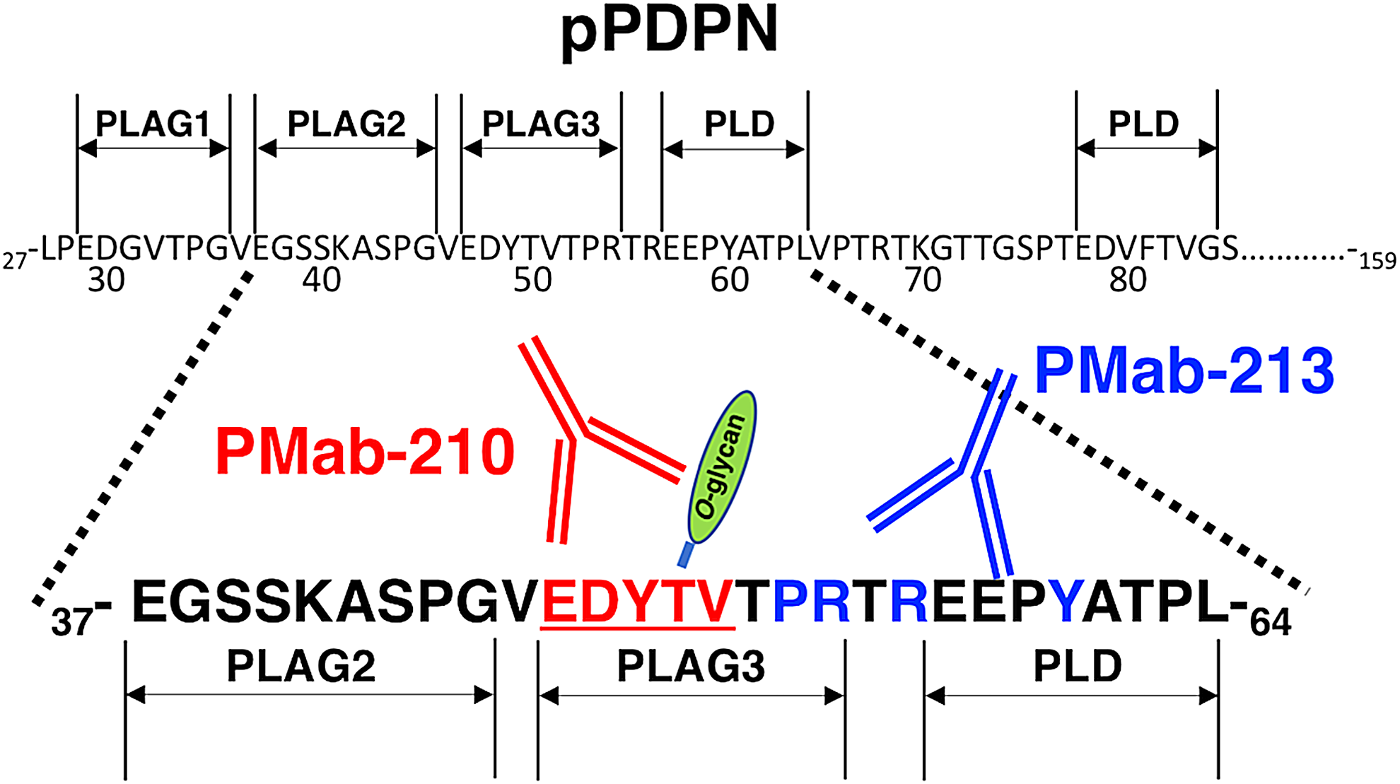
Schematic illustration of the epitope recognized by PMab-210. Underlined and red colored aa is a critical epitope of PMab-210. Blue colored aa is a critical epitope of PMab-213, which is the other anti-pPDPN mAb. aa, amino acid.
Discussion
We have demonstrated that critical epitope of PMab-210 could include Glu47, Asp48, Tyr49, Thr50, and Val51 of pPDPN using deletion mutants and point mutants of pPDPN in CHO-K1 cells. In our previous study, we developed mAb against human,(15) mouse,(16) rat,(17) rabbit,(18) dog,(19) cat,(20) bovine,(21) horse,(22) Tasmanian devil,(23) alpaca,(24) bear,(25) tiger,(26) whale,(27) goat,(28) and pig(29) PDPNs, and determined the binding epitope of those mAbs.(11,28,30–38) Both PMab-210 and PMab-213 for anti-pPDPN mAb could specifically detect pPDPN by flow cytometry and Western blotting, and stained pig tissues by immunohistochemistry. The epitope of PMab-213 (the other anti-pPDPN mAb) was identified as Pro53, Arg54, Arg56, and Tyr60 of pPDPN,(38) indicating that the epitope of both anti-pPDPN mAbs is located in the PLAG3 domain (Fig. 4). Likewise, almost all anti-PDPN mAbs react with PLAG domains or PLDs,(11,12,28,30–32,35–38) suggesting that PLAG domains and PLD were clarified to be advantageous epitopes for several applications, such as flow cytometry, Western blotting, and immunohistochemical analyses.
Using glycan-deficient CHO cell lines, such as Lec1 (N-glycan deficient), Lec2 (sialic acid deficient), or Lec8 (galactose deficient),(39) we investigated whether the epitope of PMab-210 could include not only aas but also O-glycans. Flow cytometric analysis demonstrated that PMab-210 reacts with CHO-K1/pPDPN and Lec1/pPDPN strongly and with Lec2/pPDPN weakly, but not with Lec8/pPDPN (data not shown), indicating that sialic acid and galactose might also be included in the PMab-210 epitope. Among Glu47, Asp48, Tyr49, Thr50, and Val51 of pPDPN, Thr50 could be O-glycosylated (Fig. 4). Because PDPN possesses disialyl-core 1 on many Ser/Thr residues,(40) disialyl-core 1 of Thr50 might be critical for PMab-210 binding to pPDPN.
In conclusion, Glu47, Asp48, Tyr49, Thr50, and Val51 in PLAG3 domain of pPDPN are critical for PMab-210-specific binding to pPDPN. PMab-210 did not react with glycan-deficient CHO cell lines, indicating that PMab-210 was categorized as an antiglycopeptide mAb (GpMab).(41) Several anti-hPDPN mAbs, such as LpMab-2,(42) LpMab-3,(43) LpMab-9,(44) LpMab-12,(45) LpMab-19,(46) and LpMab-21,(41) were also determined to be GpMabs. PMab-210 could be a useful tool for elucidating the pathophysiological function of pPDPN.
Footnotes
Author Disclosure Statement
No competing financial interests exist.
Funding Information
This research was supported, in part, by AMED under Grant Nos. JP19am0101078 (Y.K.), JP19am0401013 (Y.K.), JP19ae0101028 (Y.K.), and by JSPS KAKENHI Grant No. 17K07299 (M.K.K.) and Grant No. 19K07705 (Y.K.).