




Abstract
Trastuzumab is a humanized antibody against human epidermal growth factor receptor 2 (HER2) that offers significant survival benefits to patients with HER2-overexpressing breast or gastric cancer. HER2 is also known to be overexpressed in colon cancers. In this study, a novel anti-HER2 monoclonal antibody (mAb), H2Mab-19 (IgG2b, κ) was characterized for its anticancer activity in colon cancers. H2Mab-19 showed both antibody-dependent cellular cytotoxicity and complement-dependent cytotoxicity activities against Caco-2, a colon cancer cell line. Furthermore, H2Mab-19 significantly reduced tumor development in a Caco-2 xenograft model. These results suggest that treatment with H2Mab-19 may be a useful therapy for patients with HER2-expressing colon cancers.
Introduction
Humanized antibody against human epidermal growth factor receptor 2 (HER2) monoclonal antibodies (mAbs) (e.g., trastuzumab and pertuzumab) have been used for the treatment of HER2-positive breast cancer.(1–3) Treatment with trastuzumab results in significant survival benefits for these patients.(4) In comparison with trastuzumab monotherapy, the combination of trastuzumab plus pertuzumab and chemotherapy has led to significant improvements in overall survival.(5) Trastuzumab deruxtecan (DS-8201), which is a mAb that combines a novel enzyme-cleavable linker and a topoisomerase I inhibitor, was recently developed.(6) Importantly, DS-8201 shows antitumor activity in low-HER2-expressing tumors, DS-8201 has several innovative features: (i) a highly potent novel payload with a high drug-to-antibody ratio, (ii) good homogeneity, (iii) a tumor-selective cleavable linker, (iv) a stable linker-payload in circulation, (v) a cytotoxic agent with a short in vivo half-life, and (vi) bystander effect.(7)
We recently developed a novel anti-HER2 mAb (H2Mab-19; IgG2b, κ) by immunizing BALB/c mice with purified recombinant protein corresponding to the extracellular domain of HER2.(8) H2Mab-19 demonstrated both antibody-dependent cellular cytotoxicity (ADCC) and complement-dependent cytotoxicity (CDC) against a human breast cancer cell line (BT-474) as well as two human oral cancer cell lines (HSC-2 and SAS). Furthermore, H2Mab-19 significantly reduced tumor development in BT-474, HSC-2, and SAS xenografts. These results suggest that treatment with H2Mab-19 may be a useful therapy for patients with HER2-expressing breast and oral cancers. HER2 has also been reported to be expressed in colorectal cancers.(9) Based on the antitumor activities of H2Mab-19 in breast and oral cancer cell lines, we sought to investigate whether H2Mab-19 displayed similar ADCC and CDC activities in vitro in a human colon cancer cell line, and to examine its antitumor activity in vivo in a mouse xenograft model of colon cancer.
Materials and Methods
Cell lines
The LS 174T, Caco-2, HCT-116, HT-29, COLO 201, HCT-8, and SW1116 cell lines were obtained from the American Type Culture Collection (Manassas, VA), and the HCT-15, DLD-1, and COLO 205 cell lines were obtained from the Cell Resource Center for Biomedical Research Institute of Development, Aging and Cancer Tohoku University (Miyagi, Japan). COLO 201, SW1116, HCT-15, DLD-1, and COLO 205 were cultured in RPMI 1640 medium (Nacalai Tesque, Inc., Kyoto, Japan), and LS 174T, Caco-2, HCT-116, HT-29, and HCT-8 were cultured in Dulbecco's modified Eagle's medium (DMEM) medium (Nacalai Tesque, Inc.), supplemented with 10% heat-inactivated fetal bovine serum (FBS; Thermo Fisher Scientific, Inc., Waltham, MA, USA), 100 U/mL of penicillin, 100 μg/mL of streptomycin, and 25 μg/mL of amphotericin B (Nacalai Tesque, Inc.). All cell lines were incubated at 37°C in a humidified atmosphere containing 5% CO2 and 95% air.
Animals
All animal experiments were performed in accordance with relevant guidelines and regulations to minimize animal suffering and distress in the laboratory. Animal studies for ADCC were approved by the institutional committee for experiments of the Institute of Microbial Chemistry (Permit No.: 2019-066). Animal studies for antitumor activity were approved by the institutional committee for experiments of the Institute of Microbial Chemistry (Permit No.: 2019-004). Mice were monitored for health and weight every 2–5 days. Experiment duration was 3 weeks. A bodyweight loss >25% and a maximum tumor size >3000 mm3 were identified as humane endpoints. Mice were euthanized by cervical dislocation, and the death was verified by respiratory arrest and cardiac arrest.
Flow cytometry
Cells were harvested by brief exposure to 0.25% trypsin/1 mM ethylenediaminetetraacetic acid (EDTA; Nacalai Tesque, Inc.). After washing with 0.1% bovine serum albumin in phosphate-buffered saline (PBS), cells were treated with 1 μg/mL anti-HER2 mAb (H2Mab-19) for 30 minutes at 4°C, followed by incubation with Alexa Fluor 488-conjugated anti-mouse IgG (1:1000; Cell Signaling Technology, Inc., Danvers, MA, USA). Fluorescence data were collected using an EC800 Cell Analyzer (Sony Corp., Tokyo, Japan).
Determination of the binding affinity
Cells were suspended in 100 μL of serially diluted H2Mab-19 (6 ng/mL to 100 μg/mL), followed by the addition of Alexa Fluor 488-conjugated anti-mouse IgG (1:200; Cell Signaling Technology, Inc.). Fluorescence data were collected using an EC800 Cell Analyzer (Sony Corp.). The dissociation constant (KD) was obtained by fitting binding isotherms to built-in one-site binding models in GraphPad PRISM 6 (GraphPad Software, Inc., La Jolla, CA, USA).
Antibody-dependent cellular cytotoxicity
Six 6-week-old female BALB/c nude mice were purchased from Charles River (Kanagawa, Japan). After euthanization by cervical dislocation, their spleens were removed aseptically and single-cell suspensions were obtained by forcing spleen tissues through a sterile cell strainer (352360, BD Falcon; Corning, New York, NY, USA) using a syringe. Erythrocytes were lysed with a 10-second exposure to ice-cold distilled water. Splenocytes were washed with DMEM and resuspended in DMEM with 10% FBS and used as effector cells. Target cells were labeled with 10 μg/mL Calcein AM (Thermo Fisher Scientific, Inc.) and resuspended in the same medium. The target cells were plated in 96-well plates (2 × 10⁴ cells/well) and mixed with effector cells, 100 μg/mL of anti-HER2 antibodies, or control IgG (mouse IgG2b) (Sigma-Aldrich Corp., St. Louis, MO, USA). After a 4-hour incubation, the Calcein AM release of supernatant from each well was measured. Fluorescence intensity was determined using a microplate reader (Power Scan HT; BioTek Instruments, Winooski, VT, USA) with an excitation wavelength of 485 nm and an emission wavelength of 538 nm. Cytolytic activity (% of lysis) was calculated as % lysis = (E − S)/(M − S) × 100, where E is fluorescence of combined target and effector cells, S is spontaneous fluorescence of target cells only, and M is maximum fluorescence measured after lysing all cells with a buffer containing 0.5% Triton X-100, 10 mM Tris-HCl (pH 7.4), and 10 mM of EDTA.
Complement-dependent cytotoxicity
Cells were plated in 96-well plates (2 × 10⁴ cells/well), in DMEM supplemented with 10% FBS, and incubated for 5 hours at 37°C with 100 μg/mL of anti-HER2 antibodies or control IgG (mouse IgG2b) (Sigma-Aldrich Corp.) and 10% of rabbit complement (Low-Tox-M Rabbit Complement; Cedarlane Laboratories, Hornby, Ontario, Canada). To assess cell viability, an MTS [3-(4,5-dimethylthiazol-2-yl)-5-(3-carboxymethoxyphenyl)-2-(4-sulfophenyl)-2H-tetrazolium; inner salt] assay was performed using a CellTiter 96 AQueous assay kit (Promega, Madison, WI, USA).
Antitumor activity of H2Mab-19 in the xenografts of colon cancers
Sixteen 6-week-old female BALB/c nude mice were purchased from Charles River and used at 7 weeks of age. Caco-2 cells (0.3 mL of 1.33 × 10⁸ cells/mL in DMEM) were mixed with 0.5 mL BD Matrigel Matrix Growth Factor Reduced (BD Biosciences, San Jose, CA, USA). Mice were injected subcutaneously into the left flank (100 μL) with the aforementioned suspension (5 × 10⁶ cells). After day 1, 100 μg H2Mab-19 or control mouse IgG (Sigma-Aldrich Corp.) in 100 μL PBS were injected into treated and control mice through intraperitoneal injection (i.p.), respectively. Additional antibodies were then injected on days 7 and 14. Eighteen days after cell implantation, all mice were euthanized by cervical dislocation, and tumor diameters and volumes were determined as previously described.(10)
Statistical analyses
All data were expressed as mean ± standard error of the mean. Statistical analysis used ANOVA and Tukey–Kramer's test with R statistical p < 0.05 was adopted as a level of statistical significance.
Results
Characterization of H2Mab-19 against colon cancer cell lines
H2Mab-19 recognized endogenous HER2 in the 10 colon cancer cell lines tested, HCT-15, LS 174T, DLD-1, Caco-2, HCT-116, HT-29, COLO 201, COLO 205, HCT-8, and SW1116, which are HER2-positive(11) (Fig. 1A). Using flow cytometry, the binding affinity (KD) of H2Mab-19 to the Caco-2 cell line was 1.1 × 10⁻⁸ M, indicating that H2Mab-19 shows a high affinity to this HER2-expressing colon cancer cell line.

Characterization of H2Mab-19 using flow cytometry.
H2Mab-19-induced ADCC and CDC activities in a colon cancer cell line
This study examined whether H2Mab-19 induced ADCC and CDC in the HER2-expressing colon cancer cell line, Caco-2. H2Mab-19 is a mouse IgG2b subclass antibody, which possesses both ADCC and CDC activities.(8) As expected, H2Mab-19 exhibited high ADCC activity (32.6% of cytotoxicity; Fig. 2A) and high CDC activity (50.7% of cytotoxicity; Fig. 2B) against Caco-2 cells, suggesting that H2Mab-19 might exert antitumor activity in an in vivo model of colon cancer.
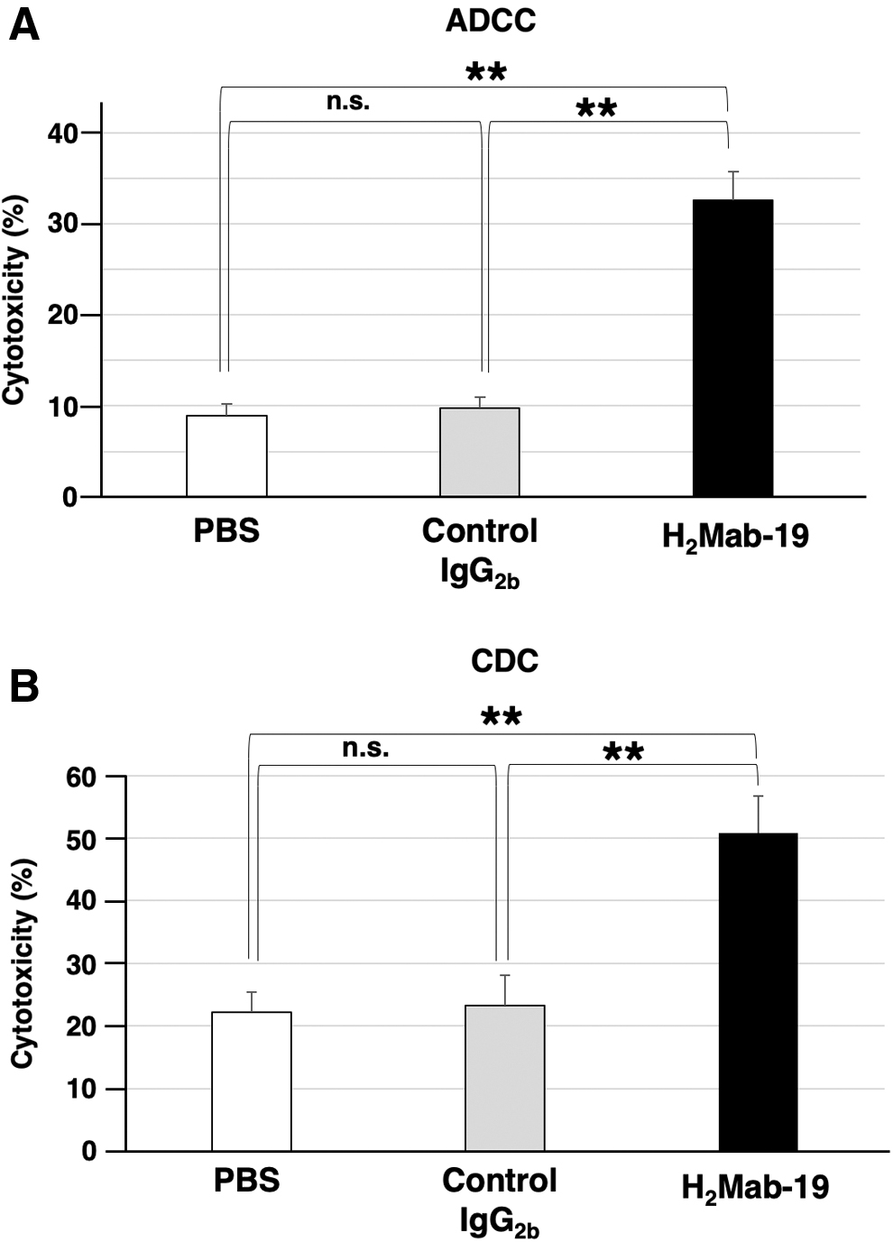
ADCC and CDC activities.
Antitumor activity of H2Mab-19 in mouse xenografts of colon cancer
To study the antitumor activity of H2Mab-19 on cell growth in vivo, Caco-2 cells were implanted subcutaneously in the flanks of nude mice. H2Mab-19 or control mouse IgG was injected i.p. three times on days 1, 7, and 14 after Caco-2 cell injection into treated and control mice, respectively. Tumor formation was observed in both H2Mab-19-treated and control groups. H2Mab-19 treatment significantly reduced tumor development compared with control mice on days 5, 7, 12, 15, and 18 of observation (Fig. 3A). Resected tumors are shown in Figure 3B. The weight of the tumors excised from H2Mab-19-treated mice was significantly less than for tumors from IgG-treated control mice (Fig. 3C). Total body weight was not significantly different between the two groups (Fig. 4).
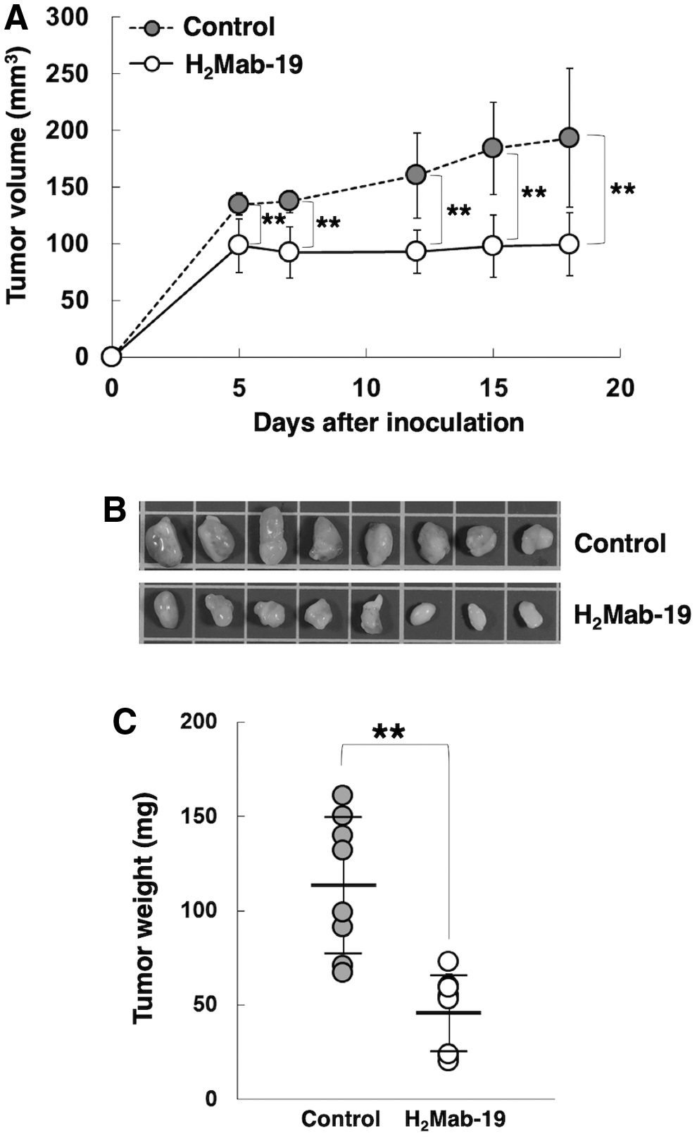
Evaluation of antitumor activity of H2Mab-19.
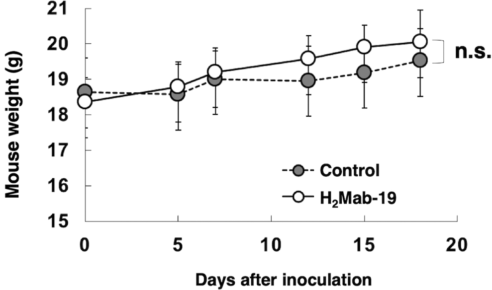
Body weights of the mice with the Caco-2 xenografts. Body weights of the mice with the Caco-2 xenografts were measured for 18 days. n.s., not significant.
Discussion
We previously developed several anti-HER2 mAbs, including H2Mab-77,(12) H2Mab-119,(13) and H2Mab-139(11) using CasMab technology.(14) For this technology, it is critical that immunogens are produced using cancer cell lines, such as LN229 glioblastoma cells, which express cancer-specific glycan-attached membrane proteins. These antibodies are useful for flow cytometry, western blot, and immunohistochemical analyses. Unfortunately, the subclass of these mAbs was determined to be mouse IgG1; therefore, they do not possess ADCC or CDC activities. Consequently, we further tried to develop an anti-HER2 mAb of either the IgG2a or IgG2b subclass using CasMab technology because both IgG2a(15) and IgG2b antibodies(16) show ADCC and CDC activity. We successfully established a novel anti-HER2 mAb (H2Mab-19) of subclass IgG2b.(8) H2Mab-19 possesses both ADCC and CDC activity in both human breast cancer and oral cancer cell lines. Furthermore, H2Mab-19 exerted antitumor activity in human breast cancer and oral cancer xenografts. HER2 has been reported to be expressed in a number of colorectal cancers.(9) Based on this data, we sought to investigate whether H2Mab-19 has ADCC and CDC activities in vitro in a human colon cancer cell line as well as determine its antitumor activity in a mouse xenograft model of colon cancer.
Using flow cytometric analysis, the binding affinity (KD) of H2Mab-19 to Caco-2 was determined to be 1.1 × 10⁻⁸ M. These results were similar to the KD of H2Mab-19 observed in BT-474 (2.3 × 10⁻⁸ M), HSC-2 (9.5 × 10⁻⁹ M), and SAS (5.5 × 10⁻⁹ M) cell lines.(8) In addition, H2Mab-19 exhibited high ADCC activity (32.6% of cytotoxicity; Fig. 2A) and high CDC activity (50.7% of cytotoxicity; Fig. 2B) against Caco-2 cells, which are compatible with the previous data obtained of ADCC and CDC activities of H2Mab-19 against human breast cancer and oral cancer cell lines: ADCC activities of H2Mab-19 against BT-474, HSC-2, and SAS were 31.5%, 26.2%, and 38.1%, respectively; CDC activities of H2Mab-19 against BT-474, HSC-2, and SAS were 45.3%, 50.2%, and 52.3%, respectively,(8) indicating that H2Mab-19 might exert high cytotoxicity against a colon cancer xenograft model.
In this study, we selected the Caco-2 cell line from 10 HER2-expressing colon cancer cell lines, which were detected by the H2Mab-19 in flow cytometry (Fig. 1A), for the colon cancer xenograft model because we previously established Caco-2 xenograft models.(17) In our previous study, H2Mab-19 treatment significantly reduced tumor development in both breast and oral cancer xenografts compared with tumor development in control mice.(8) Similar to our previous study, we found that H2Mab-19 demonstrated antitumor activity against Caco-2 xenografts (Fig. 3). Further studies using the other colon cancer xenografts should be performed in the future to confirm that H2Mab-19 could be a useful therapy for patients with HER2-expressing colon cancers.
Footnotes
Acknowledgments
We thank Ms. Akiko Harakawa for technical assistance of animal experiments.
Author Disclosure Statement
No competing financial interests exist.
Funding Information
This research was supported in part by AMED under Grant Numbers JP20am0401013 (Y.K.), JP20am0101078 (Y.K.), and JP20ae0101028 (Y.K.), and by JSPS KAKENHI Grant Numbers 17K07299 (M.K.K.) and 19K07705 (Y.K.).