
Abstract
An antisheep podoplanin (sPDPN) monoclonal antibody (mAb), PMab-256, has recently been established. PMab-256 shows positive immunostaining for lymphatic endothelial cells, lung type I alveolar cells, and kidney podocytes. PDPN possesses three platelet-aggregation-stimulating (PLAG) domains, PLAG1, PLAG2, and PLAG3, and a PLAG-like domain (PLD). The binding epitope of many anti-PDPN mAbs is located in PLAG domains or PLD. The purpose of this study is to determine the binding epitope of PMab-256. Analysis of sPDPN deletion mutants revealed that the N-terminus of the PMab-256 epitope exists between amino acids 75 and 80 of sPDPN. Furthermore, analysis of sPDPN point mutations demonstrated that the critical epitope includes Thr80 of sPDPN, indicating that the PMab-256 epitope is in the PLD of sPDPN.
Introduction
Sensitive and specific monoclonal antibodies (mAbs) against podoplanin (PDPN) of various species including human,(1) mouse,(2) and whale(3) are necessary for the analysis of expression and pathophysiological functions of PDPN. PDPN is used for discriminating lymphatic endothelial cells (LECs) from vascular endothelial cells(4) or type I alveolar cells from type II alveolar cells.(5) An antisheep podoplanin (sPDPN) mAb, PMab-256, was obtained using the Cell-Based Immunization and Screening (CBIS) method.(6–8) Like other typical anti-PDPN mAbs, PMab-256 demonstrated positive immunoreaction for LECs,(4) type I alveolar cells,(5) and kidney podocytes.(9)
The binding epitope of many anti-PDPN mAbs is located in three platelet aggregation-stimulating (PLAG) domains, PLAG1, PLAG2, and PLAG3,(10) at the N-terminus or a PLAG-like domain (PLDs)(3,11–14) in the middle of the PDPN protein. The purpose of this study was to determine the binding epitope of PMab-256.
Materials and Methods
Cell lines
Chinese hamster ovary (CHO)-K1 cells were obtained from the American Type Culture Collection (Manassas, VA). sPDPN mutation plasmids containing a RAP14 tag(15) were transfected into CHO-K1 cells using Lipofectamine LTX (Thermo Fisher Scientific, Inc., Waltham, MA). The RAP14 tag comprises 14 amino acids (aa), (DMVNPGLEDRIEDL), and is recognized by PMab-2 mAb(16) and LpMab-7.(1) Cells transiently transfected with deletion or point mutations were cultured in Roswell Park Memorial Institute (RPMI) 1640 medium (Nacalai Tesque, Inc., Kyoto, Japan), supplemented with 10% heat-inactivated fetal bovine serum (FBS; Thermo Fisher Scientific, Inc.), 100 U/mL of penicillin, 100 μg/mL of streptomycin, and 0.25 μg/mL of amphotericin B (Nacalai Tesque, Inc.) at 37°C in a humidified atmosphere of 5% CO2 and 95% air.
Production of sPDPN mutants
Synthesized DNA (Eurofins Genomics KK, Tokyo, Japan) encoding sPDPN (accession no.: XM_004013802.4) was subcloned into the pCAG vector (FUJIFILM Wako Pure Chemical Corporation, Osaka, Japan), and an RAP14 tag was added at the N-terminus. Deletion mutants of the sPDPN sequence were produced using a HotStar HiFidelity Polymerase Kit (Qiagen, Inc., Hilden, Germany) with oligonucleotides. Substitutions of aa with alanine in the sPDPN sequence were produced by QuikChange Lightning Site-Directed Mutagenesis Kits (Agilent Technologies, Inc., Santa Clara, CA). Polymerase chain reaction fragments bearing desired mutations were inserted into the pCAG vector using the In-Fusion HD Cloning Kit (Takara Bio, Inc., Shiga, Japan).
Flow cytometry
Transiently transfected CHO-K1 cells were detached by 0.25% trypsin/1 mM ethylenediaminetetraacetic acid (EDTA; Nacalai Tesque, Inc.) and collected using 10% FBS in RPMI 1640 medium. After washing with 0.1% bovine serum albumin in phosphate-buffered saline, cells were incubated with anti-sPDPN antibody (PMab-256; 1 μg/mL) and an anti-RAP14 tag antibody (PMab-2; 1 μg/mL or LpMab-7; 1 μg/mL) for 30 minutes at 4°C. Alexa Fluor 488-conjugated antimouse IgG (1:2000; Cell Signaling Technology, Inc., Danvers, MA) was added to each cell suspension and incubation continued for 30 minutes at 4°C. Fluorescence data were collected and analyzed using a Cell Analyzer EC800 (Sony Corp., Tokyo, Japan).
Results
We produced seven deletion mutants of sPDPN in CHO-K1 cells, namely, dN30, with aa 30–168 deletion; dN40, aa 40–168; dN50, aa 50–168; dN60, aa 60–168; dN70, aa 70–168; dN75, aa 75–168; dN80, aa 80–168; or wild type (WT) sPDPN, aa 27–168. All deletion mutants and WT containing the N-terminal RAP14 tag were recognized by PMab-2 or LpMab-7 (anti-RAP14 tag mAbs), indicating that the expression level of each construct is high (Fig. 1A). PMab-256 recognized dN30, dN40, dN50, dN60, dN70, and dN75, but not dN80 (Fig. 1B), suggesting that the N-terminus of the PMab-256 epitope exists between sPDPN aa 75 and 80.

Epitope mapping of PMab-256 using deletion mutations.
Next, a series of point mutants of sPDPN, G75A, E76A, D77A, L78A, P79A, T80A, A81G, E82A, S83A, T84A, T85A, and A86G, were obtained. PMab-2 and LpMab-7 reacted with all point mutants (Fig. 2A). By contrast, PMab-256 did not react with T80A (Fig. 2B). Thr80 of sPDPN is essential for PMab-256 binding (Fig. 3).
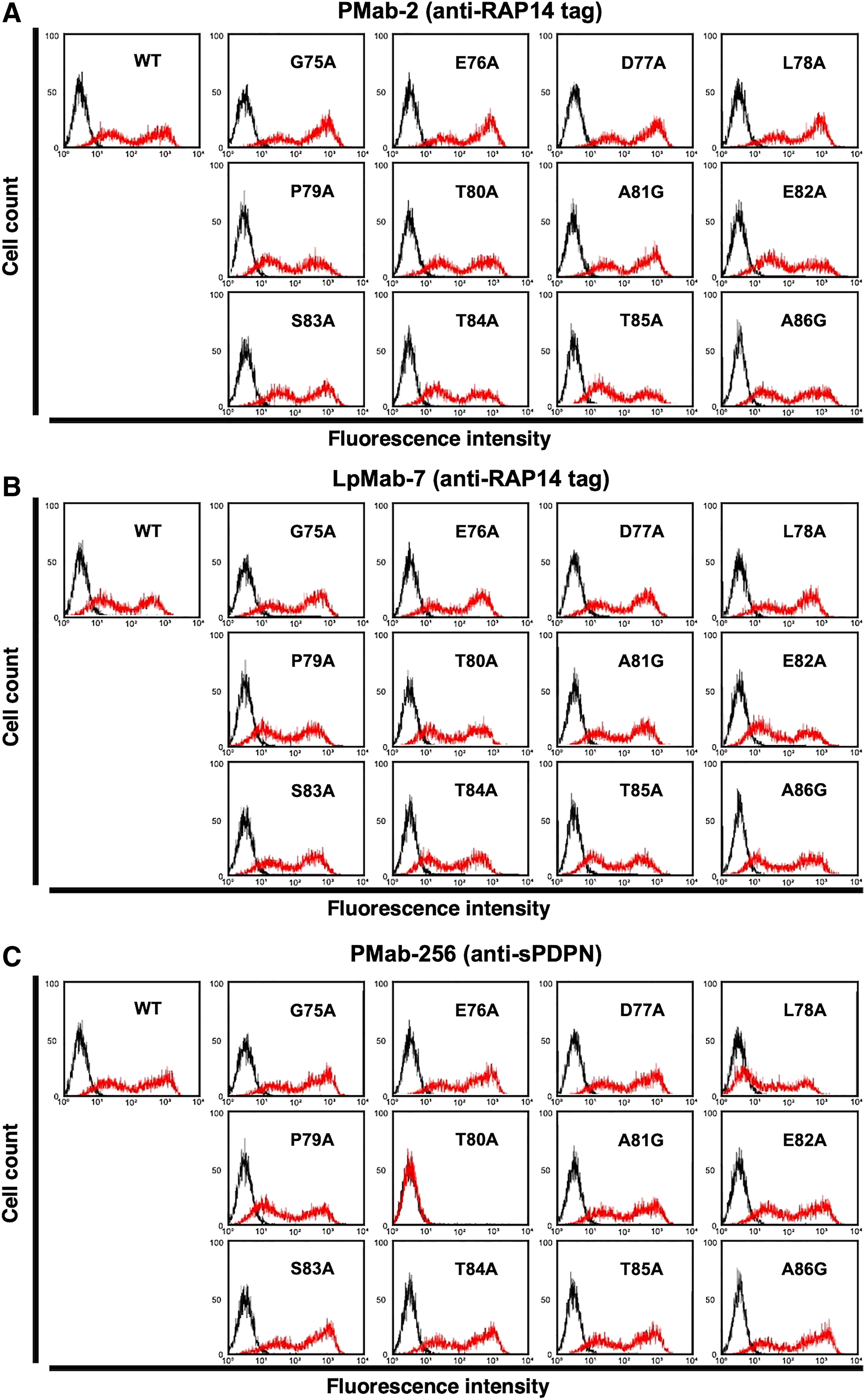
Epitope mapping of PMab-256 using point mutations.
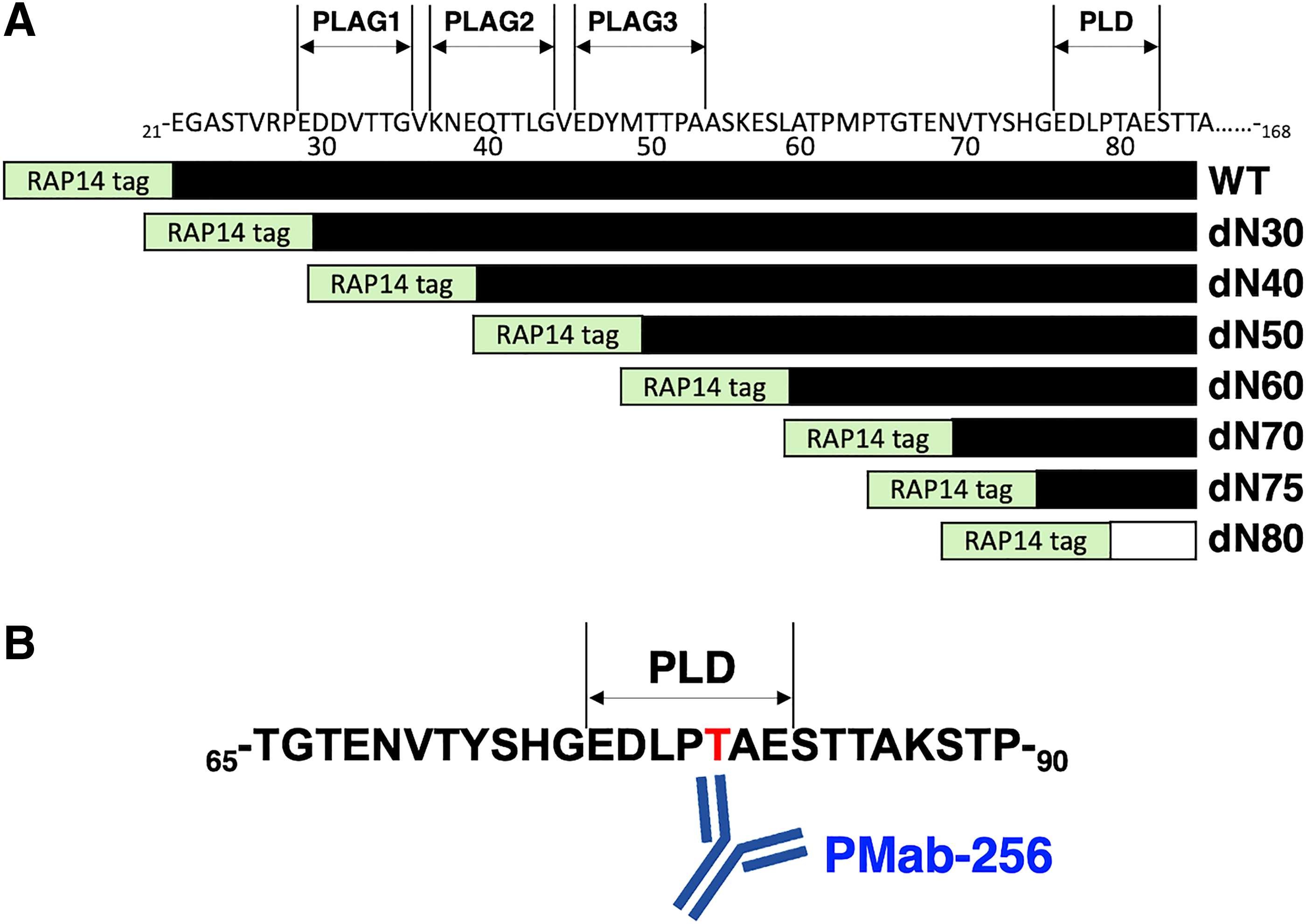
Schematic illustration of the epitope recognized by PMab-256.
Discussion
A variety of mAbs against pig,(17,18) horse,(19,20) Tasmanian devil,(21) alpaca,(22) tiger,(23) whale,(24) goat,(25,26) bear,(27,28) and sheep(29) PDPNs using the CBIS method are available.(6–8) An anti-sPDPN mAb PMab-256 is useful for immunohistochemical analyses using formalin-fixed paraffin-embedded tissues of sheep for detecting LECs of many organs, type I alveolar cells, and renal epithelial cells.(29) PMab-256 is also useful for Western blot analysis, indicating that PMab-256 recognizes sPDPN that is fixed in formalin and denatured by sodium dodecyl sulfate. Furthermore, PMab-256 detects sPDPN in flow cytometry.
PLAG and PLD domains bind C-type lectin-like receptor-2 and induce platelet aggregation and cancer metastasis.(30) Anti-PDPN mAbs, epitopes of which are located in PLAG domains or PLD, could neutralize platelet aggregation.(30) The epitope of PMab-256 is also located in PLD of sPDPN. PMab-256 is thus not only a useful mAb for research but also could be a functional mAb with the ability to neutralize induction of platelet aggregation.
Footnotes
Author Disclosure Statement
No competing financial interests exist.
Funding Information
This research was supported, in part, by AMED under Grant Nos. JP20am0401013 (Y.K.), JP20am0101078 (Y.K.), and JP20ae0101028 (Y.K.), and by JSPS KAKENHI Grant No. 17K07299 (M.K.K.) and Grant No. 19K07705 (Y.K.).