




Abstract
Overexpression of human epidermal growth factor receptor 2 (HER2) has been reported in a variety of cancer types, including breast, lung, gastric, pancreatic, and colorectal cancers. Trastuzumab, a humanized anti-HER2 monoclonal antibody (mAb), has been shown to provide significant survival benefits in HER2-overexpressing breast cancer and gastric cancer patients. Previously, an anti-HER2 mAb, H2Mab-41 (IgG2b, kappa), was developed in our laboratory and its antitumor activity was demonstrated in mouse xenograft models of human colon cancer. The present study aimed to investigate the ability of H2Mab-41 to induce antibody-dependent cellular cytotoxicity (ADCC) and complement-dependent cytotoxicity (CDC) in dog HER2 (dHER2)-overexpressed cell lines, and thus exert its antitumor activity against dHER2-overexpressed tumors in vivo. Flow cytometry results demonstrated the cross-reactivity of H2Mab-41 with dHER2. Further evaluation of interaction between H2Mab-41 and dHER2-overexpressed CHO-K1 (CHO/dHER2) cells indicated moderate binding affinity of H2Mab-41 toward dHER2, with a dissociation constant (KD) of 2.6 × 10⁻⁸ M. In vitro analysis revealed that the administration of H2Mab-41 induced high levels of ADCC and CDC in CHO/dHER2 cells. Furthermore, intraperitoneal administration of H2Mab-41 in mouse xenograft models of CHO/dHER2 resulted in significant inhibition of tumor development compared to the control mouse IgG. Thus, the findings of the present study demonstrated the in vivo safety and efficacy of H2Mab-41, highlighting its suitability to be included as a part of a therapeutic regimen for dHER2-expressing canine cancers.
Introduction
Human epidermal growth factor receptor 2 (HER2) is a type I transmembrane glycoprotein that is overexpressed in several cancer types, especially breast cancer.(1) HER2 belongs to epidermal growth factor receptor (EGFR) family, which comprises of four main members, namely EGFR (HER1/ErbB1), HER2 (ErbB2/neu), HER3 (ErbB3), and HER4 (ErbB4).(2) The EGFR family members transduce extracellular signals into intracellular signaling pathways via activation of tyrosine kinase domain. These receptor tyrosine kinases play a critical role in the regulation of cell proliferation, differentiation, survival, and migration.(3) The binding of ligand to the extracellular domain of the receptors promotes the formation of homo- or heterodimers between the EGFR family receptors.(4) Interestingly, HER2 is devoid of any ligand-binding extracellular domain, and it is represented as an activated structure in the absence of ligand binding.(5–7) Generally, HER2 is expressed in a variety of cell types, excluding the cells of hematopoietic origin.(8) The overexpression of HER2 has been reported in a variety of cancer types, including breast,(9,10) gastric,(11) pancreatic,(12) lung,(13) and colorectal cancers.(14) In HER2-positive breast cancer patients, the overexpression of this oncogene was found to be associated with poor prognosis and aggressive tumor phenotype.(9,10) Trastuzumab and pertuzumab are humanized anti-HER2 monoclonal antibodies (mAbs) that are widely used in the treatment of HER2-positive breast cancer.(15–17)
Cancer is the leading cause of death in dogs. Annual incidence rate of cancer in dogs is estimated to be ∼30%, which is similar to humans.(18,19) In fact, overexpression of HER2 has also been reported in a variety of canine tumors, including primary lung cancer,(20) osteosarcoma,(21) mammary carcinoma,(22) bladder transitional cell carcinoma,(23) and anal sac gland carcinoma.(24) The cancer therapy for veterinary oncology treatment coevolved with human cancer treatment. In the past few decades, various HER2-targeting therapeutic agents, such as Lapatinib and recombinant Listeria vaccines, have been developed and utilized for cancer treatment. Lapatinib is a low-molecular-weight tyrosine kinase inhibitor that targets EGFR and HER2. These HER2-targeted therapies have been shown to exert an antitumor effect on canine HER2 (dHER2)-positive cancers.(25,26) Thus, HER2 could be used as a potential therapeutic target and diagnostic marker for canine cancers. Previously, H2Mab-41, an anti-HER2 mAb developed in our laboratory was found to exert antitumor activity in a mouse xenograft model for human colon cancer.(27) The present study aimed to investigate the ability of H2Mab-41 to induce antibody-dependent cellular cytotoxicity (ADCC), complement-dependent cytotoxicity (CDC), and antitumor efficacy in dHER2-overexpressed cells.
Materials and Methods
Antibodies
Anti-HER2 mAb H2Mab-41 (mouse IgG2b, kappa) was developed according to the previously established procedure.(27) The resulting antibody was purified using Protein G-Sepharose (GE Healthcare Bio-Sciences, Pittsburgh, PA). Mouse IgG (Cat. No. I8765) and IgG2b (Cat. No. M1395) were purchased from Sigma-Aldrich (St. Louis, MO).
Cell lines
CHO-K1 cells were obtained from the American Type Culture Collection (Manassas, VA). Dog HER2 (dHER2; Accession No. NM_001003217)-overexpressed CHO-K1 cell line (CHO/dHER2) was established via transfection of pCAG/3xRIEDL-dHER2 into CHO-K1 cells using Neon transfection system (Thermo Fisher Scientific, Inc., Waltham, MA). Here, 3xRIEDL sequence represented 15 amino acid sequence (RIEDLRIEDLRIEDL).(28) RIEDL tag is a novel affinity tag that is used for the detection and one-step purification of membrane proteins. For cell culture, CHO-K1 and CHO/dHER2 were cultured in Roswell Park Memorial Institute (RPMI)-1640 medium (Nacalai Tesque, Inc., Kyoto, Japan), supplemented with 10% heat-inactivated fetal bovine serum (FBS; Thermo Fisher Scientific, Inc.), 100 U/mL of penicillin, 100 μg/mL streptomycin, and 0.25 μg/mL amphotericin B (Nacalai Tesque, Inc.). The cell lines were maintained at 37°C in a humidified atmosphere under 5% CO2.
Animals
To ensure minimal animal suffering and distress in the laboratory, all animal experiments were performed according to the established guidelines and regulations. Animal experiments for ADCC and antitumor activity were approved by the Institutional Committee for Experiments of the Institute of Microbial Chemistry (Permit No. 2021-019 and 2021-007 for ADCC assays and antitumor experiments, respectively). Mice were maintained under a specific pathogen-free environment, at a temperature of 23°C ± 2°C with 55% ± 5% relative humidity and 11/13 hours light/dark cycle. The animals had free access to food and water during the entire course of the study. Mice were regularly monitored for health and weight every 2 or 5 days during the 3-week period for each experiment. Loss of >25% of original body weight and/or a maximum tumor size >3000 mm3 were identified as humane endpoints for euthanasia. Mice were euthanized by cervical dislocation, and death was verified by respiratory and cardiac arrest.
Flow cytometry
For cell harvesting, CHO-K1 and CHO/dHER2 cells were treated with a solution of 0.25% trypsin/1 mM ethylenediaminetetraacetic acid (EDTA; Nacalai Tesque, Inc.) for a brief duration (1–2 minutes). Posttrypsinization, the cells were washed with blocking buffer, comprising 0.1% bovine serum albumin in phosphate-buffered saline (PBS). Further, the cells were treated with 1 μg/mL of H2Mab-41 or control blocking buffer for 30 minutes at 4°C. This was followed by addition of Alexa Fluor 488-conjugated anti-mouse IgG at a dilution of 1:1000 (Cat. No. 4408S; Cell Signaling Technology, Inc., Danvers, MA, USA). The cells were incubated for 30 minutes at 4°C. Fluorescence data were acquired using EC800 Cell Analyzer (Sony Corp., Tokyo, Japan).
Determination of the binding affinity
To evaluate the binding affinity, CHO/dHER2 cells were first suspended in 100 μL of serially diluted H2Mab-41 (0.006–100 μg/mL). This was followed by the addition of Alexa Fluor 488-conjugated anti-mouse IgG (1:200 dilution; Cell Signaling Technology, Inc.). Fluorescence data were acquired using EC800 Cell Analyzer. The dissociation constant (KD) was calculated by fitting the binding isotherms using the built-in one-site binding models in GraphPad Prism 8 (GraphPad Software, Inc., La Jolla, CA).
Antibody-dependent cellular cytotoxicity
For ADCC assay, 5-week-old female BALB/c (n = 5) nude mice (weighing 14–17 g) were purchased from Charles River Laboratories, Inc., (Kanagawa, Japan). Spleen cells were isolated from these mice and used as the source of mononuclear cells to evaluate ADCC, as previously described.(29) Mice were euthanized by cervical dislocation, and the spleens were removed aseptically. To obtain single-cell suspensions, the spleen tissues were forcefully passed through a sterile cell strainer (352360, BD Falcon; Corning, New York, NY) using a syringe. Erythrocytes were exposed to ice-cold distilled water for 10 seconds to ensure complete lysis. The resulting splenocytes were washed with RPMI-1640 and resuspended in RPMI-1640 supplemented with 10% FBS. This cell preparation was further used as effector cells. CHO/dHER2 cells were labeled with 10 μg/mL Calcein AM (Thermo Fisher Scientific, Inc.) and resuspended in the same medium. CHO/dHER2 cells were seeded in 96-well plates at a density of 2 × 10⁴ cells/well. These cells were mixed with effector cells (effector/target cells ratio, 100) and 100 μg/mL of H2Mab-41 or control mouse IgG2b, and incubated at 37°C for 5 hours. Posttreatment, the release of Calcein into the supernatant was measured in terms of fluorescence intensity in each well, measured using a microplate reader (Power Scan HT; BioTek Instruments, Inc., Winooski, VT) with excitation and emission wavelengths of 485 and 538 nm, respectively. Cytolytic activity (%lysis) was calculated using the equation %lysis = (E − S)/(M − S) × 100, where “E” is the fluorescence intensity for the combined cultures of target and effector cells, “S” represents spontaneous fluorescence of target cells alone, and “M” denotes maximum fluorescence measured in all cells postlysis. The cells were lysed using a buffer comprising of 0.5% Triton X-100, 10 mM Tris-HCl (pH 7.4), and 10 mM of EDTA.
Complement-dependent cytotoxicity
For CDC evaluation, CHO/dHER2 cells were seeded in 96-well plates at a density of 2 × 10⁴ cells/well. These cells were mixed with rabbit complement reagent (final dilution 1:10; Low-Tox-M Rabbit Complement; Cedarlane Laboratories, Hornby, Ontario, Canada) and 100 μg/mL of H2Mab-41 or control mouse IgG2b. The cells were incubated at 37°C for 5 hours, and MTS [3-(4,5-dimethylthiazol-2-yl)-5-(3-carboxymethoxyphenyl)-2-(4-sulfophenyl)-2H-tetrazolium; inner salt] assay was performed using a CellTiter 96 AQueous One Solution Cell Proliferation Assay Kit (Promega, Madison, WI).
Antitumor activity of H2Mab-41 in mice xenografts for CHO/dHER2 cells
For antitumor activity, 16 female BALB/c nude mice, aged 5 weeks and weighing 14–17 g, were purchased from Charles River Laboratories, Inc. The animals were housed in the institutional animal facility and experiments were conducted at the age of 6 weeks. For tumor inoculation, 0.3 mL of CHO/dHER2 cells (1.33 × 10⁸ cells/mL) in RPMI-1640 was mixed with 0.5 mL of BD Matrigel™ Matrix Growth Factor Reduced (BD Biosciences, San Jose, CA), and 100 μL of the resulting suspension (5 × 10⁶ cells) was injected subcutaneously into the left flanks of the mice. On day 6 postinoculation, mice received intraperitoneal injections of 100 μg of H2Mab-41 (n = 8), or control mouse IgG (n = 8) in 100 μL PBS. Additional antibody treatments were performed on days 14 and 21. At day 25 after tumor inoculation, all mice were euthanized by cervical dislocation, and tumor diameters and volumes were determined as described previously.(29)
Statistical analyses
All data are expressed as mean ± standard error of the mean. Statistical analyses for ADCC and CDC experiments were conducted using Welch's t-test. ANOVA and Sidak's multiple comparisons tests were performed for tumor volume and mouse weight. For tumor weight, Welch's t-test was conducted. All calculations were performed using GraphPad Prism 7 (GraphPad Software, Inc.). A p-Value of < 0.05 was considered to be statistically significant.
Results
Characterization of H2Mab-41 for CHO/dHER2
The cross-reactivity of H2Mab-41 for CHO/dHER2 was analyzed using flow cytometry. As shown in Figure 1A, H2Mab-41 successfully detected CHO/dHER2, however, parental CHO-K1 cells remained undetected.
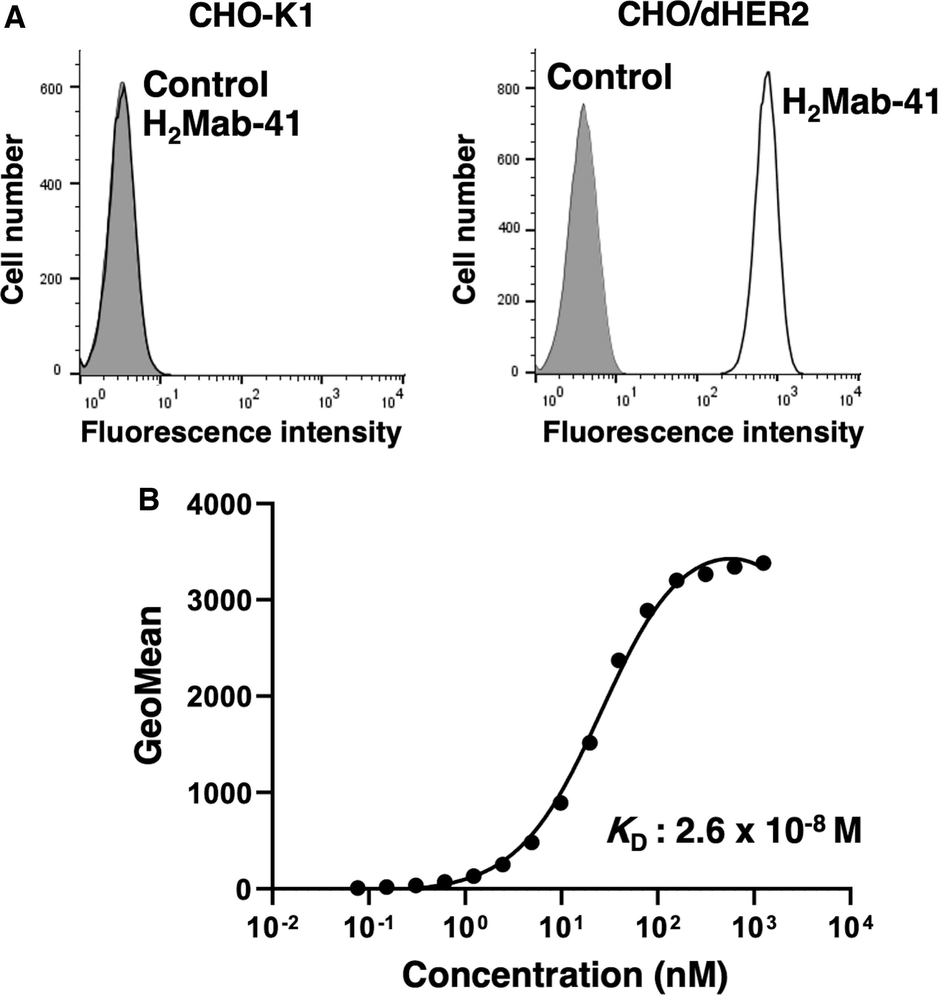
Flow cytometry analysis for interaction between H2Mab-41 and CHO/dHER2 cells.
A kinetic analysis for the interaction of H2Mab-41 with CHO/dHER2 cells was performed using flow cytometry. As shown in Figure 1B, the interaction between H2Mab-41 and CHO/dHER2 cells was characterized by a KD of 2.6 × 10⁻⁸ M. Thus, H2Mab-41 was found to have moderate affinity toward CHO/dHER2 cells.
H2Mab-41-mediated ADCC and CDC in CHO/dHER2 cells
The present study investigated the ability of H2Mab-41 to mediate ADCC in CHO/dHER2 cell line. As shown in Figure 2A, H2Mab-41 was found to exert higher ADCC activity (34% cytotoxicity; p < 0.05) against CHO/dHER2 cells compared to the control mouse IgG2b (7.2% cytotoxicity). Further, the ability of H2Mab-41 to mediate CDC against CHO/dHER2 cells was also evaluated. As shown in Figure 2B, H2Mab-41 induced a higher CDC activity (52% cytotoxicity; p < 0.05) against CHO/dHER2 cells compared to the control mouse IgG2b (18% cytotoxicity). Thus, the treatment of HER2-overexpressed cells with H2Mab-41 resulted in significantly higher levels of ADCC and CDC compared to the control mouse IgG2b.
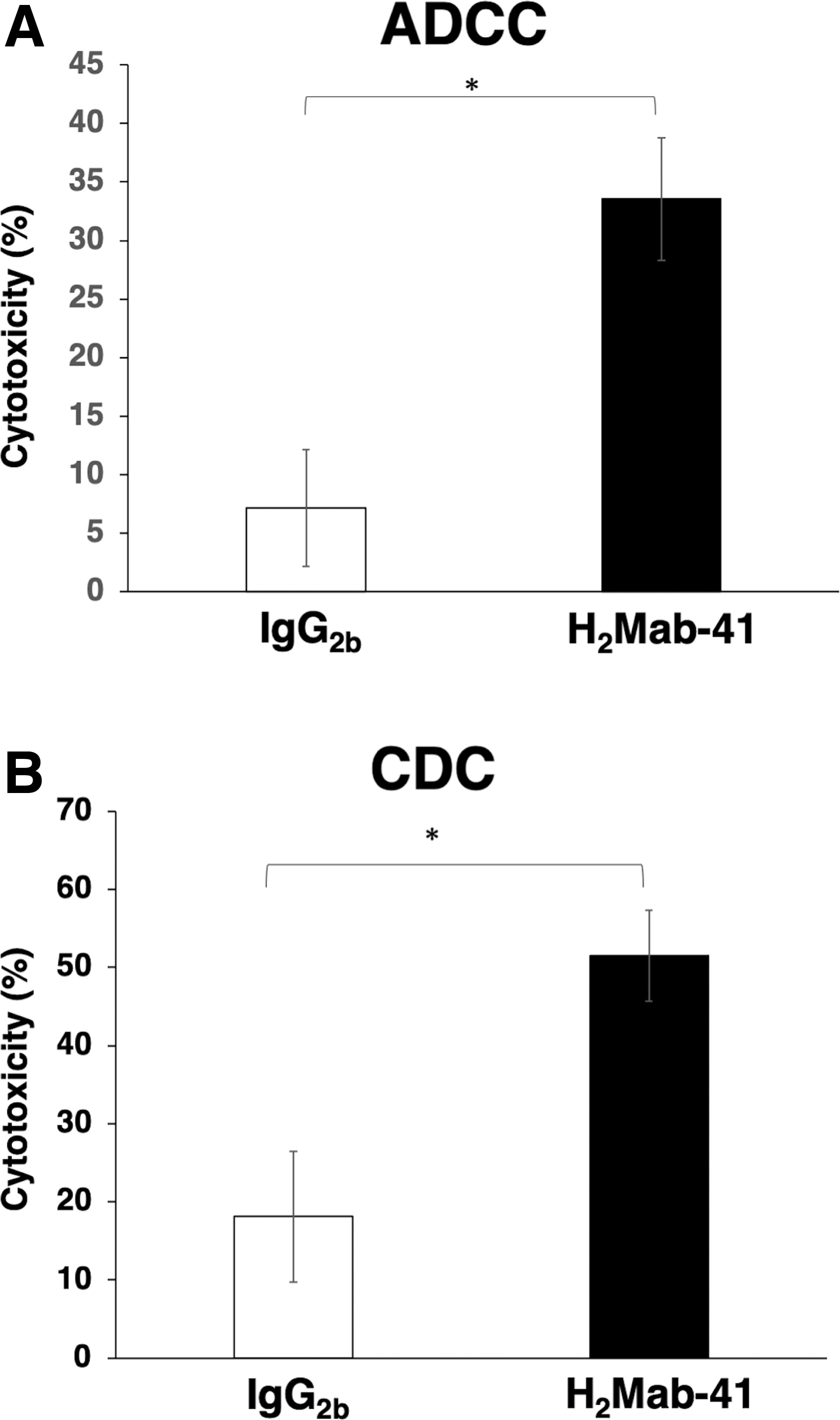
Evaluation of H2Mab-41 mediated ADCC and CDC activity in CHO/dHER2 cells.
Antitumor activities of H2Mab-41 in the mouse xenograft model for CHO/dHER2 cells
To evaluate the antitumor activity of H2Mab-41, mice xenograft models were generated by subcutaneous implantation of CHO/dHER2 cells into left flanks of female nude mice. Mice received intraperitoneal injections of H2Mab-41 (100 μg) or control mouse IgG (100 μg) at days 6, 14, and 21 posttumor inoculation. The tumor volume was measured on days 6, 11, 14, 18, 21, and 25 posttumor inoculation. The administration of H2Mab-41 resulted in a significant reduction in tumor development compared to the mice receiving control mouse IgG. The tumor reduction was particularly significant on days 18 (p < 0.01), 21 (p < 0.01), and 25 (p < 0.01), posttumor inoculation (Fig. 3A). After 25 days of tumor inoculation, 80% reduction in the tumor volume was recorded in the mice treated with H2Mab-41 compared to the mice receiving control mouse IgG. Similarly, tumor weights were also found to be significantly lower in the group treated with H2Mab-41 compared to the control group (84% reduction; p < 0.05, Fig. 3B). On day 25 posttumor inoculation, mice were euthanized and tumors were resected. The images of the isolated tumors are shown in Figure 3C. No significant differences were reported in the total body weights for the two groups (Fig. 3D). The body appearance of mice on day 25 postinoculation is shown in Figure 3E. Thus, administration of H2Mab-41 in mice xenograft for CHO/dHER2 resulted in significant reduction in tumor growth.
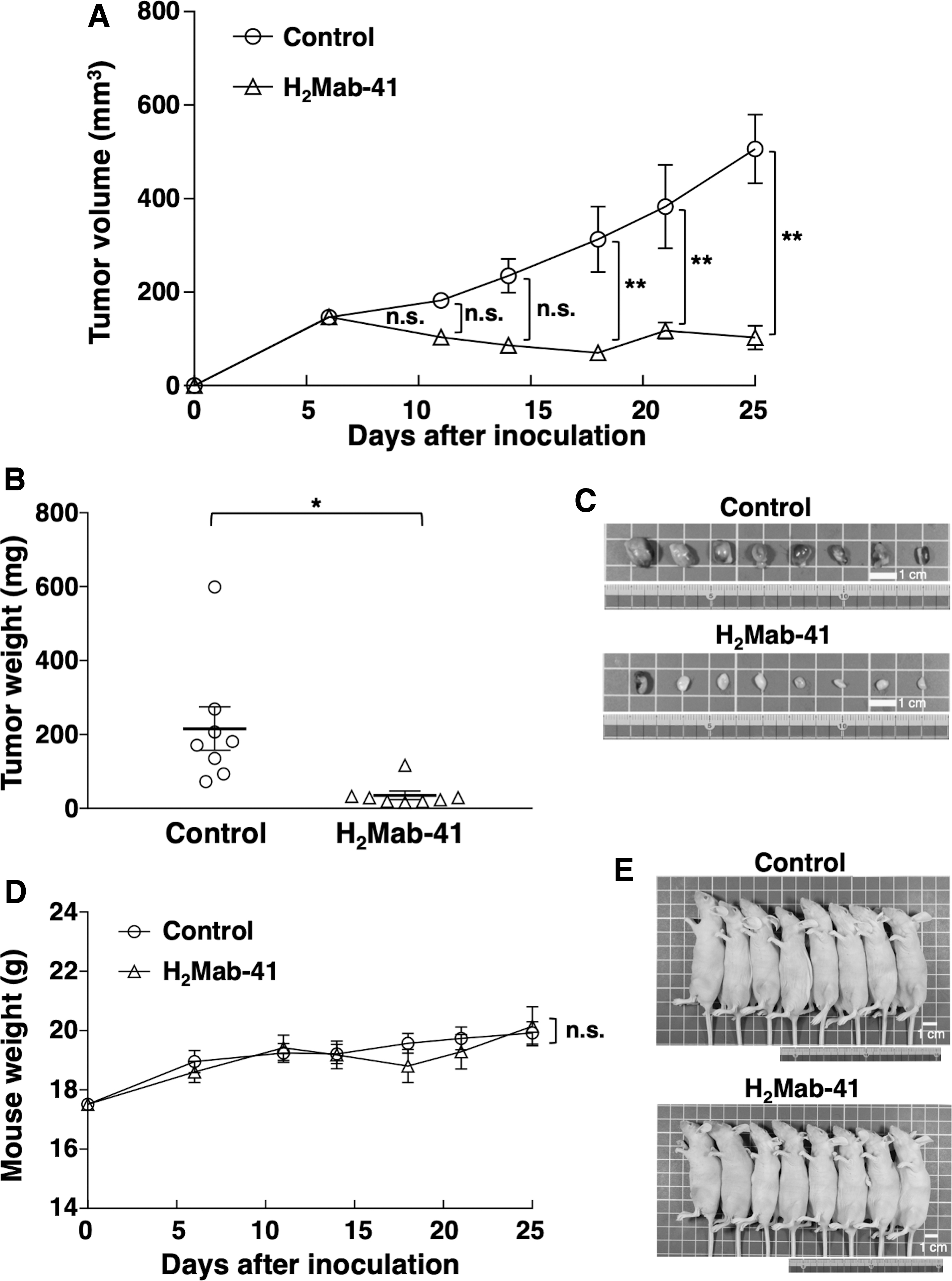
Evaluation of antitumor activity of H2Mab-41 in mice xenograft model for CHO/dHER2.
Discussion
Cancer is an evolutionary disease, which involves various genetic alterations like gene amplification and/or mutation.(30,31) Such alterations have been shown to influence patient prognosis and survival.(30–32) Some of these genetic alterations induce activation of driver mutations that result in the constitutive upregulation of signaling pathways, conferring advantages of cell proliferation, cell viability, and survival to the cancerous cells.(33,34) In the past few years, driver mutations have emerged as potential targets for molecular-targeted therapies for the management of various cancers.(35,36) Among various driver mutations, HER2 overexpression or gene amplification has been shown to be associated with poor prognosis in cancer patients.(37–39) Thus, the patients with HER2-driven signaling are considered to be suitable candidates for HER2-targeted therapy. In fact, administration of HER2-targeting agents in HER2-expressing metastatic breast cancer and gastric cancer patients resulted in dramatic improvement in the disease prognosis.(40,41)
Trastuzumab deruxtecan (T-DXd, DS-8201) is a HER2-targeting antibody-drug conjugate, comprising anti-HER2 mAb and a novel DNA topoisomerase I inhibitor.(42) Administration of T-DXd showed impressive clinical outcomes in metastatic breast cancer patients, who had received prior treatment involving multiple anti-HER2-targeting regimens.(43) These clinical outcomes encouraged the approval of T-DXd in the United States under accelerated approval for the treatment of patients with HER2-positive unresectable or metastatic breast cancer, who had previously received more than two anti-HER2-based therapies.(44) Currently, the clinical efficacy and safety of T-DXd is being evaluated in various stages of clinical trials for a variety of HER2-expressing tumors, with the aim to expand the usage of this therapeutic agent to other tumor types.
In canine tumors, the overexpression of HER2 has been reported in osteosarcoma (86% of the cell lines and 40% of the tissue samples),(21) mammary carcinoma (21% of the tissue samples),(22) bladder transitional cell carcinoma (56% of the tissue samples),(23) and anal sac gland carcinoma (80% of the tissue samples).(24) Interestingly, administration of a recombinant Listeria vaccine expressing a chimeric human HER2 resulted in the induction of HER2-specific immunity, reduction in the incidences of metastasis, and prolonged patient survival in a phase I trial for canine osteosarcoma.(45) These clinical outcomes encouraged the evaluation of anti-dHER2 mAb as a therapeutic agent for canine cancers.
One of the most important objectives of the present study was to demonstrate the cytotoxic activity of H2Mab-41 against HER2-expressing cells. In vivo administration of anti-HER2 mAb resulted in significant growth inhibition for CHO/dHER2 cells (Fig. 3). These results provided evidences to support the suitability of H2Mab-41 as a promising antibody therapy against canine cancers. Since the antitumor activity of H2Mab-41 was only established against dHER2-overexpressed cells in the present study, future studies involving various types of dHER2-expressing canine cancers are necessary to develop effective antibody therapies for canine cancers.
Footnotes
Author Disclosure Statement
No competing financial interests exist.
Funding Information
This research was supported, in part, by Japan Agency for Medical Research and Development (AMED) under Grant Numbers: JP21am0401013 (to Y.K.) and JP21am0101078 (to Y.K.), and by the Japan Society for the Promotion of Science (JSPS) Grants-in-Aid for Scientific Research (KAKENHI) Grant Nos. 21K15523 (to T.A.), 21K07168 (to M.K.K.), 20K16322 (to