
Abstract
Human epidermal growth factor receptor 2 (HER2) has been studied in many human cancer types, and its overexpression and/or gene mutation contribute to the poor prognosis. Therefore, HER2 is an important therapeutic target in various cancer types, including breast and gastric cancers. We previously developed an anti-HER2 monoclonal antibody (mAb), H2Mab-77 (mouse IgG1, kappa), which detects HER2 and dog HER2 (dHER2) with high sensitivity and specificity. In this study, we produced a defucosylated mouse-dog chimeric anti-HER2 mAb (H77Bf), and investigated the reactivity against canine osteosarcoma D-17 cells by flow cytometry. Furthermore, we showed that H77Bf exerted antibody-dependent cellular cytotoxicity and complement-dependent cytotoxicity against D-17 cells in vitro and exhibited the potent antitumor activity in vivo. These results suggest that H77Bf exerts antitumor effects against dHER2-expressing canine tumors and could be valuable as part of an antibody treatment regimen for them.
Introduction
Human epidermal growth factor receptor 2 (HER2) is a type-I transmembrane glycoprotein and member of receptor tyrosine kinases. HER2 overexpression is reported in many human cancers, including breast cancer,1,2 gastric cancer, 3 colorectal cancer, 4 lung adenocarcinoma, 5 and osteosarcoma (OS).6–8 Overexpression of HER2 results in homo- or heterodimerization of the HER family members, which leads to autophosphorylation of tyrosine residues in the cytoplasmic domains. The tyrosine phosphorylation initiates signaling transductions including Ras-MEK-ERK and PI3 kinase-AKT pathways, which play critical roles in tumor cell proliferation, invasion, and tumorigenesis.9,10
Trastuzumab, a monoclonal antibody (mAb) targeting HER2, has improved survival rates in HER2-positive advanced and early-stage breast cancer, and has been the most effective therapy for HER2-positive breast cancer.11–15 The recently developed trastuzumab deruxtecan (DS-8201) contains a novel enzyme cleavable-linker and a topoisomerase I inhibitor, deruxtecan as a payload. 16 Although there is no established regimen for patients with HER2-positive non-small cell lung cancer (NSCLC), the promising efficacy of DS-8201 in patients with HER2-mutant NSCLC has been reported. 17
HER2 is also overexpressed in multiple canine cancers, including OS, 18 anal sac gland carcinoma, 19 mammary carcinoma, 20 and bladder transitional cell carcinoma. 21 Fazekas et al conducted a clinical trial using the HER2 vaccine in a spontaneous HER2-positive canine mammary carcinoma to evaluate the safety and efficacy. 22 In a phase I study in canine OS, administration of a Listeria vaccine expressing chimeric HER2 induced dog HER2 (dHER2)-specific immunity, suppressed the development of metastases, and prolonged survival. 23 Therefore, dHER2 could be a promising therapeutic treatment of canine cancers.
We previously developed anti-HER2 mAbs, including H2Mab-19,24–27 H2Mab-41,28,29 H2Mab-77, 30 H2Mab-119, 31 H2Mab-139, 32 and H2Mab-181,33,34 which detect not only HER2 but also dHER2. This study aimed to investigate the ability of a defucosylated mouse-dog chimeric H2Mab-77 (H77Bf) to induce antibody-dependent cellular cytotoxicity (ADCC), complement-dependent cytotoxicity (CDC), and antitumor activities against a canine OS cell line, which endogenously expresses dHER2.
Materials and Methods
Cell lines
D-17, a canine OS cell line,35,36 was obtained from the American Type Culture Collection (Manassas, VA). D-17 was cultured in minimum essential media (Nacalai Tesque, Inc., Kyoto, Japan), supplemented with 10% heat-inactivated fetal bovine serum (FBS; Thermo Fisher Scientific Inc., Waltham, MA), 1 mM of sodium pyruvate, 100 U/mL of penicillin, 100 μg/mL streptomycin, and 0.25 μg/mL amphotericin B (Nacalai Tesque, Inc.). The cells were cultured at 37°C in a humidified atmosphere with 5% carbon dioxide.
Antibodies
An anti-HER2 mAb, H2Mab-77, was established in our laboratory as previously described. 30 To generate H77B, we subcloned VH cDNA of H2Mab-77 and CH of dog IgGB into the pCAG-Ble vector (FUJIFILM Wako Pure Chemical Corporation, Osaka, Japan), along with VL cDNA of H2Mab-77 and CL cDNA of dog kappa light chain into the pCAG-Neo vector (FUJIFILM Wako Pure Chemical Corporation), respectively. The vector of H77B was transfected into BINDS-09 cells (FUT8-deficient ExpiCHO-S cells)37–43 using the ExpiCHO Expression System (Thermo Fisher Scientific Inc.). The resulting mAb, H77Bf, was purified with Protein G-Sepharose (GE Healthcare Biosciences, Pittsburgh, PA). Dog IgG was commercially purchased from Jackson ImmunoResearch Inc. (West Grove, PA).
Animals
All animal experiments were carried out following relevant guidelines and regulations to minimize animal suffering and distress in the laboratory. Animal experiments for testing antitumor activity were approved by the Institutional Committee for Experiments of the Institute of Microbial Chemistry (permit no. 2021-056). Mice were kept in a specific pathogen-free environment (23°C ± 2°C, 55% ± 5% humidity) on an 11-hour light/13-hour dark cycle with food and water supplied ad libitum across the experimental period. Mice were monitored for health and weight every 2–5 days during the 3-week period of each experiment. We determined the loss of original body weight to a point >25% and/or a maximum tumor size >2000 mm3 as humane endpoints for euthanasia. Mice were euthanized by cervical dislocation; death was confirmed by respiratory and cardiac arrest.
Flow cytometry
D-17 cells were harvested by brief exposure to 0.25% trypsin/1 mM ethylenediamine tetraacetic acid (EDTA; Nacalai Tesque, Inc.). After washing with blocking buffer of 0.1% bovine serum albumin in phosphate-buffered saline (PBS), cells were treated with H77Bf or control blocking buffer for 30 minutes at 4°C. Subsequently, the cells were incubated in fluorescein isothiocyanate (FITC)-conjugated anti-dog IgG at a dilution of 1:1000 (Thermo Fisher Scientific Inc.) for 30 minutes at 4°C. Fluorescence data were collected using the EC800 Cell Analyzer (Sony Corp., Tokyo, Japan).
Determination of the binding affinity
D-17 cells were suspended in 100 μL of serially diluted H77Bf (0.006–25 μg/mL) followed by FITC-conjugated anti-dog IgG (1:200). Fluorescence data were collected using the EC800 Cell Analyzer. The dissociation constant (KD) was calculated by fitting binding isotherms to built-in one-site binding models in GraphPad Prism 8 (GraphPad Software, Inc., La Jolla, CA).
ADCC of H77Bf
Canine mononuclear cells (MNCs) were obtained from Yamaguchi University and suspended in Dulbecco's modified Eagle medium (DMEM; Nacalai Tesque, Inc.) with 10% FBS to be used as effector cells. Target cells were labeled with 10 μg/mL Calcein AM (Thermo Fisher Scientific, Inc.) and resuspended in the same medium. The target cells (2 × 10⁴ cells/well) were plated in 96-well plates and mixed with effector cells (effector/target cells ratio, 50), 100 μg/mL of H77Bf or control dog IgG. After 4.5 hours incubation at 37°C, the release of Calcein into the supernatant was measured in each well.
The fluorescence intensity was determined using a microplate reader (Power Scan HT; BioTek Instruments, Inc., Winooski, VT) with an excitation wavelength of 485 nm and an emission wavelength of 538 nm. Cytolytic activity (% lysis) was calculated as follows: % lysis = (E − S)/(M − S) × 100, where “E” is the fluorescence measured in combined cultures of target and effector cells, “S” is the spontaneous fluorescence of target cells only, and “M” is the maximum fluorescence measured after the lysis of all cells with a buffer containing 0.5% Triton X-100, 10 mM Tris-HCl (pH 7.4), and 10 mM EDTA.
CDC of H77Bf
The target cells were labeled with 10 μg/mL Calcein AM and resuspended in the medium. Then, they were plated in 96-well plates at 2 × 10⁴ cells/well with rabbit complement (final dilution 1:10; Low-Tox-M Rabbit Complement; Cedarlane Laboratories, Hornby, Ontario, Canada) and 100 μg/mL of H77Bf or control dog IgG. After 4.5 hours of incubation at 37°C, we measured Calcein release into the supernatant for each well. Fluorescence intensity was calculated as described in the ADCC section earlier.
Antitumor activity of H77Bf in mouse xenograft models of D-17 cells
A total of 16 female BALB/c nude mice (5 weeks old, weighing 14–17 g) were purchased from Charles River Laboratories, Inc. and used in experiments once they reached 8 weeks of age. D-17 cells (0.3 mL of 1.33 × 10⁸ cells/mL in DMEM) were mixed with 0.5 mL BD Matrigel Matrix Growth Factor Reduced (BD Biosciences, San Jose, CA); 100 μL of this suspension (5 × 10⁶ cells) was injected subcutaneously into the left flanks of the mice. On day 8 postinoculation, 100 μg of H77Bf (n = 8) or control dog IgG (n = 8) in 100 μL PBS were injected intraperitoneally. Additional antibody inoculations were performed on days 14 and 21. MNCs were injected surrounding the tumors on days 8, 14, and 21. The tumor volume was measured on days 7, 10, 14, 17, 21, 24, and 28 after the injection of cells. At 28 days after cell implantation, all mice were euthanized by cervical dislocation. Tumor diameters and volumes were measured as previously described.35,36,44,45
Statistical analyses
All data are expressed as mean ± standard error of the mean. All statistical analyses were carried out with Welch's t-test for ADCC, CDC, and tumor weight. Sidak's multiple comparisons tests were conducted for tumor volume and mouse weight. All calculations were performed using GraphPad Prism 8. A p-value <0.05 is statistically significant.
Results
Flow cytometric analysis against canine OS D-17 cells using H77Bf
An anti-HER2 mAb, H2Mab-77, was previously established by immunizing mice with ectodomain of HER2. 30 We next generated H77Bf, a defucosylated mouse-dog chimeric anti-HER2 mAb by combining VH and VL of H2Mab-77 with CH and CL of dog IgG, respectively. We investigated the reactivity of H77Bf against canine OS D-17 cells, and found that H77Bf could detect endogenous dHER2 in D-17 cells (Fig. 1A).

Flow cytometry using H77Bf and evaluation of ADCC and CDC.
A kinetic analysis of the interactions of H77Bf with D-17 cells was carried out using flow cytometry. The KD for the interaction of H77Bf with D-17 cells was 6.7 × 10⁻¹⁰ M (Fig. 1B), demonstrating that H77Bf has high affinity for D-17 cells.
H77Bf-mediated ADCC and CDC in D-17 cells
We investigated whether H77Bf mediated ADCC against D-17 cells. As shown in Figure 1C, H77Bf showed ADCC (10.4% cytotoxicity; p < 0.05) against D-17 cells more effectively than did the control dog IgG (3.4% cytotoxicity).
Subsequently, we investigated whether H77Bf mediated CDC against D-17 cells. As shown in Figure 1D, H77Bf showed CDC (68.4% cytotoxicity; p < 0.05) against D-17 cells more highly than did the control dog IgG (53.3% cytotoxicity). These results indicated that H77Bf promoted significantly higher levels of ADCC and CDC against endogenous dHER2-expressing D-17 cells.
Antitumor activities of H77Bf in the mouse xenograft models of D-17 cells
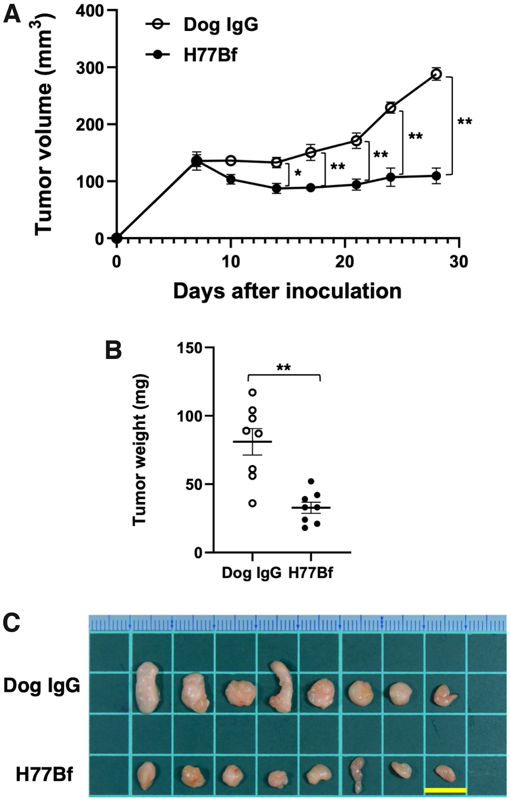
In the D-17 mouse xenograft models, H77Bf (100 μg) and control dog IgG (100 μg) were injected intraperitoneally into mice on days 8, 14, and 21, after the injection of D-17 cells. The tumor volume was measured on days 7, 10, 14, 17, 21, 24, and 28 after the injection of the cells. H77Bf treatment significantly suppressed tumor development on days 14 (p < 0.05), 17 (p < 0.01), 21 (p < 0.01), 24 (p < 0.01), and 28 (p < 0.01) compared with that of the control dog IgG (Fig. 2A). The administration of H77Bf resulted in a 62% reduction of tumor mass compared with that of the control dog IgG on day 28 postinoculation. Furthermore, tumors from the H77Bf-treated mice weighed significantly less than those from the control dog IgG-treated mice (60% reduction; p < 0.01, Fig. 2B). Tumors removed from mice on day 28 are shown in Figure 2C. Total body weights did not differ significantly among the two groups (Fig. 3A). The body appearance of mice on day 28 are shown in Figure 3B.
Evaluation of antitumor activity of H77Bf in D-17 xenograft models.

Body weights and appearance of the mice implanted with D-17 xenograft models.
Taken together, these results indicated that the administration of H77Bf effectively reduced the growth of D-17 xenografts.
Discussion
OS is the most frequent primary bone cancer in dogs. Eighty percent of dogs with OS is lethal due to the lung metastasis. 46 Selvarajah et al reported that the epidermal growth factor receptor (EGFR) expression was significantly upregulated in OS lung metastasis compared with extrapulmonary sites. 47 The rate of OS in dogs is 27 times higher than that in humans, with a 1-year survival rate of only 45%. 48 Currently, treatment options for OS in dogs include surgery (limb amputation or limb-sparing surgery), radiation therapy, and chemotherapy.49,50 However, satisfied therapeutic efficacies have not been obtained. Hence, the development of other therapeutic options for canine OS is necessary.
Antibody therapy is widely used for human diseases including cancers and immune disorders. The caninization of mAbs such as Lokivetmab 51 is essential for developing antibody therapies for dogs. However, caninized mAbs for canine cancers have not been developed yet. Thus, the development of the mAbs for canine cancer treatment is needed.
The amino acid sequences are similar between human and dog about EGFR and HER2; in contrast, low homology was observed between human and dog about CD20 and programmed-cell death ligand 1.52–54 Therefore, anti-human EGFR or anti-HER2 mAbs could be applicable to canine cancers. Indeed, cetuximab (anti-human EGFR mAb) and trastuzumab exhibited growth inhibitory effect by interfering dog EGFR (dEGFR) and dHER2 signaling, respectively. 52 We also have been investigating the cross-reactivity of our established anti-human EGFR mAb (EMab-134) to dEGFR35,36,44,45 and another anti-HER2 mAb (H2Mab-41) to dHER2. 29
Our study demonstrated that H77Bf significantly exhibited ADCC and CDC activities in canine OS D-17 cells compared with dog IgG in vitro (Fig. 1C, D). One of the most important objectives of this study was to investigate the antitumor activity of H77Bf for endogenous dHER2-expressing canine tumor cells in vivo. H77Bf administration showed the potent antitumor effect on endogenous dHER2-expressing D-17 cells without body weight loss and skin disorders (Figs. 2 and 3).
These results indicate that H77Bf could be useful for an antibody therapeutic regimen for dHER2-positive canine OS. Moreover, H2Mab-77, which is the origin of H77Bf, is applicable for immunohistochemistry 30 ; therefore, we need to investigate whether H77Bf detects endogenous dHER2 in canine OS tissues to evaluate usefulness of H77Bf as a diagnostic mAb for immunohistochemistry. Future studies are necessary to test the antitumor activity of H77Bf on other several types of endogenous dHER2-expressing canine cancers in vivo animal experimental models.
Footnotes
Author Disclosure Statement
No competing financial interests exist.
Funding Information
This research was supported in part by Japan Agency for Medical Research and Development (AMED) under Grant Numbers: JP22ama121008 (to Y.K.), JP22am0401013 (to Y.K.), JP22bm1004001 (to Y.K.), JP22ck0106730 (to Y.K.), and JP21am0101078 (to Y.K.), and by the Japan Society for the Promotion of Science (JSPS) Grants-in-Aid for Scientific Research (KAKENHI) grant nos. 22K15523 (to T.A.), 21K20789 (to T.T.), 22K06995 (to H.S.), 22K07168 (to M.K.K.), and 22K07224 (to Y.K.).