




Abstract
Human epidermal growth factor receptor 2 (HER2) is a transmembrane tyrosine kinase receptor without any known ligands and a member of the epidermal growth factor receptor (EGFR) family. It is a proto-oncogenic protein that, through signaling cascades, promotes cell proliferation and inhibits apoptosis in cancer cells through homo- and heterodimerization with other EGFR family receptors. Since several cancers, including breast cancer, overexpress HER2, it is a target of tumor therapy. Both trastuzumab and pertuzumab are recombinant humanized monoclonal antibodies (mAbs) used in clinical trials that target the extracellular domain (ECD) of HER2. Therefore, it is important to generate antibodies against various ECDs of HER2. In this study, we describe rat mAbs, which were generated against the ECD of human HER2. The human breast cancer cell line SK-BR-3 was subjected to immunofluorescence staining as it expresses HER2, and mAbs can detect both intact and endogenous HER2 within the cell line.
Introduction
The human epidermal growth factor receptor 2 (HER2), also referred to as erythroblastic oncogene B2 (ERBB2) and neu, is a member of the epidermal growth factor family of receptor tyrosine kinases. The HER2 gene encodes for a glycoprotein receptor with intracellular tyrosine kinase activity, and has no known ligand. 1 HER2 is a transmembrane receptor and consists of four extracellular domains (ECDs), a single transmembrane span, a cytoplasmic juxtamembrane linker region, an intracellular tyrosine kinase component, and a carboxyl-terminal tail.2,3 HER-mediated cell signaling is involved in regulating proliferation, differentiation, migration, and apoptosis, which are central to cancer cell survival and therapeutic resistance.4–7 This regulation occurs through homo- and heterodimerization with other receptors of the epidermal growth factor receptor (EGFR) family.
HER2 is a proto-oncogenic protein marker that promotes cell proliferation and inhibits apoptosis, leading to neoplasm. 8 In fact, overexpression of HER2 protein and/or amplification of the HER2 gene have been identified in 15%–20% of invasive breast cancers.6,9–11 In addition, its overexpression is detected in 17.9% of gastric cancer and gastroesophageal and 20%–30% of ovarian cancer.12,13 HER2-positive disease correlates significantly with a more aggressive phenotype and poorer prognosis than HER2-negative cancers.9,10,14 Therefore, HER2 is the target for selective anticancer therapies. Several antibodies directed against the ECD of HER2 are in clinical use or at advanced stages of development. The advancement in the therapy is considered a breakthrough in breast carcinoma treatment.
The humanized anti-HER2 monoclonal antibody (mAb) trastuzumab, known as herceptin, was the first targeted therapy for HER2-positive breast cancer patients at the molecular level.15,16 Herceptin binds to ECD of HER2 to inhibit HER2-mediated signaling by preventing heterodimerization, receptor internalization, degradation, inhibition of the PI3KAKT signaling pathway, and antibody-dependent cellular cytotoxicity. Patients with early- and late-stage HER2-positive breast cancer had considerably better outcomes with the addition of herceptin to chemotherapy.17,18 However, it is known that the effectiveness remains low due to resistance to the therapy. As the first of a new class of agents, pertuzumab, also known as Perjeta, is a humanized mAb against the ECD of the HER2 protein and prevents the formation of ligand-induced HER2-containing heterodimers.19,20
Furthermore, several other HER2-directed drugs, such as the antibody–drug conjugate trastuzumab–emtansine (T-DM1) and tyrosine kinase inhibitors such as lapatinib and neratinib have entered clinical use, allowing a combination therapy or sequential administration of non-cross-resistant drugs. 21 Consequently, generating various anti-HER2 antibodies that recognize the ECD of HER2 is essential. In this study, we raised novel rat mAbs against the ECD of endogenous and native human HER2, which may be relevant for studies examining the function of HER2 in cancer cells. We also suggest that these mAbs could be a new potential agent against HER2 therapy.
Materials and Methods
Cell culture
293T (human embryonic kidney, epithelial, large T transformed) and SK-BR-3 (human breast adenocarcinoma) cells were maintained in Dulbecco's modified Eagle's medium (DMEM) (Fujifilm Wako, Osaka, Japan) supplemented with 10% heat-inactivated fetal bovine serum (FBS; No. MSC1800S-500U; Medical & Scientific Equipment, Nuallié, France) under standard culture conditions (5% CO2 at 37°C) until the cells attained confluency. SP2/0-Ag14 (mouse myeloma) cells were maintained in Hybridoma-SFM (Life Technologies, Grand Island, NY) medium and were cultured as described earlier. Expi293F cells (Gibco, ThermoFisher Scientific, Waltham, MA) were cultured in HE200 medium (Gmep, Kurume, Japan) supplemented with 2 mM
Human HER2 expression vector was purchased from Promega K.K. (No. FHC11836; Tokyo, Japan). 293T cells were transiently transfected with the expression vector using ScreenFect A plus (Fujifilm Wako), according to the manufacturer's protocol.
Construction of expression vectors for recombinant proteins
The gene encoding the ECD of HER2 (HER2-ECD; Swiss-Prot accession no. P04626, amino acids 1–652) was synthesized by Eurofins Genomics K.K. (Tokyo, Japan) and was fused with the gene encoding human IgG1 Fc (Swiss-Prot accession no. P01857) at the C-terminus. The HER2-ECD/Fc fusion (HER2-ECD-Fc) gene was cloned into the pCAGGS expression vector, 22 and the plasmid was labeled as pCA–HER2-ECD–Fc.
Genes encoding the variable domains of the heavy and light chains of the humanized anti-HER2 antibody 4D5 (trastuzumab, herceptin) 23 were synthesized by Eurofins Genomics K.K. and were inserted upstream of the genes encoding the constant domains of the heavy and light chains of the human IgG1κ antibody, respectively. The resulting constructs h4D5-HC and h4D5-LC were each cloned into the pCAGGS expression vector, and the plasmids were labeled as pCA−h4D5-HC and pCA–h4D5-LC, respectively.
Expression and purification of recombinant proteins
Expi293F cells were cultured in Expi293 expression medium (ThermoFisher, Grand Island, NY) under standard culture conditions on an orbital shaker at 125 rpm to synthesize HER2-ECD–Fc. The cells were transiently transfected with pCA–HER2-ECD–Fc using PEI MAX (MW 40,000; Polysciences, Warrington, PA). After 20 hours of transfection, 1.25 mM sodium valproate (Tokyo Kasei, Tokyo, Japan) and 4 mM sodium propionate (Fujifilm Wako) were added to the medium, and the cells were cultured for 6 days. HER2-ECD–Fc recombinant protein was purified from the culture supernatant using Protein A Sepharose Fast Flow (GE Healthcare, Uppsala, Sweden).
Expi293F cells were cultured in HE400 medium (Gmep) supplemented with 5 mM
Generation of rat mAbs
Anti-HER2 rat mAbs were generated according to the rat lymph node method.24,25 Next, 0.1 mg of HER2-ECD–Fc and TiterMax Gold (Titer Max USA, Inc., Norcross, GA) were injected into the hind footpads of an 8-week-old female WKY/Izm rat (SLC, Shizuoka, Japan). After 11 days, 0.1 mg of the protein was injected into the rat tail base on both sides. After 3 days, cells from the medial iliac lymph nodes were fused with SP2/0-Ag14 cells at a ratio of 2:1 using an electroporator (No. CUY21 CS; Nepa Gene, Ichikawa, Japan).
The resulting hybridomas were plated on 96-well plates (Corning, Kennebunk, ME) and cultured in hypoxanthine–aminopterin–thymidine selection medium (Hybridoma-SFM [Life Technologies] containing 10% FBS, 100 μM hypoxanthine, 0.4 μM aminopterin, and 16 μM thymidine). At 7 days postfusion, the hybridoma supernatants were screened through immunofluorescence staining using SK-BR-3 cells. The positive clones were subcloned and rescreened; finally, the mAbs 2A12 and 2F3 were obtained. The specific immunoglobulin class of the two mAbs was determined using two rat isotyping kits (BD Biosciences, San Diego, CA, and Bio-Rad, Hercules, CA), and the analysis revealed that both mAbs were rat IgG2bκ.
Immunofluorescence staining for detecting intact antigen
SK-BR-3 cells were cultured on a cell culture coverslip (Matsunami Glass, Kishiwada, Japan) coated with poly-
Immunofluorescence staining for detecting fixed antigen
HER2-transfected/nontransfected 293T and SK-BR-3 cells were cultured on a cell culture coverslip coated with poly-
Results and Discussion
As described in Materials and Methods section, mAbs were generated against the ECD of human HER2 using the rat medial iliac lymph node method. In brief, the lymphocytes were isolated from the rat's enlarged medial iliac lymph nodes after immunization with recombinant protein of the ECD of human HER2. By fusing lymphocytes with mouse myeloma SP2/0-Ag14 cells, hybridomas were obtained, and supernatants were examined for the production of mAbs that targeted native HER2 in SK-BR-3 cells through immunocytochemistry (Fig. 1). It is known that HER2 proteins were highly expressed in SK-BR-3 cells. Next, we used immunostaining to determine if mAbs react with fixed antigens in SK-BR-3. mAbs reacted with the cell membrane of fixed SK-BR-3 cells, as shown in Figure 2.
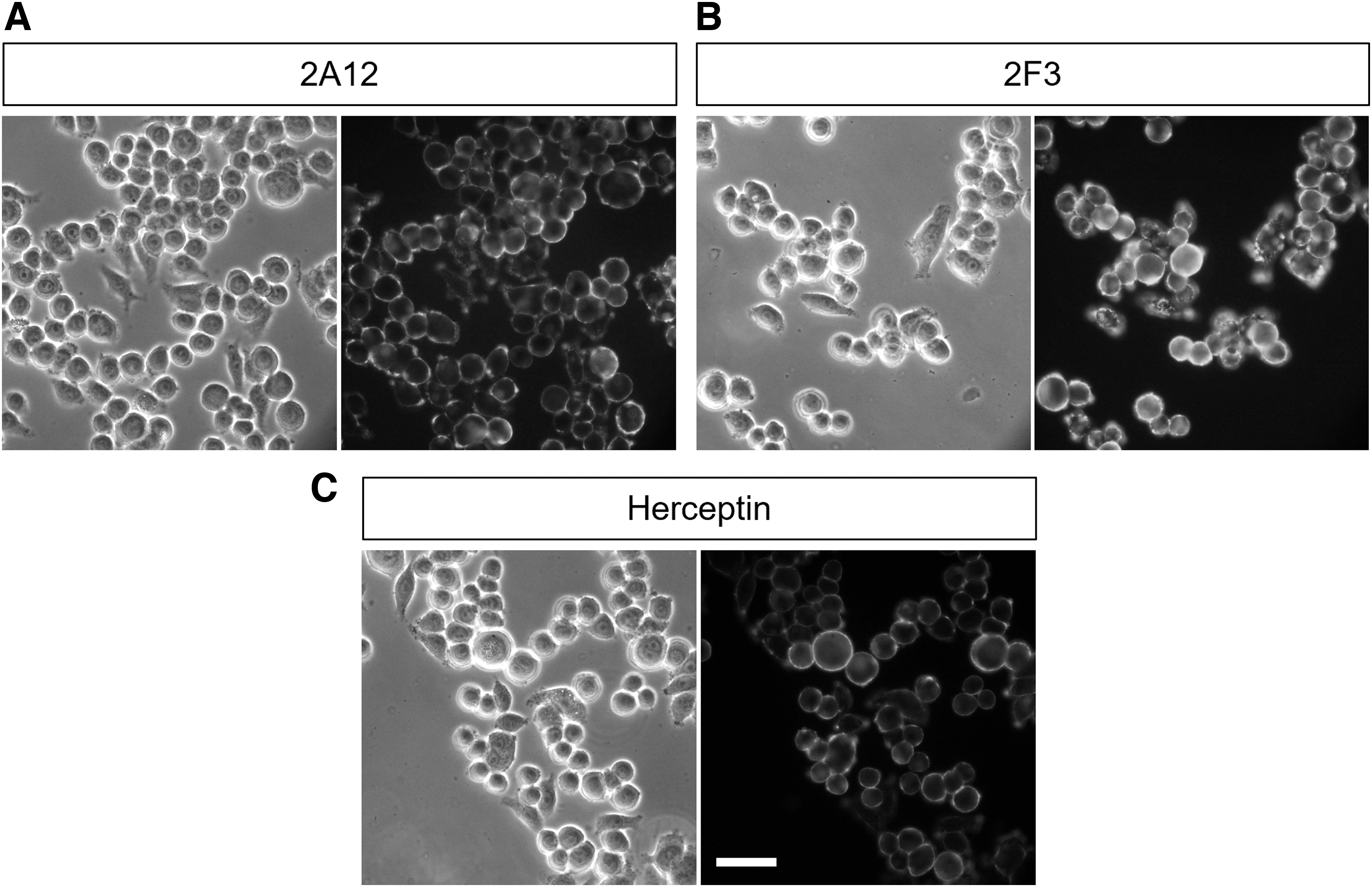
Immunofluorescence staining of rat mAbs using intact SK-BR-3 cells. SK-BR-3 cells were incubated for 1 hour at room temperature with mAb 2A12
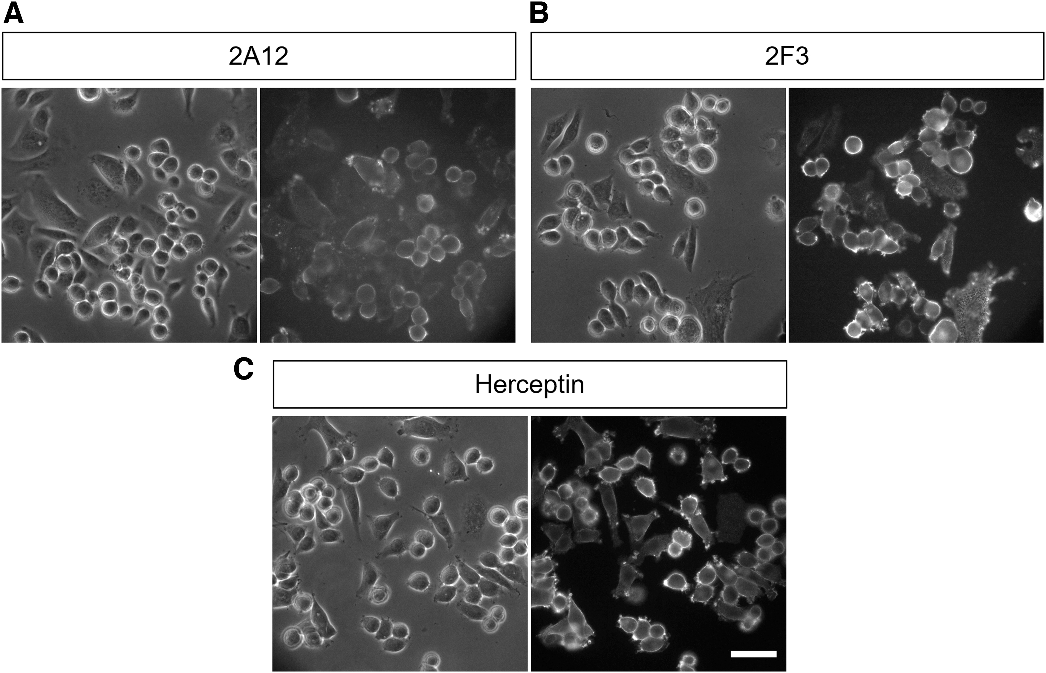
Immunofluorescence staining of rat mAbs using fixed SK-BR-3 cells. SK-BR-3 cells were fixed in 3.7% formaldehyde in PBS for 15 min at room temperature followed by permeabilization with 0.5% Triton X-100 in PBS for 5 min at room temperature. Subsequently, the cells were incubated overnight with mAb 2A12
Furthermore, we confirmed whether the antigen of the mAbs was HER2 through immunostaining using the HER2-overexpressed cells. Both rat mAbs only recognized the overexpressed cells, as shown in Figure 3. It showed that mAb 2F3 recognized intact and fixed antigens more effectively than mAb 2A12 throughout these experiments. Immunofluorescence staining was performed with herceptin and showed a similar staining pattern between rat mAbs and herceptin (Figs. 1–3). Thus, we concluded that mAbs specifically recognize the ECD of endogenous and intact HER2. Furthermore, rat mAbs recognized intact antigens as stronger than fixed antigens (Figs. 1 and 2). Otherwise, these mAbs failed to recognize the antigen in immunoblotting using a cell lysate from SK-BR-3 cells (data not shown).
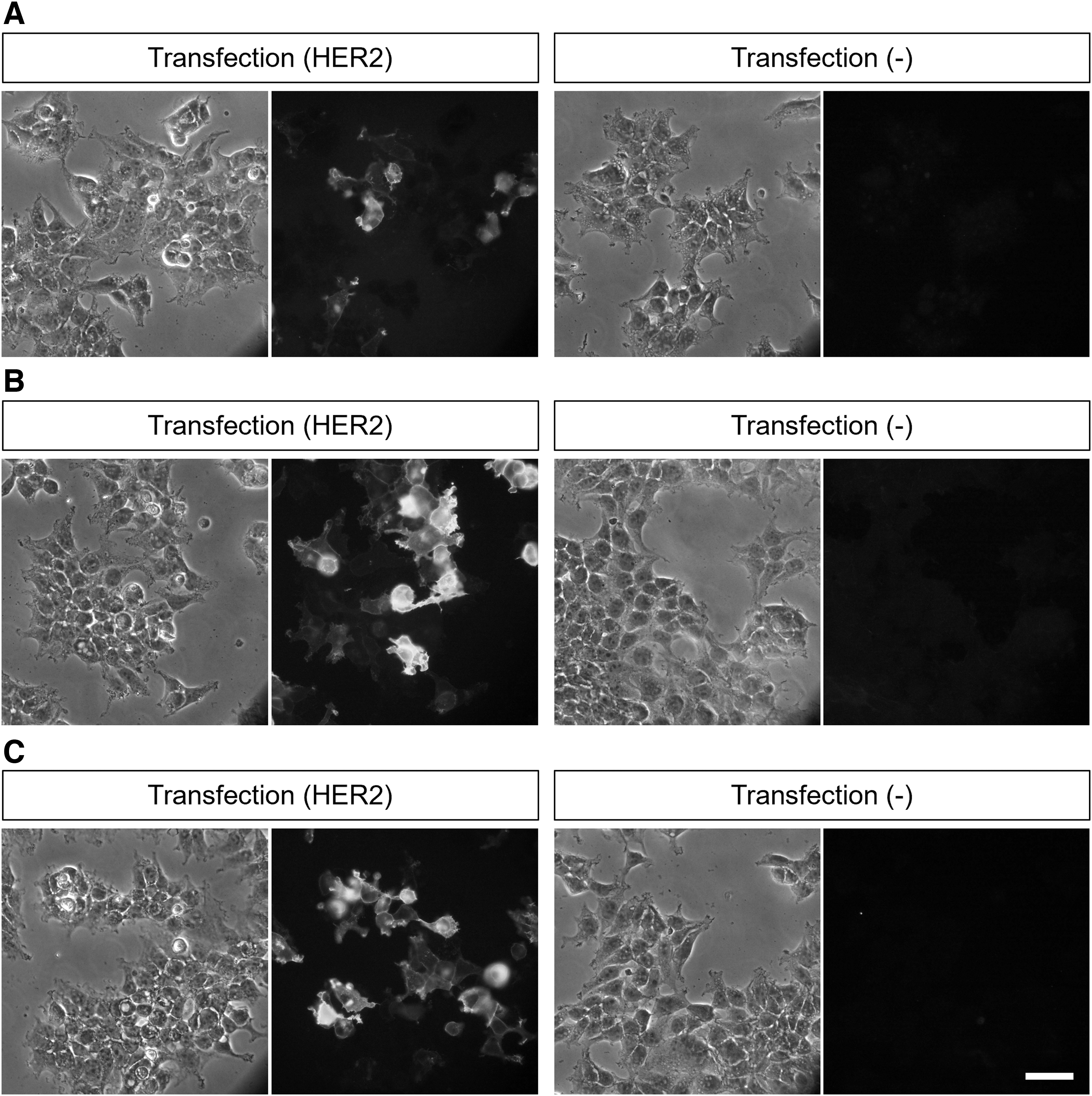
Immunofluorescence staining of rat mAbs using HER2-transfected 293T cells. HER2-transfected/nontransfected 293T cells were fixed in 3.7% formaldehyde in PBS for 15 minutes at room temperature followed by permeabilization with 0.5% Triton X-100 in PBS for 5 min at room temperature. Subsequently, the cells were incubated overnight with mAb 2A12
These findings suggest that mAbs only react with intact antigens, not denatured ones and that a three-dimensional structure is necessary for these mAbs to react. Therefore, these mAbs are useful for the analyses of HER2 in living cells, and we believe that rat mAbs may be used in the analyses of human cancer. Since it was reported that in some cases cancer cells acquire a resistance against herceptin, it is suggested a combination therapy or sequential administration of non-cross-resistant drugs. Therefore, it hopes several novel antibodies that possess various epitope in ECD of HER2 due to a development of therapies targeted HER2. Although these rat mAbs are needed to humanize in future study, we also hope that these mAbs are a new potential agent against HER2 therapy.
Footnotes
Acknowledgment
The authors would like to thank Enago for English language review.
Author Disclosure Statement
No competing financial interests exist.
Funding Information
This study was supported by expenses 3110060559 (T.T.) and 3110060567 (C.Y.) from Osaka Metropolitan University.