
Abstract
This study investigated the existence of vaginal Chlamydia infection and the prevalence of the disease in symptomatic gynecologically diseased women in Egypt. In addition, the antibiotics penicillin, tetracycline, and erythromycin were evaluated for their in vitro antichlamydial activity of the isolated strains. Vaginal swabs (n=160) were collected from females gynecologically diseased using cotton swabs. Samples were tested for Chlamydia by Vero cells tissue culture, chicken embryo, Gimenez staining, direct fluorescein-conjugated monoclonal antibody staining, and immunoperoxidase. Polymerase chain reaction (PCR) analyses conducted for the presence of chlamydial DNA was used to detect its specific DNA by the omp2 gene. PCR analyses conducted for the presence of chlamydial DNA revealed that 112/160 (70%) were positive for Chlamydiaceae. The specific DNA defined by the omp2 gene identified them as Chlamydia trachomatis (17/112, 15.2%), Chlamydophila psittaci (56/112, 50.0%), and Chlamydophila abortus (40/112, 35.7%). The antibiotics penicillin, tetracycline, and erythromycin at different concentrations were effective in inactivating the viability of Chlamydiaceae isolates.
Introduction
Public health considerations make it important to make a rapid and accurate diagnosis of chlamydiosis in order to help prevent exposure and clinical disease in humans. The diagnosis of chlamydiosis can be problematic. Isolation and identification is the gold standard for most infectious diseases, but it may be difficult to use these techniques for chlamydiosis. Hartley et al. 13 demonstrated that the omp2 gene of Chlamydiaceae is a suitable locus to which molecular detection and identification methods may be targeted. This assay has the potential to provide a simple and reliable means for the detection and identification of Chlamydiaceae from clinical specimens and cultures for the investigation of human chlamydial disease.
In Egypt, such a practice has not been adopted, and chlamydial infection in women remains undiagnosed, probably due to the lack of information on the magnitude of this problem, lack of laboratory facilities and skills, traditions, and religious beliefs. Hence, a study was undertaken, to generate evidence on genital chlamydial infection in women in Cairo, Egypt, using gold standard conventional techniques, complement fixation tests, and molecular identification. We analyzed the in vitro activities of penicillin, tetracycline, and erythromycin against the Chlamydiaceae isolates.
Methods
Study and Case participants
One hundred and sixty gynecologically diseased married women, with a mean age of 29.35±7.3 years attending the Outpatient Department of Obstetrics and Gynecology, of private hospitals in the suburbs of Cairo during 2008, were studied for Chlamydiaceae infection after ethical approval had been obtained from the Faculty of Medicine, Cairo University, Research Ethics Committee. They had one of the following complaints: The most common one was mucopurulent vaginal discharge, pain in the lower abdomen/back, recurrent abortion, and infertility. Multiple symptoms were also noted; vaginal discharge and pain in the lower abdomen or back were the frequent combination. The mean duration of vaginal discharge and pain in the lower abdomen/low backache was 42.8 and 27.3 months, respectively. The gynecologist conducted a per-speculum pelvic examination, to record the clinical signs and to collect cervical specimens, using a sterile cotton swab. Unhealthy cervix and adnexal signs suggestive of PID were seen. A history of seeking early treatment was recorded with the consultation of allopathic practitioners suggesting the development of PID due to delayed treatment.
Sampling technique
Vaginal swabs were collected from each woman after removal of the exocervical mucus and vaginal contaminants that may inhibit the polymerase chain reaction (PCR). One swab was transported in sucrose-phosphate-glutamate Chlamydia transport medium in an ice container 7 within 3 hr and stored at −20°C until processed by PCR, following a standardized PCR protocol.
Screening by cell culture
Isolation and cultivation experiments of intracellular agents were performed in Vero cells following the procedures of McElnea and Cross. 16 The cover-slip cultures were infected with the agent isolated from the vaginal sample, fixed after an incubation period of 48 hrs, and stained according to the method proposed by Giménez. 27
Inoculation to embryonated eggs
Seven-day-old embryonated chicken specific-pathogen-free eggs were inoculated with 200 μl inoculum under sterilized conditions, and their blind passages were carried out according to the procedure of Celebi and Seyyal. 4 Smears from the egg yolk sac were prepared on sterilized slides that were used for Giménez and immunoperoxidase staining. 27
Complement fixation test
Venous blood samples were obtained, and the sera were stored at −20°C until they were tested. A serological examination was conducted by a complement fixation test using reagents from Dienka Sieken Co., Ltd. (Tokyo, Japan), following the standard manufacturer's protocol. This test is specific for the family Chlamydiaceae without discriminating between different species of either Chlamydia or Chlamydophila. Microtiter plates (cold binding) with 3% amboceptor-sensitized sheep red blood cells and guinea pig complements were used. The antigen comprised Ornithose (Behringwerke, Marburg, Germany).
Molecular identification
DNA extraction
DNA was extracted from samples using a previously described method.1,13,21 Briefly, chromosomal DNA was prepared from the infected yolk sac materials. The embryo yolk sac materials were emulsified with phosphate buffer saline (PBS) pH 7.3 and were clarified by low-speed centrifugation (400 g) that produces suspensions of Elementary Bodies (EBs). The supernatant was then washed thrice with PBS, and the EBs was then sedimented by centrifugation at 14,000 g for 30 min. The pellet was suspended in 500 μl TE buffer at pH 8.0, and 200 μl of each sample were incubated for 15 min at 55°C with 200 μl of lysis buffer followed by incubation at 37°C for 45 min with an equal volume of phenol:chloroform (6:4). Then, centrifugation was performed at 10,000 g for 30 sec. The nucleic acid was precipitated by the addition of 5M NaCl and ice-cold ethanol. The dried pellet was then resuspended in 20 μl of DEPC water, and the supernatants were used in the reaction mixture for amplification.
PCR conditions
All reactions were carried out in a Perkin–Elmer type 480 thermal cycler (Perkin-Elmer, Norwalk, CT) in 50 μl reaction volumes.1,13,21 Reactions contained 1 to 10 μl of processed sample, 200 μM each of a mix of deoxynucleoside triphosphates (Amersham-Pharmacia Biotech INC, Piscataway, NJ), 5 pmol of each primer, 5 μl of 10× buffer with 15 mM MgCl2, and 1.25 U of Taq or Taq Gold (used during specificity tests with nonchlamydia isolates) polymerase (Roche Molecular Biochemicals; Roche Applied Science, Mannheim, Germany). Standard amplification conditions for primers Ch1 (5′-ATGTCCAAACTCATCAGACGAG-3′), Ch2 (5′-CCTTCTTTAAGAGGTTTTACCCA-3′) and D2 (5′-CCTTCTTTAAGAGGTTTTACC-3′) were 94°C for 4 min (or 95°C for 10 min for Taq Gold) for 1 cycle; 94°C for 1 min, 55°C for 1 min, and 72°C for 1 min for 40 cycles; and a final extension step of 72°C for 7 min for 1 cycle. Additional amplifications were performed with Ch1/Ch2 and with primer pair Ch1/D2, Taq Gold polymerase, and an annealing temperature of 45°C.
Restriction fragment length polymorphism PCR analysis was used as previously described.1,13,21 Briefly, a discriminatory restriction enzyme was sought by the analysis of cutting sites from available sequence data. Restriction with AluI was predicted as giving species-specific band lengths. Digestion was performed by incubating a 10 μl aliquot of PCR product with 1 U of enzyme (Promega, Madison, WI), 2 μl of 10×buffer, and 7 μl of water for 1 hr at 37°C. The fragments (C. trachomatis, Cp. abortus, and Chlamydophila psittaci—227 bp, 220 bp, and 140 bp, respectively) were analyzed by electrophoresis on a 4% Metaphor gel (FMC Bioproducts, Rockland, ME), stained with ethidium bromide, and compared with the predicted analysis.
Antibiotic sensitivity testing
Evaluation of the antichlamydial effect of the antibiotics in a monolayer cell-culture assay was carried out according to Perssson. 20 The bacterial isolates were divided into aliquots (200 μl of approximately 107 viable Chlamydiae) containing either no antibiotic or one of the three antibiotics at the following concentrations; erythromycin (50, 100, 200, 500, 1000, and 2000 μg/ml); penicillin G (25, 50, 100, 200, 500, and 1000 iu/ml); and tetracycline (10, 20, 50, 100, 200, and 500 μg/ml). The mixture was then incubated at 35°C for an hr in an attempt to kill the Chlamydiae. The samples were then tested for residual chlamydial viability in a cell-culture assay. Each set of antibiotic dilutions was tested in triplicate.
Statistical analysis
The data were analyzed using the Statistical Package for the Social Sciences version 18.0 (SPSS Inc, Chicago, IL) and MedCalc version 11.4.4 (MedCale Software, Inc., Mariakerke, Belgium). A multivariate analysis was done with the model of logistic regression while considering the health status of the patients (gynecologically diseased) for Chlamydiaceae as a dependent variable. The independent or explanatory variables considered in the model were those that exhibited a statistical significance p value of <0.05. Odds ratios (OR) were calculated in order to estimate the comparative risk between. In addition, the OR compared the likelihood of patients with signs of reproductive disease for Chlamydiaceae who tested positive using the different methods.
Data were also analyzed in order to determine the number of samples testing positive using each technique. A true-positive result for each species was defined as at least one of the two tests (culture or PCR) proving positive. Sensitivity and negative predictive values for each technique and Chlamydiaceae and agreement between the techniques were determined. By definition, the specificity and positive predictive values of both assays were 100%.
Results
The Gimenez stain detected Chlamydia in 60% (96/160) of the examined vaginal swab samples collected from different cases of gynecological disorders in women.
After performing immunoperoxidase stain for detecting Chlamydia inclusion bodies, in which a large number of discrete, densely labeled brown inclusion bodies was seen within the transparent background, 65% (104/160) of the examined samples were positive for Chlamydia.
The serum sample was considered positive if the titer was equal to or more than 32 unit/ml (1:32). Out of the examined 160 samples collected from human patients, 116 (72.5%) samples were positive with an antibody titer equal to or more than 32 unit/ml.
Out of the 160 vaginal swab samples examined, 112 (70%) were positive for the omp2 gene of the family Chlamydiaceae (Fig. 1). The specific DNA defined by the omp2 gene identified them as, C. trachomatis (17/112, 15.2%), Cp. psittaci (56/112, 50.0%), and Cp. abortus (40/112, 35.7%) (Fig. 2). The calculated simple odds ratios (with 95% CI) comparing the different methods used in the diagnosis are summarized in Table 1. The complement fixation test and PCR techniques indicate a significant difference from the other techniques.
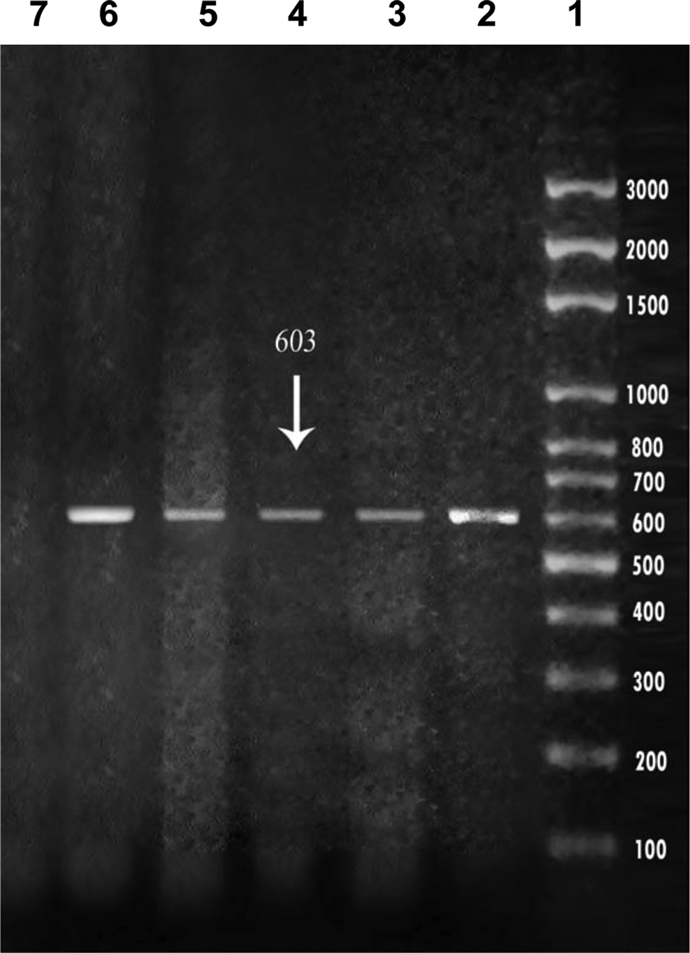
Gel electrophoresis of partial omp2 polymerase chain reaction product. Lane 1: size marker with sizes (in base pairs) of weight markers indicated at the right; lanes 2, 3, 4, and 5: family Chlamydiaceae (603 bp.) isolated from humans; lane 7: negative control (Escherichia coli ATCC 35150 serotype O157:H7).

Gel electrophoresis of omp2 PCR products of Chlamydiaceae after restriction digestion with Alu I. Lane 1: size marker with sizes (in base pairs) of weight markers indicated at the left; lanes 2, 3, and 4: Chlamydia trachomatis (77& 84& 114 & 119 and 158 bp.) isolated from human patients; lanes 5, 6, 7, and 8: Chlamydophila psittaci (140 & 220 and 227 bp.) isolated from humans; lanes 9 and 10: Chlamydophila abortus (235 & 352 bp.) isolated from humans.
CFT, complement fixation test; PCR, polymerase chain reaction; OR, odds ratio; CI, confidence interval; PPV, positive predictive values; NPV, negative predictive values.
Penicillin, tetracycline, and erythromycin at different concentrations were effective in inactivating the viability of Chlamydiae isolates (Table 2). The effect of the antichlamydial activity of penicillin (50 iU/ml), tetracycline (200 μg/ml), and erythromycin (200 μg/ml) on C. trachomatis, while their effect on Cp. psittaci was recorded at concentrations of 25 iU/ml, 100 μg/ml, and 200 μg/ml, respectively. Cp. abortus was inactivated by penicillin, tetracycline, and erythromycin at concentrations of 500 iU/ml, 50 μg/ml, and 200 μg/ml, respectively.
3+=100 to 1000 inclusions per 20 microscopic fields (×400 magnifications).
2+=21 to 100 inclusions per 20 microscopic fields.
1+=one to 20 inclusions per 20 microscopic fields.
NEG, no inclusions per 20 microscopic fields.
Discussion
Previously reported pregnancies complicated by chlamydial infection resulted from Cp. psittaci infection and Cp. abortus infection.11,14,22,23 An understanding of the modes of transmission of Chlamydophila spp. and of the infectious periods of animals is necessary in order for physicians to give good advice for the prevention of zoonotic transmission. Goats are the most likely sources of Cp. abortus infection. 17 Most of the reported cases of human infection have involved direct or/and indirect contact with infected animals. 17 However, visiting and living on or close to a farm affected by enzootic abortion or having neighbors with psittacine birds have also been indicated, in several cases, as being the only possible source of infection. 23 Zoonotic transmission is considered as occurring via the oral or respiratory route, by direct contact with infected animals or their products (e.g., bird excreta, vaginal discharge, foetal fluid, and placenta), or by inhalation of contaminated aerosols or dust. Contaminated surfaces or clothing and footwear may also be a likely vehicle for transmission. Infectious EBs can be shed in uterine secretions from goats or ewes before abortion or from ewes after abortion.18,24 Furthermore, for almost 3 years, infected ewes can shed infectious EBs from their reproductive tract 3–4 days before and after ovulation. 15 Altogether, this has led midwives in the United Kingdom to advise pregnant women on avoiding contact with lambing sheep or kidding goats; not to feed, clean, or cuddle orphan or weak lambs/kids; and not to wash or handle clothing that had been worn by their husband during lambing/kidding. 19
Cattle are an important mammalian host for Cp. psittaci, in which asymptomatic and symptomatic infection and shedding of organisms in intestinal and vaginal secretions is common. 8 In Egypt, as in Nepal, 7 fresh dung is formed into patties that are allowed to dry on the walls of the huts and then used as fuel in fires. This practice presents the potential for hand-to-body transmission of Cp. psittaci or Cp. abortus or C. trachomatis, although this particular route has not been studied to date.
If the involvement of multiple Chlamydiaceae species in genital infection is confirmed in other endemic countries, then a new approach to antimicrobial therapy may be needed. C. trachomatis infections can be successfully treated by tetracycline.15,20 Furthermore, and most relevant to our article, a number of studies have reported that various antimicrobial agents, including penicillin, chlortetracycline, erythromycin, and sulfonamides, can also induce persistence of chlamydial infection in vitro.18,20 In human patients, tetracyclines and macrolides, particularly erythromycin, are the antibiotics of choice, administered either orally or parenterally, depending on clinical severity. 5
In conclusion, this is the first article from this region showing 70% prevalence rate of Chlamydiaceae infection in symptomatic women. In addition, most clinical laboratories limit their investigations of chlamydial abortion to the detection of C. trachomatis, a policy that should be re-evaluated, thereby choosing the appropriate drug in the treatment of Chlamydiaceae infection. We have demonstrated that the in vitro activities of penicillin, tetracycline, and erythromycin are clinically suitable and useful in the treatment of genital chlamydial infection.