
Abstract
The presence of bacterial biofilm, particularly formed by Pseudomonas aeruginosa, has been considered an important factor responsible for wound chronicity. The objective of this study was to investigate the antibiofilm activity of water and ethanol extracts obtained from three traditional herbal recipes (THR-SK004, THR-SK010, and THR-SK011) on biofilm formation and on mature biofilm of a reference strain of P. aeruginosa. The effects of the extracts on the biofilm mass were evaluated by using crystal violet (CV) assay. The respiratory activity of preformed biofilm of P. aeruginosa after treatment with the extract was determined by MTT reduction assay. Scanning electron microscopy was used to furnish images of biofilm reduction after the recipe treatment. Tested ethanol extracts displayed antibiofilm activity, but the water extracts exhibited low biofilm inhibition activity at the tested concentrations. Remarkable reduction in biofilm formation of P. aeruginosa was found after treatment with the THR-SK010 ethanol extract (THR-SK010E). Treatments with this extract resulted in prevention of biofilm formation of P. aeruginosa on both polystyrene and glass surfaces. Almost 50% reduction in the bacterial metabolic activity in the preformed biofilm was seen after exposure to the extract-supplemented buffer for 12 hr. After a 24-hr treatment with THR-SK010E at 62.5 μg/ml, 97.3% of the preformed biofilms were destroyed. Promising antibiofilm activity was displayed by the THR-SK010 ethanol extract, suggesting further investigation to explore the possible utilization of the herbal recipe as an antibiofilm agent, especially for wound treatment.
Introduction
Biofilm are structured surface-associated communities of bacteria that have been well documented in the setting of a number of chronic wound states, including pressure sores, diabetic foot ulcers, and venous stasis ulcers. In addition, antibiotics and host immune responses are ineffective in combating biofilm. Previous visualization by scanning electron microscopy showed that the majority of chronic wounds (60%) had a biofilm presence, compared with only 6% of acute wounds. 18 Therefore, identification of a possible biofilm-eradicating strategy has revolutionized the research approach in medical, pharmaceutical, and biosciences.
Medicinal plants have been investigated to possess anti-Pseudomonas and antibiofilm activities. Aqueous extracts of Conocarpus erectus, Callistemon viminalis, and Bucida buceras that possessed antibiofilm-and antiquorum-sensing activities against P. aeruginosa1,3 had the ability to prevent nematode death via gut infection by this pathogen. 2 Similarly, Swietenia macrophylla used for treating wound infection caused a significant inhibition of Pseudomonas sp. virulence factors and enhanced the ability of Caenorhabditis elegans to survive Pseudomonas sp. infections. 10 Thai traditional medicine is one of the most valuable heritages handed down from ancient folk healers. In spite of increasing popularity of modern medicine, Thai traditional medicine is still widely used in taking care of health in daily life, especially among the rural Thais. 38 Some investigations on Thai herbal recipes have focused on their effects on planktonic bacteria.19,25,39 However, antibiofilm effects of Thai herbal recipes on P. aeruginosa have not been well studied. Therefore, the present study was aimed to explore the antibiofilm properties of traditional Thai herbal recipes against P. aeruginosa, which is implicated in wound chronicity. These herbal recipes were chosen on the basis of their traditional use against chronic wound and skin infections, conditions potentially caused or complicated by bacteria, including, P. aeruginosa.
Materials and Methods
Preparation of herbal recipes
Selected herbal recipes consisted of THR-SK004 prescribed by Mr. Earn Thongsongsi, a Thai folk healer, and THR-SK010 and THR-SK011 were prescribed by Mr. Somporn Chanwanisakul, a traditional Thai medical doctor at the Traditional Thai medicine hospital, Prince of Songkla University, Hat Yai, Thailand. The herbal components of the recipes were purchased from medicinal herb retailers in Songkhla, Thailand, and authenticated by a taxonomist, Dr. Katesarin Maneenoon, and the voucher specimens were deposited at the Faculty of Traditional Thai Medicine, Prince of Songkla University, Hat Yai, Songkhla, Thailand. The medicinal plants were washed with distilled water and dried at 60°C overnight. Powders of each recipe (300 g) were prepared from the dried components as described in Table 1. The ground recipe (100 g) was separately macerated at room temperature with 95% ethanol (500 ml) and distillation water (500 ml) for 7 days and 2 days, 12 respectively. After filtrations through a Whatman No. 1 paper, the aqueous filtrates were freeze-dried, and the ethanol filtrates were concentrated using a rotatory evaporator, and kept at 55°C until they were completely dry. The yields (%; w/w) of each extracts were calculated as the ratio of the weight of the extract to the weight of the recipe powder. Stock solutions (200 mg/ ml) of lyophilized water extracts (THR-SK004W, THR-SK010W, and THR-SK011W) and dried ethanol extracts (THR-SK004E, THR-SK010E, and THR-SK011E) were prepared by dissolving 0.2 g of the dried extract in 1 ml of dimethyl sulfoxide (DMSO; Merck) and stored at −20°C.
Weight (g) of the medicinal plants in 300 g of the recipes.
Inhibition of biofilm formation by the herbal recipes
A biofilm-positive strain, P. aeruginosa ATCC10145, was employed in this study to screen the antibiofilm activity of the recipe extracts.
14
Well-isolated colonies grown overnight at 37°C on tryptic soy agar were inoculated in a tryptic soy broth (TSB) supplemented with 2% (w/w)
The cultures were then diluted 200-fold in TSBGlc, divided into 100-μl aliquots, and added into a flat-bottomed 96-well polystyrene microtiter plate (Nunc). An aliquot of twofold serial dilutions (100 μl) of the extract was prepared in TSBGlc and added into the 96-well microtiter plate to final concentrations of 62.5 to 1,000 μg/ml. An additional 100 μl of TSBGlc and TSBGlc containing 1% DMSO was used as positive controls. Some wells were used for adding of an aliquot (200 μl) of each tested extract in TSBGlc to exclude a possible contamination. After incubation at 37°C for 24 hr, the effect of the extracts on the growth of P. aeruginosa was evaluated using a microplate reader (Tecan Sunrise) at optical density of 620 nm (OD620nm).
The plates were subsequently determined for the biofilm mass using a colorimetric assay adapted from the method of O'Toole. 30 Culture supernatants and from each well were decanted, and planktonic cells were removed by washing for 1 min three times with 200 μl phosphate-buffered saline (PBS; pH 7.4). The biofilm mass was stained with 200 μl of 0.1% (w/v) crystal violet (CV) solution for 30 min at room temperature. The excess dye was then washed off three times with 200 μl PBS, and the stained biofilm was resuspended using 200 μl DMSO. 11 The absorbance at 620 nm was determined using the microplate reader. The medium containing each concentration of the extracts was used as the blank control. All experiments were performed at least in triplicate.
Biofilm time-dependent prevention assay
According to potent antibiofilm inhibition, THR-SK010E was chosen for further experiment. The biofilm development of P. aeruginosa after being treated with this recipe at concentration 62.5, 125, and 250 μg/ml was observed every 12 hr for 2 days by the CV assay as described above.
Observation of biofilm formation by scanning electron microscopy
Scanning electron microscopy (SEM) images were taken to confirm the prevention of biofilm formation by THR-SK010E. 7 Briefly, this strain was allowed to form biofilm on squared glass slides (1×1 cm) placing in 24-well polystyrene plates (Greiner Bio-One) containing with 1 ml of TSBGlc supplemented with the extract at concentrations of 62.5, 125, or 250 μg/ml. Uninoculated TSBGlc containing the extract was used as a negative control, and innoculated TSBGlc containing 1% DMSO was served as a positive control. After incubation at 37°C for 48 hr, additional glass pieces were removed using sterile forceps and washed for 1 min using 2 ml PBS. The samples were initially fixed in 2.5% glutaraldehyde in a cacodylate buffer for 90 min, then washed twice with cacodylate buffer, and dehydrated for 10 min using a graded ethanol series. A critical-point drying procedure followed, and the specimens were then sputter-coated with gold. The samples were examined with an SEM (5800LV; JEOL). Beside, the biofilm glass pieces were separately placed into 0.1% (w/v) CV solution (2 ml) for 30 min at room temperature, removed using sterile forceps, and washed for an additional minute three times with 2 ml of PBS to remove nonstained CV. The attached dyed was re-eluted, and the total biomass of the biofilm was confirmed as described above.
Eradication of established biofilm by THR-SK010
To further evaluate the effect of the recipes on P. aeruginosa-preformed biofilm, the formazan-based 3-(4,5-dimethylthiazol-2-yl)-2,5-diphenyltetrazolium bromide (MTT; Sigma-Aldrich) assay was performed on preformed biofilms that were exposed to the tested extract. This assay measures enzymatic activity in actively respiring cells and is therefore a measure of cell viability and/or relative numbers of viable cells in biofilm. 43 An aliquot of 200 μl of the bacterial culture preparing as mentioned above was added into a flat-bottomed 96-well polystyrene microtiter plate. After incubation at 37°C for 24 hr, the spent medium and planktonic cells were then removed, and fresh TSBGlc (200 μl) was added. This was repeated daily for five consecutive days, and at the end of 5 days, the spent medium and the planktonic cells were gently aspirated. Thereafter, 200 μl of PBS containing different concentrations of the THR- SK010E (62.5, 125, and 250 μg/ml as final concentrations) was added into the wells.
Eradication of the preformed biofilm of P. aeruginosa by the extract was measured at selected time intervals of 1, 3, 6, 12, and 24 hr. PBS containing 1% DMSO was used a negative control. An aliquot of the MTT solution (0.2 mg/ml; 200 μL) was added to each of the prewashed wells, and the plate was then incubated for 3 hr in the dark at 37°C. After the incubation, MTT was then replaced by 200 μL of DMSO. The color intensity of DMSO-dissolved formazan was determined by a microplate reader at OD492 nm. Wells with the addition of PBS containing each concentration of the extract was served as the blank for all absorbance readings. The absorbance values for the blank were then subtracted from the tested wells to eliminate false results due to background interference. The percentage of biofilm eradication in comparison with untreated wells was calculated using the equation [(OD (at 0hr) – OD (after treatment))/OD (at 0hr)]×100.
Statistical analysis
The ODs of the amount of the CV in the destaining solution or the color intensity of DMSO-dissolved formazan, measured between the recipes treated biofilm and untreated biofilm, were compared by the paired Student's t-test by using the SPSS Win 12.0 program (SPSS, Inc.). Differences between the two groups were considered to be significant for p of 0.05.
Results
The antibiofilm ability of water and ethanol extracts of the traditional herbal recipes on P. aeruginosa ATCC 10145 is described in Fig. 1. The CV assay results illustrated that all concentrations of the ethanol extracts possessed antibiofilm activity, but the water extracts exhibited low biofilm inhibition activity at the tested concentrations. Remarkable reduction in biofilm formation of P. aeruginosa was found after treatment with THR-SK010E (62.5–1,000 μg/ml). Although, antibiofilm activities of the water extracts were lower than those ethanol extracts, a decrease in the biofilm formation was also noticed after treatment with THR-SK010W (125, 250, and 1,000 μg/ml). It has to be mentioned that at concentrations of THR-SK010E ranging 62.5–500 μg/ml affected 10% to 40% of the growth of planktonic cells in a dose-dependent manner. Moreover, 62.5, 125, and 250 μg/ml of THR-SK010E resulted in prevention of biofilm formation of the pathogen on polystyrene surfaces up to 48 hr with a weak growth inhibition effect as depicted in Fig. 2.
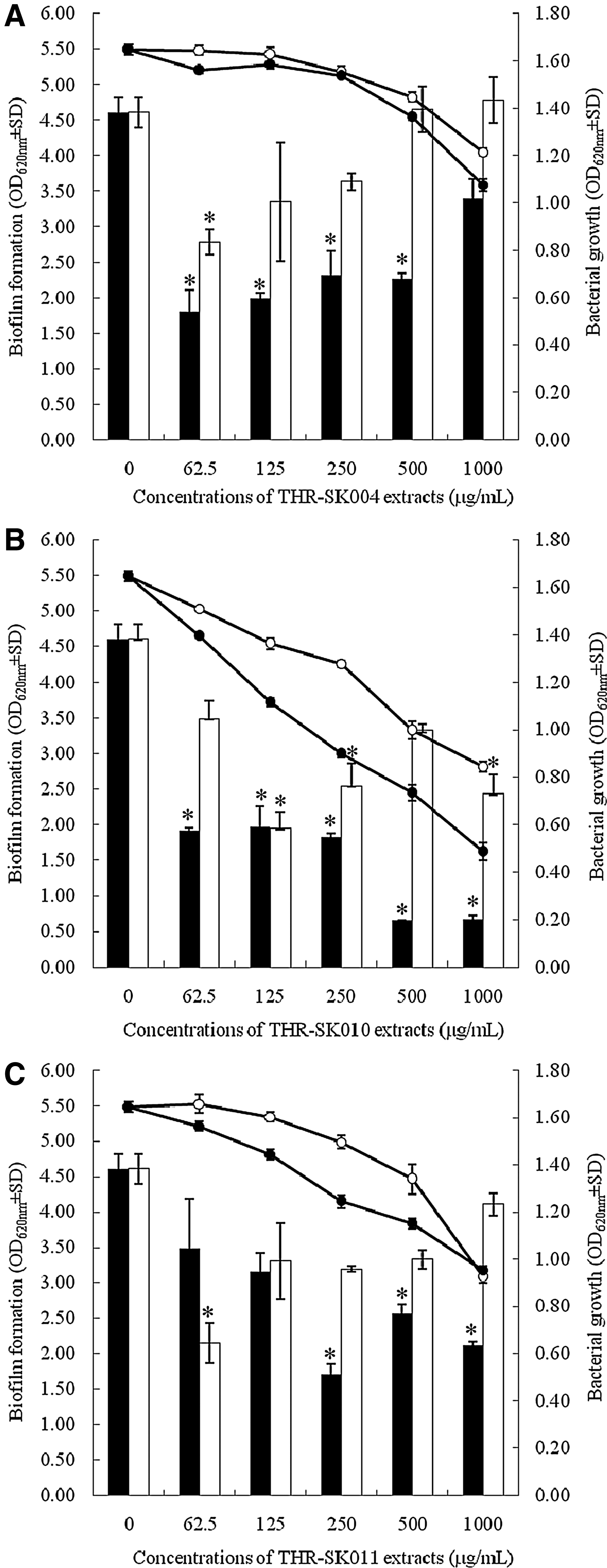
Effect of different concentrations of THR-SK004
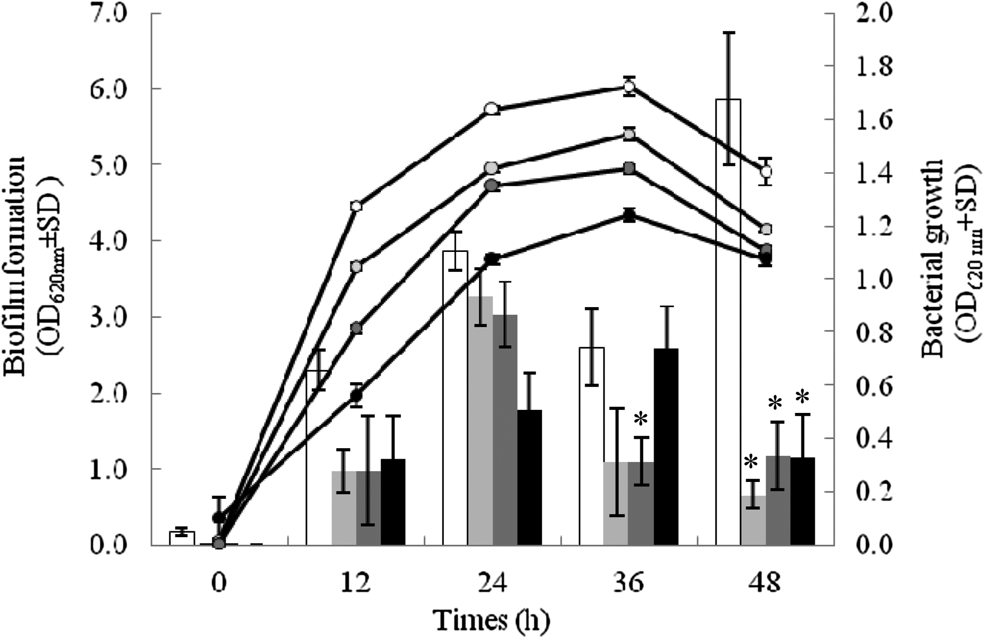
Development of P. aeruginosa ATCC 10145 biofilm (column charts) and the bacterial growth (linear charts) treated with THR-SK010 ethanol extract at 62.5 (light gray symbols), 125 (dark gray symbols), and 250 μg/ml (black symbols). Dimethyl sulfoxide (DMSO) at 0.2% (white symbols) was used as positive control. Each symbol indicates the means±SD for three independent experiments performed in duplicate. Statistical differences between the biofilm formed by recipe-treated and untreated P. aeruginosa, determined by Student's t-test, are denoted by asterisks above the error bars. *p-Value of 0.05.
Inhibition of biofilm formation on glass surfaces by THR-SK010E was additionally visualized by both SEM and the CV assay as illustrated in Fig. 3. As expected, the addition of THR-SK010E at 62.5, 125, or 250 μg/ml, which reduces the biofilm formation of P. aeruginosa on polystyrene surfaces, remarkably influenced the biofilm formation of the pathogen on the glass surfaces. Examination by SEM at a high magnification demonstrated that biofilms grown in the absence of the herbal recipe extract was dramatically thicker and have a more biofilm density and established architecture compared to biofilms grown in the presence of the extract. Biofilms of P. aeruginosa grown for 48 hr in the presence of 125 and 250 μg/ml of the extract were scattered sparsely on the glass, while biofilms grown with 62.5 μg/ml of the extract were much thicker. Scanning electron micrographs additionally revealed that the reduction in the lengths of P. aeruginosa bacilli and irregular nonuniformity of the cells with slightly rough surface were observed after 48-hr incubation in 62.5, 125, and 250 μg/ml of the extract in a dose-dependent manner, whereas regular shape and approximately equal sizes of the cells were noted in untreated cells.

SEM micrographs of P. aeruginosa ATCC 10145 biofilm formation on glass surfaces. Biofilms were grown in TSBGlc
Eradication of the 5-day-old biofilm of P. aeruginosa treated with THR-SK010E (62.5, 125, or 250 μg/ml) is presented in Fig. 4. Almost 50% reduction in the bacterial metabolic activity in the preformed biofilm was seen after exposure to the extract-supplemented buffer for 12 hr. After a 24-hr treatment with THR-SK010E at 62.5 μg/ml, 97.3% of the 5-day-old biofilm was destroyed, while the addition of the extract at 125 and 250 μg/ml caused the decrease in the bacterial metabolic activity in the established biofilm of 70.6% and 67.4%, respectively.
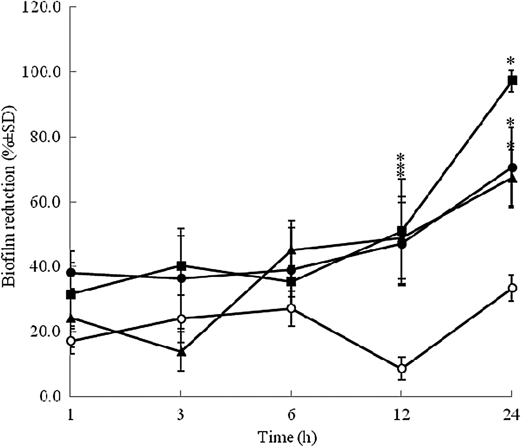
Time-dependent eradication of the mature biofilm formed by P. aeruginosa ATCC 10145 after treatment with the THR-SK010 ethanol extract at 62.5 (▪), 125 (•), and 250 μg/ml (▴). PBS containing 0.2% DMSO (o) was used as positive control. Each symbol indicates the means±SD for three independent experiments performed in duplicate. Statistical differences between the biofilm reduction of recipe-treated and untreated cells, determined by Student's t-test, are denoted by asterisks above the error bars. *p-Value of 0.05.
Discussion
Previous investigations have been pointed out that inhibition of cell attachment to a substrate is easier to achieve than inhibiting the growth of an already-established biofilm.6,36 Our data reveal that the traditional herbal recipe extract, THR-SK010E, is a potential antibiofilm agent against both the formation of biofilm and preformed biofilm. The success of the herbal recipe in inhibiting cell attachment and eradicating established biofilm as shown in this study is a promising tool for reducing P. aeruginosa colonization.
THR-SK010E displayed antibiofilm activity on P. aeruginosa on both the hydrophobic surface (polystyrene) and hydrophilic surface (glass), but it slightly affected the bacterial growth. In contrast, previous investigations documented that the biofilm formation in P. aeruginosa was induced by the subinhibitory levels of several antibiotics such as beta-lactam (imipenem) 5 and aminoglycoside (tobramycin, amikacin, streptomycin, and gentamicin). 16 Pitts et al. 33 proposed that a drug that could control ≥40% can be considered as a potent biofilm eliminator. Here, we found that THR-SK010E-treated P. aeruginosa exhibited biofilm production five to nine times lower than the production in untreated P. aeruginosa. In addition, 67%–97% of preformed biofilm of the pathogen was eradicated after treatment with THR-SK010E. Management of P. aeruginosa biofilm-infected wounds is limited. Information on biofilm elimination of ulcer isolates P. aeruginosa using near-infrared laser treatment has recently been investigated. This treatment did not affect the biomass and the cell viability of the biofilm. 4 In contrast, evidence on biofilm inhibition of the pathogen in pulmonary infections of a cystic fibrosis model has been continuously documented. Natural compounds, furanone and ginseng extract, possessed antibiofilm properties and subsequently reduced the bacterial pathogenicity in a mouse pulmonary model,15,40 while curcumin-treated P. aeruginosa exhibited inhibition of biofilm initiation and reduced its pathogenicity in Arabidopsis thaliana/Caenorhabditis elegans model. 35
THR-SK010 consists of Oryza sativa, Curcuma longa, Areca catechu, and Garcinia mangostana. All of the medicinal plants either possessed wound-healing activities or other related biological properties that are beneficial in overall wound care. Previous reports have shown that Curcuma longa,13,21,34 Areca catechu, 22 Oryza sativa, 9 and Garcinia mangostana,8,20 which are herbal components of THR-SK010 possessing anti-inflammatory and antioxidant activities. With the exception of Oryza sativa, the herbal components of THK-SK010 displayed antibiofilm-related biological activities. Active constituents of Curcuma longa were well recorded for antibiofilm and antiadherence potencies on Candida albicans, 23 Streptococcus mutans, 26 Vibrio vulnificus, 27 Helicobacter pylori, 31 and P. aeruginosa. 35 The study has demonstrated that curcumin, a major constituent of Curcuma longa, has an antibiofilm activity against P. aeruginosa, but exhibit a weak antipseudomonal activity. Pericarp of Garcinia mangostana has been well established as a traditional Thai medicinal plant for the treatment of abdominal pain, diarrhea, dysentery, infected wound, suppuration, and chronic ulcer. 28 Xanthones (α- and β-mangostins) are major bioactive compounds of the Garcinia mangostana pericarp displayed a broad-spectrum antibacterial activity against clinically important pathogens, including P. aeruginosa. 32 However, there is very limited information on their antibiofilm capability. Inhibition of biofilm formation by α-mangostin has been reported only against oral streptococci. 29 Areca catechu seeds have been known for a long time for their antidepressant property, and the alkaloid arecoline was recorded as a major constituent. 17 Although, there is no information on its antibiofilm activity, the capability of Areca catechu to inhibit the quorum-sensing system that regulates biofilm formation of bacteria has been proposed. 24 Further studies are necessary to clarify the antibiofilm activity of the herbal components and their interaction in this activity.
The abilities of the THR-SK010 ethanol extract that is regularly applied for wound treatments to prevent biofilm formation and to eradicate preformed biofilm of the biofilm-producing P. aeruginosa strain were clearly defined in this study. Antibiofilm activity against clinically isolated P. aeruginosa is therefore warranted and currently being pursued in our laboratory. Mechanisms of biofilm inhibition such as inhibitions of quorum sensing, bacterial motility, and molecular mechanisms of this promising antibiofilm agent are still unknown and need further studies.
Footnotes
Acknowledgments
This work was supported by the grants from The Agricultural Research Development Agency (Public Organization) (No. CRP5605010130). We are thankful to Miss Stefania Vignotto for editing the manuscript.
Disclosure Statement
No competing financial interests exist.