
Abstract
We examined the prevalence and mechanism of fosfomycin resistance in CTX-M-producing Escherichia coli isolates from healthy Japanese individuals. One hundred thirty-eight CTX-M-producing E. coli isolates were subjected to fosfomycin susceptibility testing. The presence of acquired fosfomycin resistance genes such as fosA, fosA3, and fosC2 was explored, and the transmissibility of fosfomycin resistance, replicon type of plasmid, and genetic environment of fosA3 were investigated. Eight isolates (5.8%) showed resistance to fosfomycin, five of which harbored fosA3, which was in genetic linkage with blaCTX-M. The replicon types of the five transferred fosA3-carrying plasmids were as follows: IncI1 (n=2), IncN (n=1), and IncFII (n=2). Each fosA3 gene was located close to the blaCTX-M gene and was flanked by IS26 elements. These genetic environments of fosA3 in E. coli from healthy individuals were quite similar to those observed in the clinical and veterinary settings. Our results indicate that fosA3 genes possibly inserted by small mobile genetic elements flanked by two IS26 elements have already spread throughout the plasmids along with the blaCTX-M genes of commensal E. coli colonizing in healthy Japanese people.
Introduction
Meanwhile, fosfomycin-resistant bacterial strains have been occasionally observed in clinically isolated E. coli. Fosfomycin resistance in E. coli is mainly attributable to the depression of fosfomycin influx transporters, glycerol-3-phosphate transporter (GlpT) and/or hexose phosphate transporter (UhpT), and the substitution of amino acids in UDP-N-acetylglucosamine enolpyruvyl transferase (MurA), which is the target enzyme to which fosfomycin binds. 23 In addition, the acquired fosfomycin resistance determinants, namely fosA3 and fosC2, which inactivate fosfomycin by exerting glutathione-S-transferase activities, were identified in E. coli clinical isolates. 26 These resistance determinants were located along with the blaCTX-M genes on transferable plasmids. 26 The presence of fosA3 with genetic linkage to blaCTX-M was subsequently revealed in E. coli isolates from human, food animal, and domestic animal sources exclusively in East Asian countries.9–11,13,14
We recently investigated the fecal carriage of ESBL-producing E. coli in healthy individuals and found that most of the isolates resistant to cefotaxime were categorized as CTX-M-type ESBL producers. Moreover, it has been reported that CTX-M-producing E. coli from clinical samples and domestic animals often carry the fosA3 gene in its plasmids.10,12,14 These findings, thus, raise the possibility that the CTX-M-producing E. coli dwelling in the intestinal tract of healthy individuals have also acquired fosA3. Therefore, in this study, we investigated the prevalence and mechanisms of fosfomycin resistance in CTX-M-producing E. coli isolates from the stool samples of healthy individuals.
Materials and Methods
Bacterial isolation and pulsed field gel electrophoresis
A total of 4,314 stool specimens were collected from 2,563 healthy adult volunteers at a City Public Health Center in Japan from January to August 2010. Written informed consent was provided to all study participants, and the study was conducted with the approval of the ethics committee of the Nagoya University Graduate School of Medicine. The stool specimens were directly inoculated onto MacConkey agar plates (Eiken Chemical Co., Ltd, Tokyo, Japan) supplemented with 1 μg/ml of cefotaxime (CTX-MacConkey). The three to four colonies growing on the CTX-MacConkey plates were picked up and identified in terms of bacterial species by using the API-20E system (SYSMEX bioMérieux, Tokyo, Japan). These isolates were further subjected to pulsed field gel electrophoresis (PFGE) analysis. Plugs containing whole genomic DNA were digested with XbaI (Takara Bio., Inc., Tokyo, Japan), and electrophoresis was performed using a CHEF-DR III system (Bio-Rad Laboratories, Hercules, CA) with pulses ranging from 2.2 to 54.2 sec at a voltage of 6 V/cm at 14°C for 19 h. A dendrogram showing genetic relatedness among the isolates was constructed with Fingerprinting II software (Bio-Rad Laboratories). In the isolates from each volunteer, when ≥85% genetic similarity was observed, these were classified as a clone with a common genetic background, and one representative isolate was further studied. When <85% genetic similarity was observed, these were considered a different clone and separately treated in this study.
ESBL screening and antimicrobial susceptibility test
ESBL screening was performed on the basis of the double-disk synergy test by using three different commercially available discs: ceftazidime (30 μg), cefotaxime (30 μg), and amoxicillin/clavulanic acid (20 μg/10 μg; all three from Eiken). 22 An antimicrobial susceptibility test was performed using the agar dilution method as recommended by the Clinical and Laboratory Standards Institute (CLSI), 4 and the interpretation of minimal inhibitory concentration (MIC) results was in accordance with CLSI criteria in document M7-A9. The antimicrobial agents were obtained from the following sources: cefotaxime, fosfomycin, gentamicin, minocycline, sulfamethoxazole, trimethoprim, rifampin, and ciprofloxacin from Wako Pure Chemical Co., Inc. (Osaka, Japan); amikacin and chloramphenicol from Sigma-Aldrich JAPAN (Tokyo, Japan); and florfenicol from LKT Laboratories, Inc. (St. Paul, MN). E. coli ATCC 25922 was used as the control strain.
Detection of antimicrobial resistance genes
The presence of CTX-M-type β-lactamase genes was determined by polymerase chain reaction (PCR) using the CTX-M-1 group, CTX-M-2 group, CTX-M-8 group, and CTX-M-9 group-specific primers. 5 The TEM- and SHV-type β-lactamase genes were detected by PCR, and their genotypes were further determined by sequencing analysis. 28 The presence of acquired fosfomycin resistance genes such as fosA, fosA3 and fosC2 was determined by PCR using specific primer sets (Table 1). In addition, for fosA3-harboring isolates, the genotypes of CTX-M-type β-lactamases were characterized by PCR and nucleotide sequencing analysis.15,22 The floR gene was amplified using specific primer sets (Table 1).
Used in a pair with IS26&IS903-F.
Used for sequencing analysis.
PCR, polymerase chain reaction.
Multilocus sequence typing and serotyping
Multilocus sequence typing (MLST) of the E. coli isolates harboring the fosA3 gene was performed by analyzing seven housekeeping genes (adk, fumC, gyrB, icd, mdh, purA, and recA) according to the protocol of the E. coli website (http://mlst.ucc.ie/mlst/dbs/Ecoli).
Serotyping was performed using E. coli antisera “SEIKEN” Set 1 (Denka Seiken, Tokyo, Japan) for the O-antigen and Set 2 (Denka Seiken) for the H-antigen, according to the manufacturer's instructions.
Transfer of plasmid and replicon typing
The conjugation experiments were performed with rifampin-resistant E. coli CSH-2 (metB F−, nalidixic acid and rifampin resistant) as the recipient strain using the broth mating method. Transconjugants were selected on Luria–Bertani (LB) agar plates supplemented with fosfomycin (40 μg/ml), glucose-6-phosphate (G6P) (Wako; 25 μg/ml), and rifampin (100 μg/ml). Transformation by electroporation was performed using E. coli DH10B as the host strain, and the transformants were selected on LB agar plates containing fosfomycin (40 μg/ml) and G6P (25 μg/ml).
Replicon types of plasmids were analyzed using the PCR-based replicon-typing scheme described by Carattoli et al. 3 In addition, the plasmid multilocus sequence typing (pMLST) of IncF, IncN, and IncI1 plasmids were carried out as previously described.7,8,24 The alleles or sequence types (STs) were assigned by submitting the amplicon sequence to the pMLST website (www.pubmlst.org/plasmid).
Determination of genetic environment mediating fosA3
To determine the genetic environment of fosA3, plasmids carrying the fosA3 gene were extracted from the transconjugants and used as a DNA template for PCR mapping experiments. All the primers were designed based on the sequences of GenBank Accession nos. JQ343849, JQ343850, and JQ343851, which encompassed the surrounding region of fosA3 of clinically isolated E. coli strains from Korea 14 (Table 1).
Nucleotide sequence accession numbers
The nucleotide sequences of the genetic environments of fosA3 presented in this study have been deposited in GenBank under Accession nos. AB778291 and AB778503.
Results
Bacterial isolates
Of the 4,314 stool specimens, ESBL producers were recovered from 197 stool samples, which included more than one sample recovered at a different time point from a participant. PFGE was performed to determine the genetic relationships of the isolates (three to four colonies grown on CTX-MacConkey plates) collected from one participant. As a result, 145 nonduplicate ESBL producers were identified, of which 138 E. coli isolates harbored blaCTX-M genes (30 blaCTX-M-1 group, 19 blaCTX-M-2 group, 7 blaCTX-M-8 group, 81 blaCTX-M-9 group, 1 blaCTX-M-1 group and blaCTX-M-9 group). The remaining seven E. coli isolates harbored the blaSHV-5 genes. Finally, 138 nonduplicate CTX-M-producing E. coli isolates were subjected to further analyses.
Assay of fosfomycin susceptibility
Of the 138 CTX-M-producing E. coli isolates tested, 129 isolates (93.5%) were classified as susceptible to fosfomycin (MIC ≤64 μg/ml); whereas 1 (0.7%) showed intermediate resistance (MIC, 128 μg/ml), and 8 (5.8%) showed resistance to fosfomycin (MIC ≥256 μg/ml) under the presence of G6P that induces production of fosfomycin influx transporters in bacterial inner membrane. The MIC50 and MIC90 were 1 and 4 μg/ml, respectively, which were far below the clinical breakpoints of fosfomycin in the CLSI and the European Committee on Antimicrobial Susceptibility Testing (EUCAST) guidelines.
Detection of acquired fosfomycin resistance genes
The mechanism underlying fosfomycin resistance were evaluated in eight isolates showing resistance to fosfomycin (MIC ≥256 μg/ml). It was reported that fosfomycin resistance in clinically isolated CTX-M-producing E. coli strains is attributable to the presence of horizontally acquired fosfomycin resistance genes such as fosA, fosA3, and fosC2. 26 Our analysis showed that five of the eight isolates carried fosA3, whereas no isolates harbored fosA and fosC2 (Table 2).
The korA gene of the IncN plasmid was not amplified. Resistance phenotype transferred to recipient cells by conjugation is underlined.
OUT, O-antigen untypable; HUT, H-antigen untypable; MINO, minocycline; CPFX, ciprofloxacin; CP, chloramphenicol; FLO, florfenicol; ST, trimethoprim/sulfamethoxazole; GM, gentamicin; ESBL, extended-spectrum β-lactamase; MIC, minimal inhibitory concentration; pMLST, plasmid multilocus sequence typing; ND, not determined.
Conjugation and transformation experiments
Conjugation experiments were performed to confirm the transmissibility of the fosfomycin resistance in eight resistant isolates. The results showed that the fosfomycin resistance of five fosA3-harboring isolates was successfully transferred to the recipient cells along with the cefotaxime resistance phenotype (Table 2). Transformation by electroporation was performed to transfer the fosfomycin resistance gene of the remaining three isolates; however, these attempts were unsuccessful. In the five fosA3-harboring transconjugants, the blaCTX-M genes (i.e., three blaCTX-M-14, one blaCTX-M-3, and one blaCTX-M-55), which were the same as those found in the parental clinical isolates, were also detected. The chloramphenicol and florfenicol resistance phenotype was observed in only one transconjugant, thus confirming the presence of the floR gene.
Genetic background of five fosA3-harboring E. coli isolates
The phenotypic and genotypic characteristics such as serotypes, MLST types, and plasmid replicon types of the five fosA3-carrying plasmids were determined (Table 2). The serotypes of the five isolates varied, and they were also assigned to different STs: ST155, ST224, ST3502, ST3503, and ST3504. STs 155 and 224 were previously reported,2,16,18 whereas the latter three are newly described for the first time in this article. The replicon types of the fosA3-carrying plasmids were IncI1 (n=2), IncN (n=1), and IncFII (n=2). The pMLST types of IncI1 plasmids were ST71 and ST97, and those of IncFII plasmids were FII:2 and FII:33. However, we could not determine the pMLST type of the IncN, because the korA gene could not be amplified.
Genetic environments of fosA3
The genetic environment of the fosA3 genes located on the three plasmids belonging to different incompatibility groups (i.e., IncN, IncI1, and IncFII) was determined (Fig. 1). In the IncN plasmid of E. coli no. 48, truncated IS903, blaCTX-M-14, and truncated ISEcp1 were found upstream of fosA3. This structure was 99% identical to that found in the plasmid of ECO021TF (Accession no. JQ343849), which harbored the fosA3 gene. On the other hand, the downstream sequences of fosA3 showed 78% genetic identity with a part of the chromosomal nucleotide sequence of Klebsiella pneumoniae 342. These regions were flanked by two IS26 elements that were oriented in opposite directions. The surrounding region of fosA3 in the IncI1 plasmid of E. coli no. 559 was identical to that in the IncN plasmid of E. coli no. 48. In the IncFII plasmid of E. coli no. 558, a structure comprising a truncated blaTEM-1, orf477, blaCTX-M-55, and truncated ISEcp1 was found upstream of fosA3. This structure was flanked by two IS26 elements, and was 100% identical to that in the plasmid of ECO141TF (Accession no. JQ343851), which harbored the fosA3 gene. Downstream of fosA3, an IS26 element and a region that had 79% nucleotide identity with a part of the chromosomal sequence of K. pneumoniae 342 were observed. These genetic environments surrounding the fosA3 were 98% −100% in sequence identity compared with those reported from clinical and veterinary settings.11,14,26
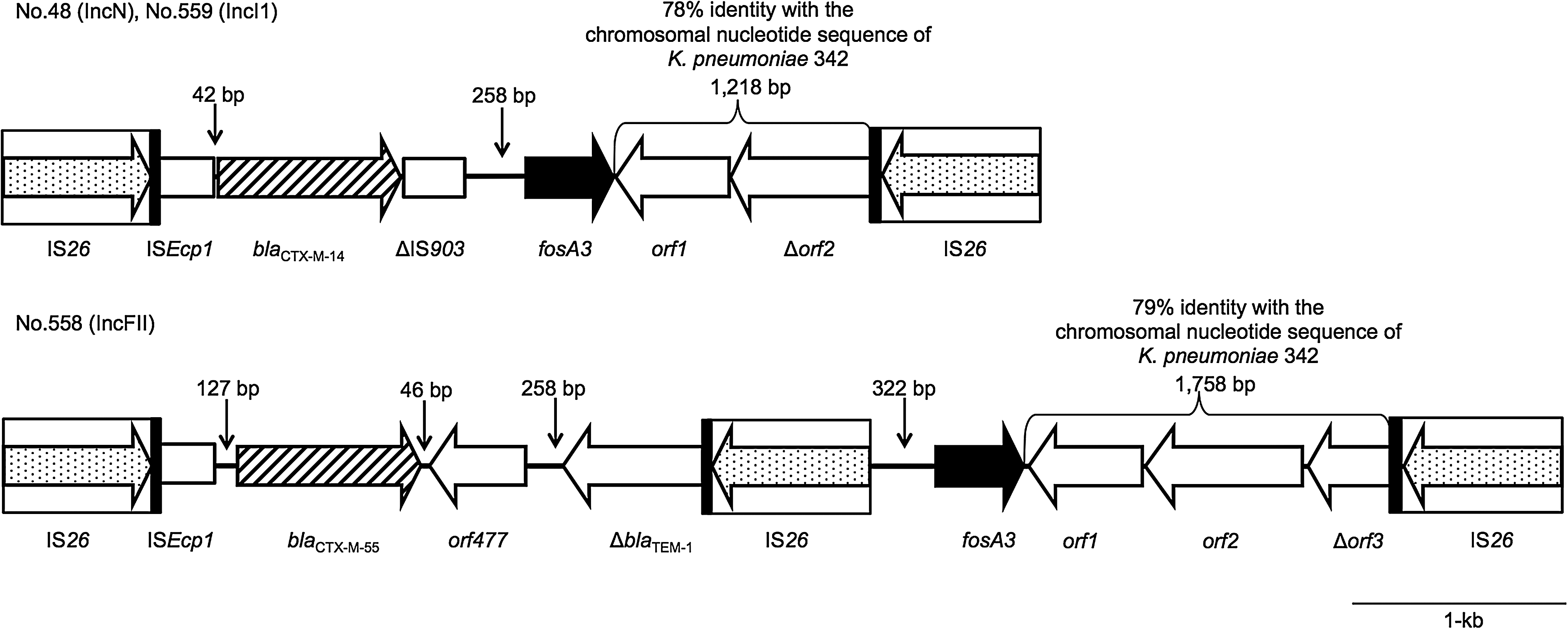
Schematic representation of the genetic environments carrying the fosA3 gene.
Discussion
This study demonstrated the presence of fosfomyin-resistant CTX-M-producing E. coli from the gut of healthy individuals who were not exposed to any antimicrobial agents, including fosfomycin and third-generation cephalosporins in this investigation period (January–August 2010). The rate of fosfomycin resistance was 5.8%, which is similar to that reported in clinical isolates from Japan (3.6%), 26 Spain (9.1%), 19 and Korea (4.2%). 14 No significant difference in the rates of fosfomycin resistance between CTX-M-producing E. coli from healthy individuals and those from clinical specimens were observed. Recently, similar E. coli strains were detected in the gut of domestic animals in China, 12 indicating that fosfomycin resistance in CTX-M-producing E. coli may have occurred early on in a variety of settings.
The most prevalent mechanism of fosfomycin resistance in CTX-M-producing E. coli is the horizontally acquired fosA3.11,14,26 As expected, the carriage of fosA3 was identified at a high rate (62.5%, 5 out of 8) among fosfomycin-resistant CTX-M-producing E. coli isolates, and the fosA3 genes were commonly linked to the blaCTX-M genes, although the genotypes of CTX-M β-lactamases were diverse (i.e., three blaCTX-M-14, one blaCTX-M-3, and one blaCTX-M-55). These results indicate that fosA3-harboring E. coli isolates associated with cefotaxime resistance that was related to CTX-M β-lactamase production naturally dwell in the intestinal flora of healthy individuals as well as in clinical and veterinary settings.13,14,26 As previously reported, the blaCTX-M gene could have widely spread across the members of family Enterobacteriaceae in various settings17,21; fosA3 may have, thus, disseminated across these CTX-M-type ESBL producers. However, reports of fosA3-harboring CTX-M-type ESBL producers have so far been limited to East Asian countries such as Japan, 26 China, 11 and Korea 14 ; these organisms have not yet been reported from other countries in Europe or the United States of America. A worldwide surveillance program is, thus, essential to precisely elucidate the prevalence of acquired fosfomycin-resistance genetic determinants such as that for FosA3.
The resistance determinants of the three fosA3-negative fosfomycin-resistant isolates could not be fully characterized in the present study. Fosfomycin resistance is largely influenced by mutations in specific chromosomal loci such as glpT and murA 23 ; these determinants may well be, therefore, partly involved with the resistance phenotype of the three isolates.
We determined the phenotypic and genotypic characteristics of serotypes, MLST types, and plasmid replicon types of five fosA3-harboring isolates (Table 2). The serotypes of the five isolates varied and were assigned to different STs. The STs of the fosA3-harboring isolates differed from those found in Korea. 14 In addition, the replicon types of the plasmids carrying fosA3 were also diverse. The fosA3-carrying plasmids with the replicon of FII:2 and FII:33 have been earlier identified in fosfomycin-resistant E. coli isolates from humans, food animals, and pets.11–14 These results suggested that fosA3 was incorporated into plasmids of varying incompatibility groups, namely FII:2 or FII:33, and may have been disseminated across genetically different E. coli clones.
As shown by conjugation experiments, fosA3 could be often co-transferred along with the other antibiotic resistance genes. Currently, the second most common antibiotic resistance gene with genetic linkage to fosA3 is the aminoglycoside-resistant 16S rRNA methyltransferase gene rmtB.9,13 In this study, the chloramphenicol and florfenicol resistance phenotype, which was conferred by floR (chloramphenicol and florfenicol efflux transporter), along with fosA3, was transferred in only one of the five E. coli isolates. Hereafter, fosA3 may accumulate in association with a variety of antimicrobial resistance genes in plasmids belonging to different incompatibility groups.
Contrary to the diversity of plasmids carrying fosA3 and in STs of the parent E. coli isolates, the genetic environment of fosA3 showed common patterns regardless of origin, one of which is that it is flanked by IS26 elements.11,13,14 This observation suggests a possibility that fosA3 is commonly associated with mobile genetic units that are driven by the IS26 elements distributed among plasmids with varying replicon types and have spread across various settings such as hospitals, communities, and livestock. The IS26 element would thus be the main vehicle accelerating the spread of small genetic units mediating fosA3, because it usually locates upstream and/or downstream of fosA3. In fact, IS26 is genetically combined with a variety of antibiotic resistance genes and this governs its transfer.20,25,27
In conclusion, we report the first identification of E. coli isolates harboring both fosA3 and blaCTX-M from healthy individuals. The fosA3 gene could have already spread across CTX-M-producing E. coli in various settings such as the clinics, as well as the veterinary and community areas, and could eventually have contributed to their fosfomycin resistance. Although fosfomycin has been recently reconsidered a potent therapeutic option for infectious diseases such as community-acquired UTIs caused by CTX-M-producing E. coli, 6 these bacteria have already been armed with new mechanisms such as fosA3 to hamper fosfomycin. Indeed, the prevalence of fosfomycin resistance among CTX-M-producing E. coli is still less than 10% in most settings at present, but it is of great concerns that fosA3 would further spread with the increase of fosfomycin consumption in both clinical and veterinary settings, thereby decreasing its efficacy in therapeutics.
Footnotes
Acknowledgment
The authors thank the Okazaki City Public Health Center for collecting the E. coli isolates.
Disclosure Statement
This study was supported by the Ministry of Health, Labor, and Welfare of Japan (Grant nos. H21-Shinkou-Ippan-008 and H24-Shinkou-Ippan-010).