
Abstract
Novel mdfA gene variants were identified simultaneously from 3 of 13 positive isolates of PCR amplification in Escherichia coli from patients. These 13 positive isolates showed resistance to chloramphenicol, tetracycline, and erythromycin. The 3 mdfA gene variants were of the same genotype and all the 13 positive isolates were investigated by conjugation experiment, EcoRI restriction, and gene mapping. Conjugation experiments demonstrated that the novel mdfA variant and mdfA genes were located on plasmids that were restricted by EcoRI for ∼8.2 kb-length, which was also validated by gene mapping. Further study indicated three types of genetic structures (A, B, and C) in the recombinant plasmids harboring mdfA and surrounding genes, and structure B was first reported in the article. Structure A comprises two partial-length and six full-length genes, including the mdfA gene variant in the recombinant plasmid; structure B comprises four full-length genes, the mdfA, ybjG, dacC, and ybjI; structure C comprises two full-length genes, the mdfA and dacC. These results suggested that the mdfA gene can function as transporter responsible for multidrug resistance and also mediated the synergistic function with its surrounding genes in conjugative plasmids.
Introduction
A
In addition to its role as a multidrug resistance transporter, MdfA also mediated alkaline resistance. In deletion mutation experiments, mdfA mutants were sensitive even to mild alkaline conditions, and the wild-type phenotype was restored fully by MdfA expressed from a plasmid. MdfA expressed from a multicopy plasmid was found to confer extreme alkaline pH resistance, allowing the growth of cells under conditions that were close to those used normally by alkaliphiles (up to pH 10). Inverted vesicle fluorescence studies demonstrated that MdfA catalyzes Na+- or K+-dependent proton transport. 11
In this study, we described characteristics of the mdfA genes (novel mdfA variant and mdfA genes) in recombinant plasmids from 13 positive isolates of polymerase chain reaction (PCR) amplification, including isolates EC2347, EC2341, and EC2415 of E. coli by EcoRI restriction experiments, which exhibited multidrug efflux activities with an unusually broad pattern of drug specificities.
Materials and Methods
Bacterial strains
Sixty-seven unique multidrug-resistant isolates of E. coli were collected from hospitalized patients from February 2010 to November 2011 in Taizhou Municipal Hospital affiliated with Taizhou University. The patients were distributed in six clinical units, including the ICU ward (17/67), infection unit (15/67), neurology surgery (12/67), respiratory unit (10/67), urology surgery (8/67), and neurology unit (5/67). All the strains were selected from unrelated E. coli isolates recovered from different inpatient units and isolated from sputum (35/67), blood (15/67), urine (12/67), and venous cannula (5/67) in Taizhou Municipal Hospital affiliated with the Taizhou University. EC2347, EC2341, and EC2415 were three of 13 positive isolates of PCR amplification in E. coli. All isolates were identified as E. coli using a Vitek GNI+ card (bioMérieux), simultaneously, species identification was confirmed by sequence analysis of the16S-28S rRNA gene intergenic spacer region.
Conjugation and susceptibility tests
Conjugation experiments were carried out in a Luria-Bertani (LB) broth with E. coli J53 AzideR (a strain with resistance to sodium azide) as the recipient, and PCR amplification-positive strains were used as the donor strains. 23 Cultures of donor and recipient cells in the exponential phase (0.5 ml of each) were added to 4 ml of fresh LB broth and incubated at 37°C overnight without shaking. Transconjugants were selected on trypticase soy agar plates supplemented with 300 mg/L sodium azide and 0.03 mg/L ciprofloxacin, and incubated for 18–24 hr at 35°C. The susceptibility was assessed by minimum inhibitory concentration (MIC) values. To determine if the plasmids harboring the mdfA gene were transferred, the MICs of antibiotics for the donor, recipient, and transconjugant strains were compared. Antimicrobial agent used susceptibility plate of Microscan's broth dilution methods for the following antibiotics: amikacin, aztreonam, cefazolin, cefotetan, ceftazidime, ceftriaxone, chloramphenicol, ofloxacin, erythromycin, gentamicin, imipenem, tetracycline, and tobramycin. The MICs were determined by broth dilution and interpreted according to the Clinical and Laboratory Standards Institute guidelines. 4
PCR amplification and restriction
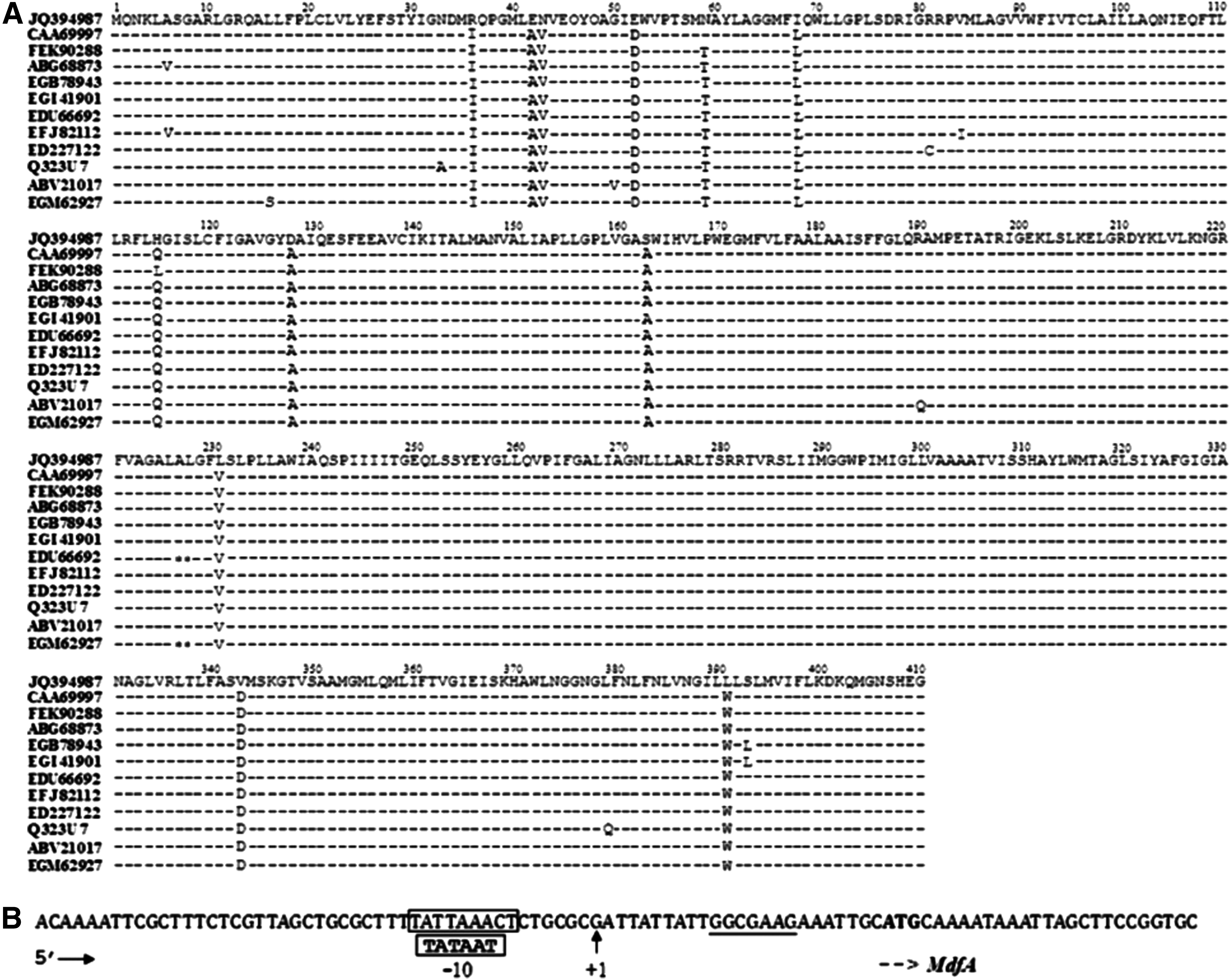
The study was to analyze genetic characteristics and expressions of the mdfA variant and surrounding genes in 13 positive isolates of PCR amplification, including isolates EC2347, EC2341, and EC2415. The PCR systems contained 10×PCR buffer (2.5 μl), dNTP mix (25 mM each nucleotide) (0.2 μl), primer mix (25 pmol/μl each primer) (0.4 μl), Taq DNA polymerase (0.2 μl), genomic DNA template (100 ng/μl) (1.0 μl), and filtered water (pH 7.0) (20.7 μl); the total reactive volume was 25 μl. The final concentrations of the 10×PCR buffer contained 50 mM KCl, 10 mM Tris-HCl, and 1.5 mM MgCl2. PCR amplifications were performed using the following cycling conditions: 3 min at 94°C and 30 cycles of 1 min at 94°C, 53°C, or so and 72°C, respectively, followed by a final elongation step of 10 min at 72°C. The total reactive volume was 25 μl. The primers used to amplify the gst, dacC, deoR, ybjG, MdfA, ybjH, ybjI, and ybiJ genes are listed in Table 2. The results of genetic structures associated with the mdfA variant and surrounding genes for E. coli are listed as Fig. 2. To study promoter structures for the mdfA gene variant, we detected the pre-mdfA gene sequences using primers pre-mdfA, as mentioned in Table 2 and analyzed as shown in Fig. 1. The amino acids of MdfA were compared by BLASTp, as shown in Fig. 1.
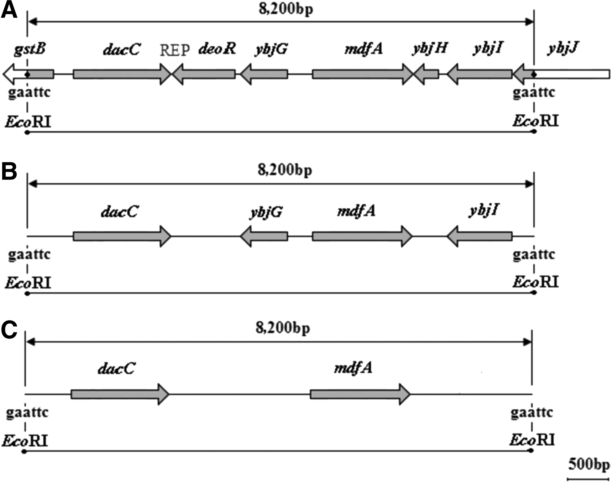
The diagram of genetic structures for the mdfA and surrounding genes in recombinant plasmids by EcoRI retriction (gray region). EcoRI restrictions are indicated as well as the corresponding sites at gaattc. The recombinant plasmids are ∼8.2 kb length. Three types of genetic structures
Genetic structures of the mdfA variant and surrounding genes in isolates of E. coli were cloned by restricting total DNA with EcoRI, ligating it into EcoRI-restricted plasmid pBKCMV, and transforming the recombinant plasmids into E. coli DH10B. Recombinant plasmids were selected on trypticase soy agar plates containing amoxicillin (50 μg/ml) and kanamycin (30 μg/ml). The cloned DNA fragments of several recombinant plasmids were sequenced and analyzed as specification.
Plasmid mapping
To study the plasmids harboring mdfA and surrounding genes, the plasmid DNAs were extracted (Axygen kit) and separated by 0.6% agarose gel electrophoresis (60 V, 90 min) (Promega), and different sizes of plasmid DNA fragments were then recovered. PCR amplification of mdfA and surrounding genes was carried out to determine their position in the recombinant plasmids. The recombinant plasmid DNAs were then sequenced and analyzed. 23
Novel nucleotide sequence accession number
The mdfA gene variant was deposited in GenBank as accession number JQ394987.
Results
Bacteria source
The majority of 67 isolates of E. coli came from the ICU (25.4%), infection unit (22.4%), neurology surgery (17.9%), and most of the strains were isolated from sputum (52.2%), blood (22.4%), and urine (17.9%) in Taizhou Municipal Hospital affiliated with Taizhou University of China. The isolates of EC2347, EC2341, and EC2415 were three representative strains of E. coli, which were isolated simultaneously from sputum in the ICU, blood in urology surgery, and sputum in the infection unit.
Antibiotic susceptibility
All of the 67 isolates of E. coli were sensitive to amikacin, aztreonam, cefazolin, ceftazidime, ceftriaxone, ofloxacin, gentamicin, imipenem, and tobramycin, but 13 of the 67 isolates of E. coli, including isolates EC2347, EC2341, and EC2415, were resistant to chloramphenicol, erythromycin, and tetracycline. Their MIC values were similar to the isolates EC2347, EC2341, and EC2415 (Table 1). The results of susceptibility testing showed that the MIC values of transconjugants for chloramphenicol, erythromycin, and tetracycline were higher than those of E. coli J53 AzideR (the strain was resistant to Azide) (Table 1). Conjugation experiments also indicated that the resistance to chloramphenicol, erythromycin, and tetracycline was conferred by the plasmids.
MIC, minimum inhibitory concentration.
Genetic environment and amino acid analysis for the mdfA genes
There were 13 positive isolates by PCR amplification in 67 isolates using primers listed in Table 2. By sequencing and analyzing, we discovered the same genotype of mdfA variant in isolates EC2347, EC2341, and EC2415, simultaneously, and other mdfA genes in other 10 isolates.
The promoter study of the mdfA variant showed that sequences had two overlapping hexameric sequences at −7 (TAAACT) or −10 (TATTAA), both with four of six matches to the consensus TATAAT, which may constitute a functional −10 promoter region, and a no obvious −35 region was apparent in the mdfA variant. Partial alignments of putative amino acid sequences for mdfA genes deposited in GenBank are shown in Fig. 1 (the JQ394987 was a novel GenBank accession number of mdfA in the study).
Recombinant plasmids and gene mapping
To realize characteristics of these mdfA genes, we studied the plasmids harboring the mdfA and surrounding genes by EcoRI restriction. The structure of recombinant plasmids was also identified with gene mapping. The recombinant plasmids were obtained from 13 positive isolates of PCR amplification (including isolates EC2347, EC2341, and EC2415). The results showed that there were ∼8.2 kb-length fragments harboring the mdfA and surrounding genes (Fig. 2).
Three types of genetic structures (A, B, and C) were also identified in the recombinant plasmids of the 13 positive isolates (including isolates EC2341, EC2415, and EC2347) (Fig. 2). Six isolates (including isolate EC2341) were of genetic structure A, which comprised two partial-length and six full-length genes, including the mdfA, as well as the dacC, mdfA, and other genes (Fig. 2A). Five isolates (including isolate EC2415) were of genetic structure B, which comprised four full-length genes, the ybjG, dacC, mdfA, and ybjI (Fig. 2B). Two isolates (including isolate EC2341) were of genetic structure C, which comprised two full-length genes, the dacC and mdfA (Fig. 2C).
Discussion
Membrane proteins belonging to the MFS transport molecules across the membrane are known to be associated with drug resistance. The MdfA gene is one of the MFS major multidrug efflux pumps, whose overexpression is frequently linked to resistance to tetracycline, erythromycin, and chloramphenicol, and much higher MIC values to ofloxacin and gentamicin compared with recipients (E. coli J53) in conjugation experiments. The E. coli Mdr transporter MdfA is able to transport differentially charged substrates in exchange for protons. This includes neutral compounds, namely, thiamphenicol, chloramphenicol, and lipophilic cations such as ethidium and tetraphenylphosphonium. 12 Our study also demonstrated that the mdfA gene can function as transporter responsible for multidrug resistance. Chloramphenicol resistance is often associated with the presence of chloramphenicol acetyltransferases, which inactivate chloramphenicol, 15 and it may also be due to the efflux of chloramphenicol through specific membrane-associated transporters. 2 Both of these genes encoding chloramphenicol acetyltransferases and specific exporters were often located in plasmids, transposons, or gene cassettes. Tetracycline resistance was most often due to the acquisition of new genes or for a protein. 3 Erythromycin displayed a bacteriostatic activity especially at higher concentrations, but the mechanism was not fully understood. However, the resistance to tetracycline, chloramphenicol, and erythromycin were primarily caused by the mdfA gene in this study.
The mdfA gene variant had more than 98% nucleotide identity with the canonical mdfA gene. Promoter structures of the mdfA variant from the isolates EC2347, EC2341, and EC2415 suggested different mechanisms for the expression of mdfA gene variant compared with conventional means. As shown previously, the mdfA gene did not contain a classical promoter, 17 which was also identified in our study (Fig. 1B), its expression and regulation mechanism was currently unknown. Meanwhile, the putative amino acid sequences of MdfA had 12 sites of difference and showed more than 96% homology with others deposited in GenBank. Varied codons included Iie 36 Arg, Ala 42 Glu, Va l43 Asn, Asp 52 Glu, Thr 59 Asn (except for GenBank accession no. CAA69997), Leu 68 Iie, Gln 115 Met (except Leu 115 His for GenBank accession no. FKK90288), Ala 128 Asp, Ala 163 Ser, Val 231 Leu, Asp 343 Val, and Trp 391 Leu (Fig. 1). Mdr (MdfA) transporters were remarkably promiscuous in various basic aspects of active transport, 12 were able to extrude a large number of chemically dissimilar compounds, and many Mdr transporters also differed in their charge. Moreover, the remarkable versatility of these transporters was exemplified further by their ability to catalyze dissimilar transport reactions. 12
Our results showed that three types of genetic structures (A, B, and C) were identified in the three isolates EC2347, EC2341, and EC2415 and other 10 positive isolates of PCR amplification by EcoRI restriction, and the 8.2 kb-length fragments in recombinant plasmids were identified with gene mapping. The structure A isolated from six isolates, including isolate EC2341, comprised two partial-length and six full-length genes, harboring the mdfA variant and surrounding genes. The C-terminal domain of the MdfA protein (ybjH, ybjI, and ybiJ) was not required for the function of MdfA. Five isolates, including isolate EC2415, contained the sequence of genetic structure B, with four full-length genes, the mdfA, ybjG, dacC, and ybjI, in the recombinant plasmid, and two isolates, including isolate EC2347, contained the sequence of genetic structure C, with two full-length genes, the mdfA variant and dacC genes, in the recombinant plasmid. To our knowledge, genetic structure B, harboring novel mdfA variant and canonical mdfA genes, was first discovered in the study.
Genetic structure A was of MFS. One of the GstB proteins was a glutathione S-transferase that was able to dehalogenate the non-natural toxic chemical bromoacetate. 6 A GstB mutant was hypersensitive to bromoacetate. Overexpression of GstB does not result in resistance to bromoacetate, but decreases its minimum inhibitory concentration. 6 One of the deoR belonged to the DeoR family of transcriptional regulators, the transcriptional repressor DeoR, for “Deoxyribose Regulator,” was involved in the negative expression of genes related to transport and catabolism of deoxyribo-nucleoside nucleotides. 5 The YbjG gene in genetic structures B and C, and its protein, YbjG, was similar to a bacitracin resistance protein BcrC of Bacillus licheniformis. Disruption of ybjG caused increased bacitracin sensitivity, and overexpression caused increased resistance to bacitracin. 7 PBP6, synonymous of dacC protein, 10 existed in genetic structures A, B, and C, was a penicillin-binding protein that was required for proper cell morphology and provides some resistance to penicillin. 21 To genetic structures A, B, and C, deletions in dacC were viable and had no obvious growth defects, although they were slightly penicillin sensitive, and expression of dacC was induced upon entry into a stationary phase by BolA. 8 These suggested that the novel mdfA variant and mdfA gene played leading roles for multidrug resistance in the genetic structures.
Multidrug resistance is most frequently due to active transporters that pump a broad spectrum of chemically distinct, cytotoxic molecules out of cells. Overcoming or circumventing multidrug resistance in a clinical setting has been successful. Recent structural and biochemical data for several multidrug transporters now have provided mechanistic insights into how they work. 21 More and more studies will demonstrate the mechanism of transmembrane proteins such as Cmr in bacteria. In this study, we identified the mdfA gene variant and canonical responsible for multidrug resistance, and study is undergoing for mechanisms of drug resistance.
Footnotes
Acknowledgments
This study was supported by grants from the Zhejiang Natural Science Foundation (Y2100248), the Foundation of Department of Science and Technology of Zhejiang Province (2009C33155), the Foundation of Zhejiang Health Department (2009A218), the Foundation of Taizhou Science and Technology Bureau (081KY30, 102KY15, 1201KY22, and 1301KY36), the Zhejiang Province Chinese Medicine Study Foundation (2011ZA113), and the Foundation of Jiaojiang Science and Technology Bureau of Taizhou (83041 and 112071), China.
Disclosure Statement
No competing financial interests exist.