
Abstract
Serratia marcescens IOMTU115 has a novel 6′-N-aminoglycoside acetyltransferase-encoding gene, aac(6′)-Ial. The encoded protein AAC(6′)-Ial has 146 amino acids, with 91.8% identity to the amino acid sequence of AAC(6′)-Ic in S. marcescens SM16 and 97.3% identity to the amino acid sequence of AAC(6′)-Iap in S. marcescens WW4. The minimum inhibitory concentrations of aminoglycosides for Escherichia coli expressing AAC(6′)-Ial were similar to those for E. coli expressing AAC(6′)-Ic or AAC(6′)-Iap. Thin-layer chromatography showed that AAC(6′)-Ial, AAC(6′)-Ic, or AAC(6′)-Iap acetylated all the aminoglycosides tested, except for apramycin, gentamicin, and lividomycin. Kinetics assays revealed that AAC(6′)-Ial is a functional acetyltransferase against aminoglycosides. The aac(6′)-Ial gene was located on chromosomal DNA.
Introduction
T
Serratia marcescens is well recognized as a nosocomial pathogen and has been responsible for outbreaks in neonatal and pediatric intensive care units 21 and outbreaks of bloodstream infection associated with intravenous drug solutions. 15 Moreover, respiratory colonization with S. marcescens has been a risk factor for outbreaks of bloodstream infection. 18 Drug-resistant S. marcescens isolates have been emerging in medical settings; these isolates produce aminoglycoside modification enzymes [AAC(6′)-Ib, AAC(6′)-Ib-cr, AAC(6′)-Ic, AAC(6′)-Iid, and ANT(3")-Ii],3,4,13,19 16S rRNA methylases (ArmA and RmtB),1,2 and metallo-β-lactamases,9,11,23 although S. marcescens is naturally resistant to tetracycline, amoxicillin, amoxicillin-clavulanate, cephalothin, and colistin. 14
Materials and Methods
Bacterial strains, plasmids, and antimicrobial susceptibility
S. marcescens IOMTU115 was isolated from an endotracheal aspirate of a patient in a medical ward of a hospital in Nepal in 2012. S. marcescens WW4 was originally isolated from environmental water samples in a paper factory in Taiwan. 6 Escherichia coli DH5α (Takara Bio, Shiga, Japan) and E. coli BL21-CodonPlus (DE3)-RIP (Agilent Technologies, Santa Clara, CA) were used as hosts for recombinant plasmids and proteins, respectively. Plasmids pSTV28 (Takara) and pQE2 (Qiagen, Tokyo, Japan) were used for cloning of aac(6′)s and purification of recombinant AAC(6′)s, respectively, as described previously. 16 Minimum inhibitory concentrations (MICs) for S. marcescens IOMTU115 and E. coli transformants with aac(6′) genes were determined using the microdilution method. 8
Genome sequencing and cloning
Genomic DNA of S. marcescens IOMTU115 was extracted using the DNeasy Blood and Tissue kit (Qiagen) and sequenced with MiSeq (Illumina, San Diego, CA). More than 20-fold coverage was achieved for IOMTU115. A new 6′-N-aminoglycoside acetyltransferase variant was designated as aac(6′)-Ial. aac(6′)-Ial and its genetic environment (315,403 bp) was deposited in GenBank with the Accession Nos. AB871481 and AB894481, respectively. A putative aac(6′) gene of S. marcescens WW4 was designated as aac(6′)-Iap and deposited in the GenBank with the Accession No. AB979699.
The aac(6′)-Ic gene was synthesized by Funakoshi, Tokyo, Japan. aac(6′)-Ic, aac(6′)-Ial, and aac(6′)-Iap genes were cloned into the multiple cloning site of pSTV28, as described previously. 17 E. coli DH5α was transformed with pSTV28-aac(6′)-Ic, -aac(6′)-Ial, and -aac(6′)-Iap to determine the MICs for aminoglycosides.
Amino acid sequence alignment was calculated using the CLUSTAL W2 program. Genetic environments surrounding drug-resistant genes were annotated and compared using the BLAST database (http://blast.ncbi.nlm.nih.gov/Blast.cgi?CMD=Web&PAGE_TYPE=BlastHome).
Analysis of aminoglycoside acetyltransferase activity
The open reading frames (ORFs) of aac(6′)-Ic, aac(6′)-Ial, and aac(6′)-Iap were cloned into the pQE2 expression vector with the same primer sets as described above. Purification of the recombinant AAC(6′)-Ic, AAC(6′)-Ial, and AAC(6′)-Iap proteins and thin-layer chromatography analysis were performed as described previously. 17
Results
Drug resistance profile of S. marcescens IOMTU115
The MICs of S. marcescens IOMTU115 were as follows: ampicillin, 32 μg/ml; ampicillin–sulbactam, 16 μg/ml; arbekacin, 8 μg/ml; amikacin, 8 μg/ml; aztreonam, <0.125 μg/ml; ceftazidime, 0.25 μg/ml; cephradine, 1,024 μg/ml; cefepime, 0.25 μg/ml; cefotaxime, 0.25 μg/ml; cefoxitin, 32 μg/ml; ciprofloxacin, <0.125 μg/ml; colistin, >1,024 μg/ml; fosfomycin, 32 μg/ml; gentamicin, 2 μg/ml; imipenem, 8 μg/ml; kanamycin, 16 μg/ml; meropenem, <0.125 μg/ml; penicillin, >1,024 μg/ml; tigecycline, 2 μg/ml; and tobramycin, 4 μg/ml.
Amino acid sequence of AAC(6′)-Ial enzyme
AAC(6′)-Ial consists of 146 amino acids. Multiple sequence alignments among AAC(6′) enzymes revealed that AAC(6′)-Ial had 97% identity to AAC(6′)-Iap from S. marcescens, 6 92% identity to AAC(6′)-Ic from S. marcescens, 13 38% identity to AAC(6′)-Iag from Pseudomonas aeruginosa, 5 and 37% identity to AAC(6′)-Iy from Salmonella enterica. 7 Both AAC(6′)-Ial and AAC(6′)-Iap are newly described in this study, although AAC(6′)-Iap was deposited as a putative protein in a complete genome sequence of S. marcescens WW4 (Accession No. NC020211). Biological properties of AAC(6′)-Iap had not been analyzed. As shown in Fig. 1, amino acid substitutions existed at positions 4, 18, 23, 24, 25, 26, 28, 56, 64, 84, 86, 95, and 122 among the three AAC(6′) variants.
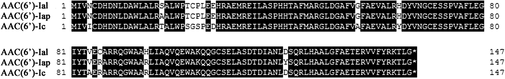
Alignment of AAC(6′)-Ic, AAC(6′)-Ial, and AAC(6′)-Iap amino acid sequences. Identical residues are marked with black boxes.
Aminoglycoside susceptibility of E. coli transformants
As shown in Table 1, a vector control of E. coli DH5α/pSTV28 was susceptible to all the aminoglycosides tested, whereas E. coli DH5α/pSTV28-aac(6′)-Ic, aac(6′)-Ial, and aac(6′)-Iap were resistant to all aminoglycosides, except for apramycin, gentamicin, and lividomycin, with 2- to 64-fold higher MICs than those of the vector control. The MICs of apramycin, gentamicin, and lividomycin in E. coli expressing aac(6′)-Ic, aac(6′)-Ial, and aac(6′)-Iap were the same as those in the vector control. The MIC profiles were essentially similar among the E. coli expressing aac(6′)-Ic, aac(6′)-Ial, and aac(6′)-Iap (Table 1). E. coli expressing aac(6′)-Ial and aac(6′)-Iap were resistant to dibekacin, with MICs 64-fold higher than those of the other aminoglycosides.
MICs of aminoglycosides for E. coli DH5α transformants with aac(6′)-Ic, aac(6′)-Ic, and aac(6′)-Ic were determined using the microdilution method. 8
MICs for E. coli strains were determined with Mueller–Hinton broth preparations and individual aminoglycosides.
ABK, arbekacin; AMK, amikacin; APR, apramycin; DIB, dibekacin; GEN, gentamicin; ISP, isopamicin; KAN, kanamycin; LIV, lividomycin; MIC, minimum inhibitory concentration; NEO, neomycin; NET, netilmicin; SIS, sisomicin; SPM, spectinomycin; STM, streptomycin; TOB, tobramycin.
TLC analysis and kinetics against aminoglycosides
The results of TLC analysis of aminoglycosides acetylated by AAC(6′) enzymes were shown in Fig. 2. Lividomycin was used as a negative control because it has a hydroxyl group instead of an amino group at the 6′ position and, therefore, cannot be acetylated by AAC(6′). In total, 9 out of the 12 aminoglycosides tested were acetylated by AAC(6′)-Ic, AAC(6′)-Ial, and AAC(6′)-Iap, with the exception of apramycin, gentamicin, and lividomycin (Fig. 2). Kinetics parameters of AACs(6′) against the nine aminoglycosides were determined, and the results are shown in Table 2. The profiles of the kinetics parameters were essentially similar among the three enzymes. The kcat/Km values against dibekacin were relatively high compared with those against other aminoglycosides, whereas the values against amikacin were relatively low (Table 2). The substrate specificity of AAC(6′)-Ial is similar to that of AAC(6′)-Ic and AAC(6′)-Iap. The kinetics parameters of the three enzymes against dibekacin were relatively high among the three enzymes, whereas those against amikacin were relatively low (Table 1). These differences in substrate specificity will be associated with an amino group at position 1 of ring I. Dibekacin has an amino group, whereas amikacin has a hydroxyl group at position 1 of ring I.

TLC analysis of acetylated aminoglycosides by AAC(6′)-Ic
The proteins were initially modified by a His-tag, which was removed after purification.
Km and kcat values represent the means of three independent experiments±standard deviations.
Genetic environments surrounding aac(6′)-Ial and aac(6′)-Iap
As shown by the genome rearrangement map in Fig. 3, a genetic region of S. marcescens IOMTU115 from nucleotide (nt) 1 to 315,403 (Accession No. AB894481) had 97% identity to a region of S. marcescens WW4 from nt 4,490,655 to 4,842,041 (Accession No. CP003959) (Fig. 3). Bacteriophage-derived genes with 33,097 bp were inserted from nt 4,583,432 to 4,616,528 in WW4. The downstream region of the genetic environment surrounding aac(6′)-Ial was similar to the one surrounding aac(6′)-Iap (Fig. 3). The downstream region of the genetic environment surrounding aac(6′)-Ial contained several housekeeping genes, including dnaG, rpoD, and mug (Fig. 3), indicating that aac(6′)-Ial was located in the chromosome.

A genome rearrangement map between Serratia marcescens IMOTU115 (Accession No. AB894481) and S. marcescens WW4 (Accession No. CP003959), and the comparison of the genomic environments surrounding aac(6′)-Ial in IOMTU115 and aac(6′)-Iap in WW4. The genetic map from nt 1 to 315,403 in S. marcescens IMOTU115 had 97% identity compared to that from nt 4,490,655 to 4,842,041 in S. marcescens WW4. The upstream region of genomic environment surrounding aac(6′)-Ial was similar to that surrounding aac(6′)-Iap. The genomic environment surrounding aac(6′)-Ial contained several housekeeping genes, including dnaG, rpoD, and mug.
Discussion
Amikacin and isepamicin share a characteristic structure of an
These intrinsic aac(6′) variants in S. marcescens may be evolved while mutating in their unique ways in the conserved genetic environment, because these genes were not observed in other organisms. Our study suggests that aac(6′) variants in S. marcescens have potential to become more resistant to aminoglycosides by causing further genetic selection. Shaw et al. reported that expression of the AAC(6′)-I aminoglycoside resistance profiles can be due to (1) mutation of a gene encoding a negative regulator, (2) insertion upstream or within the 5′ coding region of the aac(6′)-I gene in S. marcescens, or (3) point mutations that create a new promoter. 13
The aac(6′) genes may contribute to aminoglycoside resistance in S. marcescens. The aac(6′)-Ic, aac(6′)-Ial, and aac(6′)-Iap were detected in clinical and environmental isolates of S. marcescens.6,13 Therefore, further monitoring and investigation of aminoglycoside-resistant S. marcescens are necessary to understand the diversity and evolution of S. marcescens intrinsic aac(6′) genes.
Footnotes
Acknowledgments
This study was approved by the Institutional Review Board of the Institute of Medicine at Tribhuvan University (6-11-E) and the Biosafety Committee of the Research Institute of the National Center for Global Health and Medicine (Approval Nos. 26-D-088 and 26-D-089). This study was supported by a grant from Japan Agency for Medical Research and Development (AMED), a grant (26-A-103) from International Health Cooperation Research, and a grant from Kanae Foundation for Asia-Oceania Collaborative Research.
Disclosure Statement
No competing financial interests exist.