
Abstract
The purpose of this study was to assess the antimicrobial resistance profiles of several strains of Lactobacillus and Bifidobacterium spp. of probiotic interest. The International Organization for Standardization method was used to determine the minimal inhibitory concentrations of the antibiotics recommended by the European Food Safety Authority (EFSA). As a result, some of the tested microorganisms showed a visible growth up to the microbiological cutoff values indicated by EFSA guidelines in 2012. We were not able to categorize these strains as susceptible or resistant on the basis of antimicrobial resistance phenotypic testing as EFSA document does not explicitly deal with such a phenotypic condition where strains grow at antibiotic concentrations up to the established cutoff value. Although a few strains have been analyzed for this study, our findings highlight a potential challenge in accurately determining the antibiotic resistance in specific strains relevant for human and animal health.
Introduction
T
Antimicrobial susceptibility testing can be performed at two different levels: phenotypic and genotypic. Many methods of phenotypic testing for antimicrobial susceptibility are currently available, including E test, agar disc diffusion, and agar and broth dilution. 7 The international standard published by the International Organization for Standardization (ISO) and the International Dairy Federation (IDF) 8 recommends broth microdilution as the standard method following the results of the EU funded Assessment and Critical Evaluation of Antibiotic Resistance Transferability in Food Chain (ACE-ART) project, which was devoted to studying drug resistance in nonpathogenic food-related bacteria. Many factors need to be considered when choosing the right method, but reproducibility, reliability, and accuracy are of major importance. 9
A second key issue for assessing resistance is the availability of suitable and clear criteria for interpreting test results to promote uniform application of guidelines and draw unbiased conclusions.
This study, aimed to assess antibiotic resistance in eight potentially probiotic strains of LAB and Bifidobacterium, shows the importance of applying appropriate criteria in the interpretation of antimicrobial susceptibility testing results.
Materials and Methods
Bacterial strains and culture conditions
A total of eight strains from the collection of the Microbiology Institute of Università Cattolica del Sacro Cuore (Piacenza) were analyzed. The strains used in this study included five lactobacilli (Lactobacillus plantarum, Lactobacillus paracasei, Lactobacillus delbrueckii subsp. bulgaricus, Lactobacillus acidophilus) and three Bifidobacterium strains (two Bifidobacterium breve, Bifidobacterium animalis subsp. lactis). The taxonomic identity of all strains was confirmed previously by species-specific polymerase chain reaction (PCR).10–15
Cryopreserved cultures of all Lactobacillus strains were inoculated on de Man, Rogosa, Sharpe (MRS) (Difco)/agar (Oxoid) plates. The MRS for Bifidobacterium included 0.05%
Antibiotic susceptibility testing
The phenotypic resistance of all strains to antimicrobial agents was determined using the broth microdilution method following procedures set by the ISO 10932/IDF 223 standard. 8
All assays were carried out in sterile 96-well microtiter plates (Sterilin) containing appropriate concentrations of antibiotics as indicated by the ISO/IDF protocol. 8 The antibiotics used for each species were based on EFSA recommendations (gentamicin, kanamycin, streptomycin, tetracycline, erythromycin, clindamycin, ampicillin, vancomycin, and chloramphenicol). The plates were incubated for 48 hours at 37°C under anaerobic conditions. At the end of the incubation period, the samples were read with a microplate photometer (Multiskan EX; Thermo Scientific). Each assay was performed in triplicate.
PCR detection of antibiotic resistance genes
Strains displaying growth until the cutoff value were subsequently analyzed for the presence of antibiotic resistance genes. The following primers were used: Catfw/Catrev for cat (chloramphenicol resistance), 16 erm(B)-1/erm(B)-2 for clindamycin, 17 and the primers described by Ouoba et al. 18 for aminoglycosides.
Reverse transcriptase-polymerase chain reaction
ErmB PCR-positive strains were grown in MRS with or without clindamycin (0.25 μg/ml for L. acidophilus and 0.5 μg/ml for L. plantarum). Harvested cells were treated with RNA protect Bacteria Reagent (Qiagen) to stabilize the RNA and stored at −80°C. Total RNA was purified using the RNeasy Mini Kit with on-column DNAse I digestion according to the manufacturer's instructions. Any residual DNA was removed by DNAse I digestion (Ambion) in solution after RNA purification. RNA was qualitatively and quantitatively analyzed using a BioPhotometer (Eppendorf) and the Experion automated electrophoresis system (Biorad). The expression of ermB was determined by reverse transcriptase-polymerase chain reaction (RT-PCR) using the Verso-1 Step RT-PCR kit (Thermo Scientific) and previously described primers. Positive control (RNA from E. faecalis UC7251), no-template control (water), and an RT negative control (no RT enzyme) were included in each run. The reference gene 16S rRNA was amplified using primers Uni331F/Uni797R 19 and served as an internal control. RT-PCR products were observed under UV light on a 2% agarose gel after electrophoresis.
Results
The results were analyzed using the cutoff values indicated by EFSA,
1
categorizing the strains as follows:
• Susceptible (S): a bacterial strain is defined as susceptible when it is inhibited at a concentration of a specific antimicrobial equal or lower than the established cutoff value (S ≤ x mg/L). • Resistant (R): a bacterial strain is defined as resistant when it is not inhibited at a concentration of a specific antimicrobial higher than the established cutoff value (R > x mg/L) (EFSA
1
).
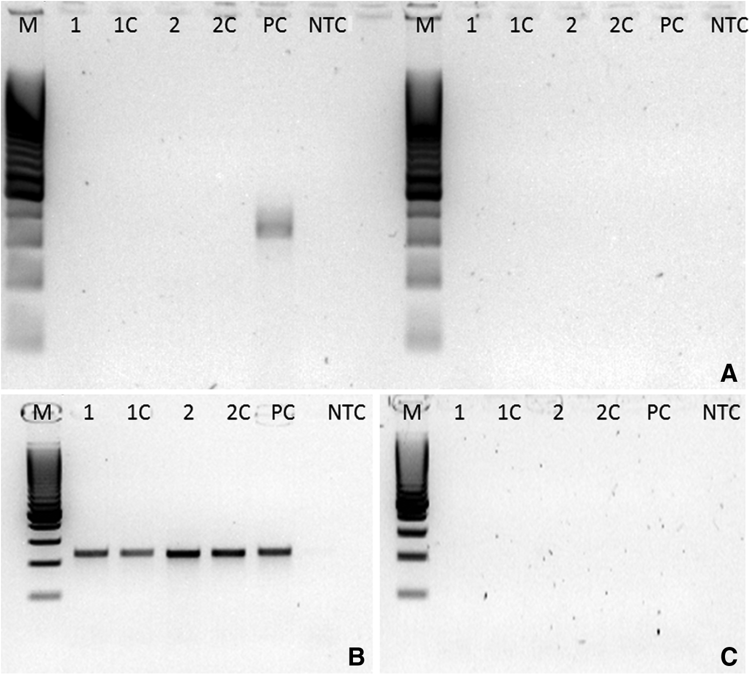
Following these indications, L. acidophilus and L. paracasei were susceptible to all tested antibiotics (data not shown). The resistance of individual strains is provided in Table 1. Notably, we found that almost every strain grew until the cutoff value for at least one tested antibiotic, as reported in Table 2. PCR was performed on the strains that displayed growth until cutoff values were negative for cat and aminoglycoside genes, whereas the ermB gene was detected in both L. acidophilus and L. plantarum strains (data not shown). Thus, we tried to assess gene expression using RT-PCR. No RT-PCR amplification products were detected in RNA samples extracted from L. acidophilus and L. plantarum cultures under noninducible and inducible growth conditions (Fig. 1).
MIC, minimal inhibitory concentration.
A band of the expected size (639 bp) was present only for RNA isolated from Enterococcus faecalis UC7251 cells, indicating no detectable mRNA expression of ermB in the bacterial strains under investigation. The quality of RNA extraction, reverse transcription, and amplification reactions was confirmed by successful amplification of a 466 bp fragment of the 16S rRNA gene from all RNA samples. No product was seen in the RT negative control, indicating that the band observed in the positive control was not derived from contaminating genomic DNA.
Discussion
Phenotypic analysis of antimicrobial resistance is the very first step in the safety assessment of bacterial strains intended for probiotic or nutritional use. EFSA guidance gives the indications for the interpretation of results and provides strict definitions for the classification of resistant and susceptible strains, based on their growth below or above established cutoff values. In our study, a problem with interpreting the results arose when growth of the strains occurred at the cutoff value. For example, L. plantarum displayed growth at the cutoff value for clindamycin but was inhibited at higher concentrations. On the basis of the EFSA definitions, this strain cannot be categorized as susceptible to this antibiotic because it is not inhibited at the cutoff value. In contrast, for the same definitions, this strain cannot be considered resistant because it should not be inhibited at concentrations above the cutoff value. Therefore, the definitions do not clearly take into consideration the possibility of bacterial growth at the antibiotic cutoff value.
The EFSA document uses the EUCAST document as a reference. The EUCAST document introduced the concept of epidemiological cutoff values indicating the upper limit of the wild-type minimal inhibitory concentrations (MICs) distribution of each antimicrobial for a bacterial population. Wild-type strains are defined by the absence of acquired and mutational resistance mechanisms. Microbiological cutoff values were established on the basis of a huge amount of data collected from the literature and from national and European monitoring programs. Our values of MICs were obtained by testing a few isolates only, so it is clear that a larger number of isolates should be evaluated to corroborate these findings. Anyway, our study documents the presence of several bacterial strains whose susceptibility testing produces results that oscillate between resistant and susceptible. Since strains displaying such behavior cannot truly be classified as wild-type, they should be suspected of having potentially transmissible resistance mechanisms.
The use of appropriate methods for antibiotic resistance testing and accurate interpretation of results should always be in mind, given that antibiotic resistance does not respect geographic or biological borders. The use of antibiotics in one sector, setting, or country affects the spread of resistance in others. 20 The inappropriate interpretation of antibiotic resistance results in the food chain may have grave consequences.
Footnotes
Disclosure Statement
No competing financial interests exist.