
Abstract
The aim of this study was to characterize virulence determinants and antibiotic resistance profiles in enterococci obtained from various clinical sources in the northwest of Iran. A total of 160 enterococcal clinical isolates from various wards of University Teaching Hospitals were collected and specified by biochemical test, from September 2014 to July 2015. Identification of enterococci was confirmed by multiplex PCR in the genus and species level. Antibiotic resistance properties and virulence determinants were examined by phenotypic and molecular methods. Of 160 enterococcal isolates, 125 (78.12%) and 35 (21.88%) isolates were identified as Enterococcus faecalis and Enterococcus faecium, respectively. The most common antibiotic nonsusceptible pattern observed was resistance toward rifampicin [n = 122 (76.25%)] followed by erythromycin [n = 117 (73.12%)]. Among all isolates, gelE [n = 140 (87.5%)], cpd [n = 137 (85.6%)], and asa1 [n = 118 (73.8%)] were the most prevalent virulence genes studied. Thirty isolates (11 E. faecalis, 19 E. faecium) were found to be resistant to vancomycin, with minimum inhibitory concentration of ≥256 μg/ml. Twenty-seven isolates carried the vanA gene, whereas none of the isolates carried vanB. E. faecalis had a considerable ability to show virulence genes and drug resistance. Emergence of antibiotic-resistant enterococci and the high prevalence of virulence traits in our study could be regarded as an alarming situation.
Introduction
E
Since multidrug-resistant microorganisms can impose many problems for healthcare systems, including increased length and expenses of hospitalization, and also, new data about virulence and antibiotic resistance of enterococci circulating species in northwest of Iran are still scarce, this study aimed to determine the antibiotic susceptibility patterns and virulence determinants of the isolates and also, to characterize vancomycin-resistant (VR) E. faecalis and E. faecium from different human clinical specimens.
Materials and Methods
Sampling
A total of 160 enterococcal isolates obtained from different human clinical specimens, including urine, blood, body fluids, wound, and catheter, were collected from inpatients and outpatients at the University Teaching Hospitals in Urmia and Tabriz, northwest of Iran, during the time period September 2014 to July 2015.
Identification of Enterococcus spp.
The enterococcal isolates were identified to the species level according to standard biochemical tests 8 later confirmed by PCR. 9
Antibiotic susceptibility testing
Three methods were used to study the antimicrobial susceptibility patterns of all enterococcal isolates according to the Clinical and Laboratory Standards Institute (CLSI) guidelines 10 : (1) Kirby-Bauer disk diffusion technique, (2) screening agar method for gentamicin and vancomycin, and (3) determination of the minimum inhibitory concentration (MIC) by the E-test method.
Kirby-Bauer standard disc diffusion method
The enterococci isolates were subjected to antibiotic sensitivity testing on Mueller-Hinton agar (Merck, Co., Germany) by Kirby-Bauer, standard disc diffusion method, according to CLSI. 10 Susceptibility of Enterococcus isolates was tested for the following antibiotics (Mast Group Ltd., Merseyside, UK): ampicillin (AMP, 10 μg), vancomycin (VAN, 30 μg), erythromycin (ERY, 15 μg), doxycycline (DXT, 30 μg), gentamicin (GM, 120 μg), streptomycin (S, 300 μg), linezolid (LZD, 30 μg), ciprofloxacin (CIP, 5 μg), teicoplanin (TEC, 30 μg), penicillin G (PG, 10units), rifampicin (RP, 5 μg), and quinupristin/dalfopristin (Qui/Dal, 15 μg). For urine isolates, susceptibility testing for nitrofurantoin (NI, 300 μg) was also done. For quality control, E. faecalis ATCC 29212 was used.
Vancomycin screening agar test
For each isolate which showed intermediate susceptibility to vancomycin in the disc diffusion test (DDT), vancomycin (6μg/ml) screening agar test was performed according to CLSI guidelines. 10 E. faecalis ATCC 29212 was used as a negative control, and VR E. faecium OE-3 was used as a positive control collected in an earlier work on enterococci. 11
Gentamicin screening agar test
Based on the results of the disc diffusion test (DDT), each isolate, which showed a diameter of the zone of inhibition of 7–9 mm to high level gentamicin (GM, 120 μg), indicated that the test was inconclusive; therefore, gentamicin (500 μg/ml) agar screening test was performed according to the recommendations of CLSI. 10 E. faecalis ATCC 29212 was used as the negative control and previously isolated gentamicin-resistant strain was taken as the positive control. 11
MIC determination
The MICs of linezolid, quinupristin/dalfopristin, and daptomycin were determined for vancomycin-resistant enterococci (VRE) strains as alternative therapeutic options using the E-test (Liofilchem, Italy) method, based on the manufacturer's instructions and CLSI guidelines. 10 For quality control, E. faecalis ATCC 29212 was used as the susceptible control.
Molecular assay
Detection of genus and species
Multiplex PCR was performed to further confirm isolates at the genus and species level by using rrs (16SrRNA) gene and genes encoding D-alanine-D-alanine ligases specific to E. faecalis (ddlE. faecalis) and E. faecium (ddlE. faecium). Total DNA extraction and multiplex PCR assay were done as previously described.9,12 The primer sequences and the expected sizes of amplicons are presented in Table 1.
Detection of virulence factors and antibiotic resistance genes
Genes encoding virulence factors (gelE, esp, ace, asa1, cpd, hyl, cylA), gentamicin resistance gene (aac(6′)Ie-aph(2″)Ia), and vancomycin resistance determinants (vanA and vanB) were investigated by multiplex PCR, using specific primers and PCR conditions as previously described.11–13 The primer sequences and the expected sizes of amplicons for each PCR assay are shown in Table 1. The amplicons were analyzed by electrophoresis in 1.5% agarose gels using the Tris/Borate/EDTA buffer. The agarose gels were stained with ethidium bromide and visualized on a ultraviolet transilluminator.
Statistical analyses
The values are presented as percentages. Pearson's chi-square test was used for comparison of the frequency. All statistical analyses were performed by using SPSS22 software. p Values of <0.05 were assumed statistically significant.
Results
A total of 160 isolates were collected; 114 (71.25%) were obtained from inpatients and 46 (28.75%) from outpatients. Table 2 displays the demographic data of patients. Sixty-seven (41.87%) isolates were isolated from males, while 93 (58.13%) were from females. The age of the patients ranged between <1 and 95 years, with the maximum number of isolates obtained in the age group of 45–60 years (n = 42; 26.25%) (Table 2). The majority of strains (78.12%) were E. faecalis; while E. faecium accounted for 21.88%. E. faecalis covered a greater percentage of isolates from urine and blood cultures, in comparison to wound, body fluids, and catheter cultures (Table 2).
ICU, intensive care unit.
Table 3 shows the antibiotic susceptibility profile of enterococcal species. The most common resistance was observed for rifampicin 122 (76.25%), which was followed by erythromycin 117 (73.12%). Table 4 depicts the multiple antibiotic resistance patterns in E. faecalis and E. faecium isolates. The most common patterns were ampicillin resistant-gentamicin resistant (28 isolates), which were followed by vancomycin resistant-teicoplanin resistant (27 isolates). Among 30 E. faecalis and 2 E. faecium isolates, which showed intermediate susceptibility to vancomycin in DDT, 4 isolates exhibited resistance in the vancomycin screening agar test. According to the in vitro antibiotic susceptibility testing by E-test method, totally, 30 (18.75%) isolates (11 E. faecalis, 19 E. faecium) were identified as VRE. All VRE strains showed vancomycin MIC ≥256 μg/ml, and 27 isolates carried the vanA gene, whereas none of the isolates carried vanB. The highest prevalence of the vanA gene was observed in E. faecium strains isolated from Tabriz with a frequency of 47.82% (n = 11). On the contrary, four E. faecalis isolates showed intermediate susceptibility to gentamicin in DDT, with all of them showing resistance in the gentamicin screening agar test. Totally, 81 (50.63%) isolates were of high-level gentamicin resistant (HLGR).
AMP, ampicillin; CIP, ciprofloxacin; DXT, doxycycline; ERY, erythromycin; GM120, gentamicin120; LZD, linezolid; NI, nitrofurantoin; PG, penicillin G; Qui/Dal, quinupristin/dalfopristin; RP, rifampicin; S, streptomycin; TEC, teicoplanin; VAN, vancomycin.
The results of the MIC using linezolid, quinupristin/dalfopristin, and daptomycin showed 46.66%, 100%, and 66.66% resistance in VRE isolates, respectively.
The cpd, gelE, and asa1 were the most common virulence genes in E. faecalis isolates; the prevalences of these genes were 100%, 88%, and 74.4% respectively. In E. faecium isolates, gelE (85.7%), hyl (77.1%), and asa1 (71.4%) were the most common detected genes (Table 5). Among E. faecalis isolates, asa1+, gelE+, and cpd+ genotype were the most common virulence gene patterns; in contrast, among E. faecium gelE+, aac(6′)Ie-aph(2″)Ia+ genotype was the most common (Table 6). Seventy-three (45.62%) out of 160 Enterococcus spp. isolates tested (56 E. faecalis and 17 E. faecium isolates) showed gelE+, aac(6′)Ie-aph(2″)Ia+ genotype (Table 6). In this study, gelE virulence gene was detected in E. faecalis strains isolated from Urmia with the frequency of 94.11% (n = 64). In addition, hyl gene was the least virulence gene detected among E. faecalis isolates in Tabriz (n = 1; 1.75%).
Figures 1 and 2 show PCR results for the detection of virulence factors and vancomycin resistance gene in E. faecium and E. faecalis isolates, respectively.

Detection of asa1, esp, and gelE genes in Enterococcus isolates by multiplex PCR. Lane 1, 50 bp size marker; Lane 2, positive control for asa1, esp, and gelE virulence genes; Lanes 3 and 4, Enterococcus strain isolated from clinical specimens; Lane 5, negative control without DNA.
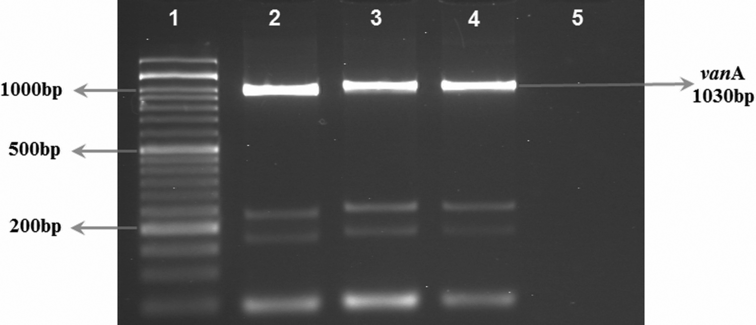
PCR detection of vanA gene in isolates. Lane 1, 50 bp size marker; Lane 2, positive control for vanA gene; Lanes 3 and 4, vanA gene in Enterococcus strain isolated from clinical specimens; Lane 5, negative control without DNA.
Discussion
This study investigated the prevalence of virulence factors and antibiotic resistance profiles in Enterococci spp. isolated from clinical specimens.
According to multiplex PCR results, all 160 enterococci isolates were identified as E. faecalis and E. faecium, and the prevalence rate of E. faecalis was more than that of E. faecium. Most of Enterococcus spp. isolates were recovered from outpatients (n = 46; 28.75%), which were followed by internal ward (n = 37; 23.12%). The prevalence rate of E. faecalis isolates was higher in Urmia hospitals (54.4%) than Tabriz hospitals (45.6%); however, this difference was not statistically significant (p = 0.053). The observations on the prevalence of enterococcal species were similar to those of other authors, noting that the isolation rate of E. faecalis was more than that of E. faecium among clinical enterococci isolates.14,15
Antibiotic resistance among enterococci is a global problem. 14 Although some clinical isolates of enterococci showed antimicrobial activity against closely related species, the presence of multiple potential virulence factors and resistance to clinically relevant antibiotics, especially the emergence of VRE strains, 16 and the ability of transferring van genes to other bacteria, such as Methicillin-resistant Staphylococcus aureus, have put them in the spotlight of the researchers. 17 Depending on the DDT results, the current study revealed that the E. faecium isolates showed a higher rate of antibiotic resistance than E. faecalis; this was in accordance with the findings of other studies, showing the high prevalence of antibiotic resistance among clinical E. faecium isolates.14,15 Beta-lactams (ampicillin and penicillin) are important drugs for the treatment of enterococcal infections. The relatively high frequency of ampicillin-resistant E. faecium (n = 30; 85.7%) and E. faecalis (n = 12; 9.6%) isolates have been observed in this study. It is an alarming condition for treatment of severe enterococcal infections such as endocarditis, since it limits the synergistic therapy with a cell wall-active antibiotic such as ampicillin plus an aminoglycoside such as gentamicin. Similar results have been reported by Gangurde et al. 18 However, long-term use of these antimicrobials in hospitals, community, and veterinary practice in Iran may be the primary reason for the presence of penicillin-resistant and ampicillin-resistant isolates.
Furthermore, rifampicin and erythromycin resistance in this study was quite high in accordance with findings reported by Sharifi et al. 19
In our study, 30 (18.75%) isolates were found to be resistant to vancomycin, with E. faecium (63.3%) showing higher resistance than E. faecalis (36.7%). In this study, the prevalence of VRE (18.5%) was higher than that of another report on the prevalence of VRE (12%) in Tehran, Iran. 15 A similar enhancement in the prevalence of VRE, especially E. faecium, has been noticed earlier in several studies reported from different countries.20,21 Of all 30 VRE isolates, 27 (90%) carried the vanA gene, while the vanB gene was not detected in any of them, as reported by another study carried out in Tehran, Iran. 15 The progressive increase in VRE prevalence in Iran is disturbing; therefore, vancomycin cannot serve as the drug of choice for treating multidrug-resistant gram-positive cocci infections in near future.
As reported, the selection of antibiotics for the treatment of VRE infections was very limited and isolates were resistant to most of the common drugs. In this investigation, VRE strains were tested for alternative therapeutic options using the E test. According to the obtained results, the rates of resistance among VRE strains to linezolid, quinupristin/dalfopristin, and daptomycin were 46.66%, 100%, and 66.66%, respectively. This was in contrast with a previous study from the northwest of Iran, which reported that all VRE isolates were susceptible to linezolid and daptomycin. 22
The high-level resistance to aminoglycoside in enterococci is usually encoded by aac(6′)Ie-aph(2″)Ia gene. 23 In our study, the frequency of HLR to gentamicin among the enterococcal isolates was 50.63%, with a significant difference between E. faecalis (76.54%) and E. faecium (23.45%) isolates (p < 0.05). Furthermore, the PCR results of isolates confirmed that all HLGR isolates contained aac (6′) Ie-aph (2″) Ia gene. These results were similar to those of another report from Iran, showing the prevalence rate of HLGR to be 52%; furthermore, all of them contained aac (6′) Ie-aph (2″) Ia gene. 15
To consider the collection of the isolates from two cities revealed the frequent occurrence of VRE (vanA+) and HLGR strains among the E. faecalis isolates from Urmia compared with those from Tabriz. However, the major differences in the occurrence of resistance between the two regions were most likely due to the differences in the usage of therapeutic drugs in hospitals.
The presence of virulence factors associated with enterococci enhanced their pathogenicity. 6 We found that the prevalence of virulence determinants in E. faecalis and E. faecium differed significantly, which was consistent with the findings of previous studies.19,24 Moreover, it was found that E. faecalis strains had a larger number of virulence determinants, in comparison with E. faecium, as shown in a previous study. 19
The present study revealed the higher frequency of the gelE, asa1, cpd, and ace virulence genes among E. faecalis isolates, while the frequency of the esp gene was higher among E. faecium isolates. Our results were similar to those of another study from Iran and other parts of the world, reporting the high prevalence of these virulence genes among E. faecalis and the high prevalence of the esp gene among E. faecium isolates.13,19,25 The high frequency of esp gene among E. faecium isolates may explain the role of this gene in the emergence of resistance to the tested antibiotics.
The cylA gene was not detected in any of the 35 E. faecium isolates, which was in agreement with the results reported by other investigators testing E. faecium strains for the presence of cylA gene.13,26 The hyl gene was detected in 18.1% of all tested isolates, with E. faecium (77.1%) showing higher prevalence than E. faecalis (1.6%). This was in accordance with the findings of another study identifying the hyl gene in about 17% of E. faecium strains. 13
Overall, in this study the distribution of virulence genes was more common in E. faecalis than E. faecium strains, and the high frequency of multiple pathogenesis determinants could potentially contribute to bacterial colonization and pathogenesis of E. faecalis in the human community.
To conclude, E. faecalis was found to be more common than E. faecium among our isolates; also, it had a considerable ability to show virulence genes and drug resistance. Thirty (18.75%) isolates (11 E. faecalis and 19 E. faecium) were identified as VRE, and 27 isolates carried the vanA gene. Likewise, 81 (50.63%) of isolates were high level gentamicin resistant (HLGR). Emergence of antibiotic-resistant enterococci, especially to high level aminoglycoside and vancomycin, and the high prevalence of virulence traits in our study could be regarded as an alarming situation.
Footnotes
Acknowledgments
This article is a report of a database from the PhD thesis of Ali Jahansepas registered in Tabriz University of Medical Sciences. This work was supported fully by Infectious and Tropical Diseases Research Centre (Grant No. 93.5-7.17), Tabriz University of Medical Sciences, Tabriz, Iran.
Disclosure Statement
No competing financial interests exist.