
Abstract
Aim:
To evaluate the antibacterial efficacy of double antibiotic paste (DAP), silver nanoparticle (AgNP) gel, and tailored amorphous multiporous bioactive glass (TAMP-BG) in concentrations suitable for regenerative endodontics (RE) against 3-week-old Enterococcus faecalis biofilms after 24 hours and 7 days.
Results:
Radicular human dentin specimens were prepared and inoculated with E. faecalis to form 3-week-old biofilms. DAP (1 mg/mL), AgNPs 0.02%, and TAMP-BG 100 mg/mL, in methylcellulose gel formulations, were used as antimicrobials. The biofilm disruption assay was done followed by quantification of bacterial colony-forming units and scanning electron microscopic analysis. Results showed that 1 mg/mL of DAP or AgNPs 0.02% provided significant antibiofilm effects at both time intervals. Both DAP and AgNPs significantly reduced bacterial counts and biofilms after 7 days compared with 24 hours. Furthermore, 100 mg/mL of TAMP-BG had a comparable antibiofilm effect, but it was less potent than DAP and AgNPs at both time intervals.
Conclusions:
DAP (1 mg/mL), 0.02% AgNPs, and TAMP-BG (100 mg/mL) can significantly reduce E. faecalis biofilms. However, complete elimination was only possible with DAP and AgNPs.
Introduction
One of the most critical clinical considerations for regenerative endodontic procedures (REPs) is disinfection of the root canal system as infection prevents regeneration, repair, and stem cell activity. 1 Chemical disinfection of the root canal system represents two main challenges as it is not only solely dependent on bactericidal/bacteriostatic properties of the antibacterial agents but also these irrigants/medicaments should not damage the survival and proliferative capacity of the patient's stem cells. 2 In addition, the lack of mechanical instrumentation during (REPs) keeps the bacterial biofilm on the root canal walls intact without enough access to chemical irrigants and medication. 3
The current concepts in endodontic microbiology assert endodontic disease as a biofilm-mediated infection, 4 which is considered the most recalcitrant type of chronic root canal infection. 5 It is understood that no single mechanism may account for the general resistance of bacterial biofilm to antimicrobial agents as different mechanisms act in concert within the biofilm to present it with considerably high resistance to antimicrobials. 6 Enterococcus faecalis is the predominant microorganism and occasionally the only species detected in root canals of teeth associated with persistent periradicular lesions. 7 It is a resisting microbe that possesses certain virulence factors, including lytic enzymes, cytolysin, aggregation substance, and lipoteichoic acid. 8
E. faecalis is able to invade dentinal tubules and remain viable within them for a prolonged period of time. 9 It can also resist intracanal disinfectants and survive in harsh conditions within root-filled teeth. 7 Therefore, many previous studies used biofilm models that use this bacterium to test the efficacy of different antibacterial agents.10–12 According to the American Association of Endodontists recommendations, triple antibiotic paste (TAP) or double antibiotic paste (DAP) should be used at concentrations ranging from 0.1 to 1 mg/mL for REPs13,14 as these concentrations are conducive with stem cell survival and proliferation 13 and higher ones would be cytotoxic to recruited stem cell populations. 15
Unfortunately, these antibiotic combinations are associated with the possibility of staining of tooth structure 16 and the potential development of antimicrobial resistance, which are limitations for the use of TAP and DAP. 17 This is also in addition to the fact that, at such low concentrations, these formulations become more fluid and lose their pasty consistency requiring the development of vehicle systems for their delivery.18,19 Some of these vehicles are difficult to remove from the dentin surface and may require additional appointments and increased chance of contamination. 20
Moreover, a recent study found that 1 mg/mL of methylcellulose (MC)-based TAP caused a significant reduction in dentinal microhardness when compared with the control group. 21 This finding was attributed to the low pH of these drug combinations when suspended in water or saline, leading to demineralization of radicular dentin. 22
Recently, silver nanoparticles (AgNPs) have been recommended as an antibacterial agent for endodontic disinfection because of their broad-spectrum antibacterial properties 23 and lesser liability to induce microbial resistance when compared with antibiotics. 24 Hypothetically, this antibacterial effect may be attributed to the fact that positively charged nanoparticles electrostatically interact with the negatively charged bacterial cells, resulting in deterioration of cell permeability, leakage of intracellular components, and killing of bacteria. In addition silver ions released from AgNPs penetrate inside the cell membranes interacting with sulfur- and phosphorus-containing compounds such as proteins and DNA, inhibiting bacterial cell replication and ultimately resulting in its death. 23
Furthermore, they have been shown to have acceptable biocompatibility when used at low concentrations. 25 Bioactive glasses (BAGs) have also received considerable interest in root canal disinfection due to their antibacterial properties 26 and biocompatibility, as well as the ability to promote hard tissue regeneration. 27 The antibacterial mechanism of BAGs has been attributed to several factors acting together; high pH due to release of ions in an aqueous environment, osmotic effects due to increase in osmotic pressure above 1%, inhibitory for many bacteria and calcium/phosphate (CaP) precipitations as this induces mineralization on the bacterial surface. 28
Therefore, the rationale of this study was to compare the antibacterial efficacy of tailored amorphous multiporous bioactive glass (TAMP-BG), which is considered a novel formula of BAG, AgNPs, and DAP intracanal medicaments against E. faecalis biofilms after 24 hours and 7 days of application, using the biofilm disruption assay followed by quantification of bacterial colony-forming units (CFUs) as well as scanning electron microscopic (SEM) analysis.
Materials and Methods
Preparation of radicular dentin specimens
Extracted human single-rooted teeth due to periodontal reasons from patients with age range from 25 to 40 years were collected from the Oral Surgery Department at the Faculty of Dentistry, Alexandria University, clinics and hospitals in Alexandria governorate following appropriate informed consent and stored in physiological saline at 4°C. The crowns and 3 to 4 mm of the apical portions of the teeth were sectioned off by using a diamond bur (Dentsply Maillefer, Ballaigues, Switzerland). The roots were then vertically sectioned along the midsagittal plane into two halves. One hundred twenty dentin sections 4 × 4 × 1 mm (width × length × height) were obtained from roots using a diamond disc (MICRODONT, Lda, Brazil) with continuous water cooling. The smear layer was removed by placing the dentin sections in an ultrasonic bath of 5.25% sodium hypochlorite and 17% ethylenediaminetetraacetic acid for 4 minutes each. Finally, all the dentin sections were rinsed in sterile water for 1 minute. The dentin samples were placed in a brain/heart infusion (BHI) broth (Becton, Dickinson and Company, Sparks, MD) and autoclaved for 20 minutes at 121°C and then incubated for 24 hours at 37°C. Evidence of no growth indicated sterility of the samples. 12 An additional (negative) control sterile dentin group (n = 12) was included.
Preparation of bacterial suspension
A standard strain of Enterococcus faecalis bacteria (29212 ATCC) was subcultured on blood agar plates (Oxoid, Ltd., Basingstoke, England) and incubated aerobically for 24 hours at 37°C. A suspension of the bacterial colonies was prepared in sterile water and then it was vortexed to obtain a homogenous suspension of bacterial solution. For standardization, the bacterial suspension was adjusted compared with 0.5 McFarland turbidity standards (1.5 × 10⁸ CFUs/mL). All the microbiological steps were performed under aseptic conditions in a class II biosafety cabinet (Shanghai, Lishen Scientific Equipment, Co., Ltd., Shanghai, China) to prevent airborne bacterial contamination.
Biofilm growth on dentin specimens
Dentin samples were placed individually in separate wells of a sterile 96-well plate with the pulpal side facing upward. Then, 10 μL of E. faecalis suspension (1.5 × 10⁸ CFUs/mL) was added to each specimen followed by 190 μL of fresh BHI growth media using micropipettes. Using a Gas-Pak system (Oxoid, Ltd.), dentin samples were incubated anaerobically for 3 weeks at 37°C, the growth medium and bacterial suspension were replenished every 7 days to confirm the purity of the formed E. faecalis biofilm. 29 At the end of incubation period, the specimens were removed from the wells aseptically and gently rinsed with 5 mL sterile phosphate-buffered saline to remove the culture medium and nonadherent bacteria. Four dentin sections were selected randomly and observed by SEM to verify the thickness, homogeneity, and presence of an extracellular polymeric substance of E. faecalis biofilms. Negative control group samples were subjected to the exact experimental conditions as all other groups with the exception of addition of the bacterial cell suspension.
Antimicrobial preparation
Three antimicrobials were tested in this study. A commercially available AgNP gel in MC 0.02% (Nanotech, Cairo, Egypt) was used as well as DAP (1 mg/mL) used after loading into a vehicle system (MC) to create a pasty consistency that could be applied clinically using commercial endodontic tips (NaviTip; Ultradent). 18 The preparation of DAP medicament was performed by dissolving 50 mg of equal portions of metronidazole and ciprofloxacin (Pharco, Alex, Egypt) in 50 mL of sterile water. Then, 4 g of MC powder (Methocel 60 HG; Sigma-Aldrich) was gradually added to the diluted DAP solution under vigorous stirring using a digital homogenizer (PRO25D; PRO Scientific, USA) at room temperature to obtain homogenous paste with 1 mg/mL concentration of DAP.18,19 TAMP-BG was prepared as previously documented. In brief, polyethylene oxide was dissolved into 0.05 N acetic acid solution, to which tetramethyl orthosilicate and calcium nitrate tetrahydrate were added.
After vigorous stirring for 10 minutes 2.5 vol.% hydrofluoric acid (a gelation catalyst) was added, and then the sol was cast into wells of tissue culture plates. Samples were aged at 40°C for 1 day and then soaked in ammonia solution for 3 days before they were dried and thermally stabilized at 700°C. 30 TAMP-BG was ground to a fine powder and sieved to achieve 180–300 μm particles. The preparation of (TAMP-BG) hydrogel was performed by the same method as for DAP where 5,000 mg of (TAMP-BG) powder was added to 50 mL of sterile water and then 4 g of MC powder was added to obtain TAMP-BG with 100 mg/mL concentration. A placebo MC paste for the control group was also prepared by the same recipe but with no DAP or TAMP-BG powder.
Characterization of antimicrobials
Synthesis of silver particles in nanoscale was verified by ultraviolet visible spectroscopy that monitored the absorption spectra of the formed AgNPs at a wave length range of 300–700 nm with maximum wave length equal to 410 nm, which coincided with the optical properties of nanoparticles. 31 Also, the size and shape of AgNPs were identified through transmission electron microscopy examination, which indicated the spherical shape of particles with average size 25 ± 5 nm (Fig. 1).

Transmission electron microscopy image of AgNPs showing their spherical shape and size range (25 ± 5 nm). AgNPs, silver nanoparticles.
The microstructure and size of the synthesized tailored amorphous BAG particles were detected using SEM, which confirmed the multiscale porous nature of the TAMP-BG particles (Fig. 2).

SEM image of TAMP-BG showing the particle size range (180–300 μm) and multiscale porosity. SEM, scanning electron microscopic; TAMP-BG, tailored amorphous multiporous bioactive glass.
Verification of antimicrobial sterility
Sterility of the three prepared antibacterial agents and MC was confirmed by spiral plating of 10 μL of each agent on blood agar and then incubation for 48 hours. Absence of growth confirmed their sterility.
Grouping and treatment of dentin samples
At the end of the incubation period, the dentin samples were randomized into three equal groups according to the antimicrobial agent to be used, and control group (n = 30 per group). Group (1) was treated with DAP, group (2) treated with AgNPs, and group (3) treated with TAMP-BG. Each group was divided into two subgroups (n = 15), according to time of assessment (24 hours and 7 days). The samples were transferred into individual wells of 96-well plate containing 100 μL of BHI growth media. The pulpal sides (biofilm growth sides) were treated with 50 μL of each medicament. The same experimental setting was also used to treat the control group with placebo paste (MC only). The additional negative control group samples (n = 12) were also subjected to a further incubation period corresponding to the experimental time intervals (24 hours and 7 days) without any treatments. This group was then also subjected to both SEM examination and CFU assays. All samples were stored at 37°C and 100% humidity.
Biofilm disruption assay and quantification of bacterial colonies per milliliter
At the end of each time point, samples from each subgroup were gently washed with 5 mL of sterile saline for 1 minute to remove the antibacterial agents. 32 Each specimen was then immersed in a sterile screw capped plastic test tube containing 1 mL of sterile saline and vortexed for 30 seconds to detach the biofilm cells. Calibrated disposable loops were used to obtain 10 μL of diluted bacterial suspension from each screw capped tube and spiral plated on the corresponding blood agar plates, then incubated for 24 hours in 5% carbon dioxide at 37°C. After the incubation period, the grown CFUs were counted using a digital colony counter (Stuart Scientific SC6, Keison, UK). 33
Preparation of the samples for SEM evaluation
Microscopic evaluations were done after 24 hours and 7 days for each subgroup to analyze them for the presence of E. faecalis biofilm on sample surfaces. For preparation of the samples, randomly selected sections from each subgroup per time interval (n = 5) were immersed in 4% glutaraldehyde in 0.1 mol/L sodium cacodylate buffer, dehydrated through ascending grades of ethanol, dried by a critical point dryer, and sputter-coated with gold in a vacuum evaporator. 12 Samples were then viewed under the SEM (JEOL JSM-5300 scanning electron microscope).
Statistical analysis
The sample size was calculated using G. Power software. The mean differences among groups were compared by the Kruskal–Wallis test using SPSS software. A p-value <0.05 was considered statistically significant.
Results
Biofilm verification
SEM images showed a thick, uniform mat-like biofilm structure covering the whole dentin surface of randomly selected E. faecalis inoculated samples before treatment. Extracellular polymeric matrix was also observed at higher magnifications (Fig. 3).
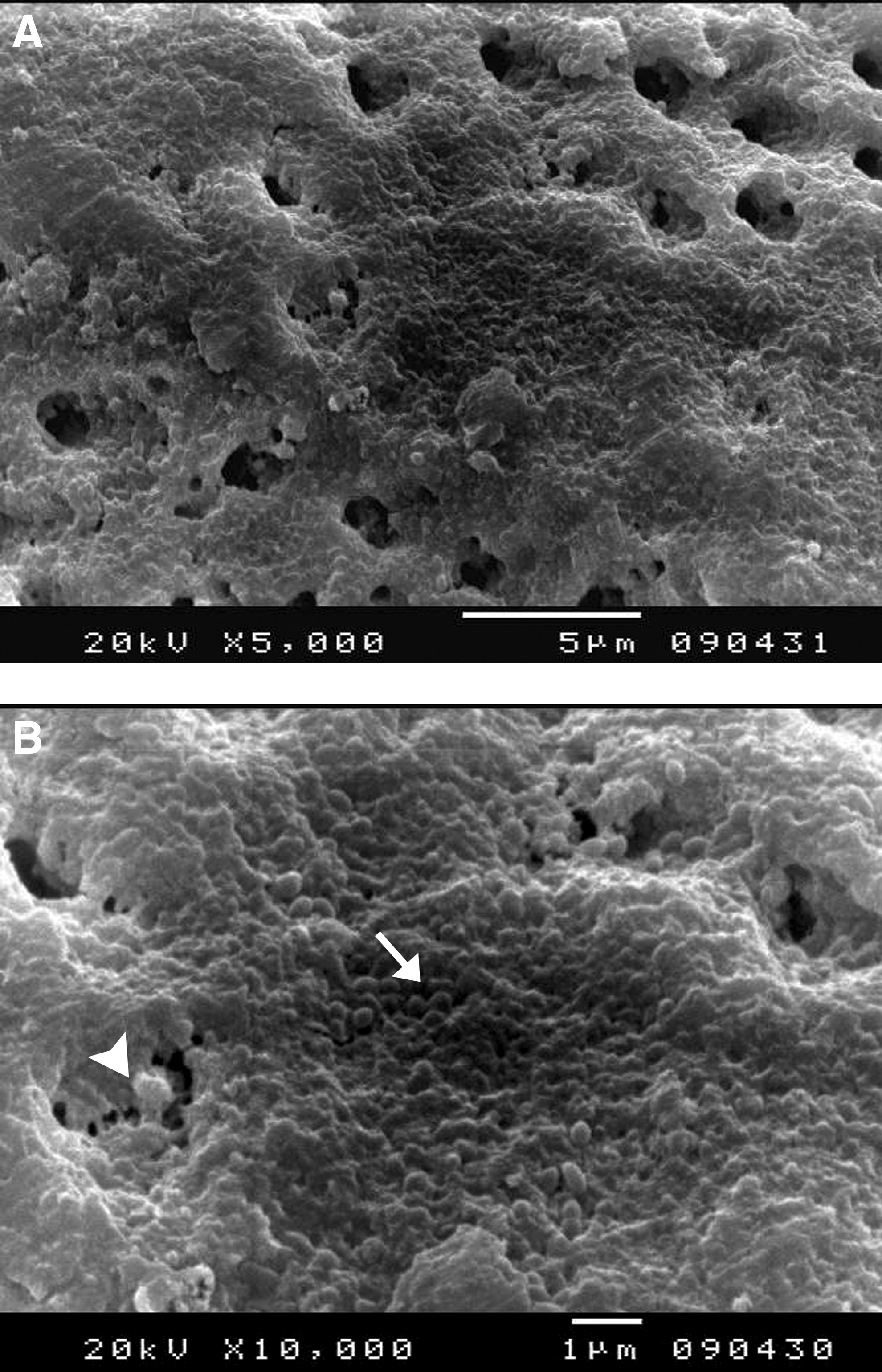
SEM images of 3-week-old Enterococcus faecalis biofilm showing a thick mat-like structure encrusting the dentin surface (arrow) and mostly occluding dentinal tubules (arrow head) × 5,000 OM in
For the negative control group samples, results showed the absence of any bacterial growth in all of the tested negative control samples for both assessment methods and time intervals confirming the absence of any cross-contamination of the samples during conduction of the experiment (Fig. 4).

Analysis of negative control dentin group (sterility test) after 4 weeks of incubation plus 24 hours and 7 days.
Antibiofilm effect of antimicrobials (CFU/mL test)
The CFUs of E. faecalis after 24 hours and 7 days of treatment with DAP, AgNPs, and TAMP-BG showed a significant reduction in the number of bacterial counts in comparison with the corresponding control subgroups that were treated with MC. Also, there was a significant difference in reduction of the bacterial count between the samples that were treated with the three antibacterial agents for 7 days when compared with the samples treated with them for 24 hours. It was found that there was no significant difference in reduction of bacterial count between DAP (1 mg/mL) and AgNP gel (0.02%) at both time intervals, but there was a significant difference between both DAP (1 mg/mL) and AgNP gel (0.02%) in comparison with BAG (100 mg/mL) at both time intervals (Fig. 5; Table 1).

Graph demonstrating the antibiofilm effects of the antimicrobials represented as the mean of CFU/mL × 10 2 . CFU, colony-forming units; DAP, double antibiotic paste.
The Mean of Colony-Forming Units per Milliliter × 10 2 ± Standard Deviation After Application of the Antimicrobial Agents for 24 Hours and 7 Days
H: H for Kruskal–Wallis test, pairwise comparison between each two groups was done using post hoc test (Dunn's for multiple comparisons test) = 33.214*.
p: p-value for comparing between the four groups in both time intervals (<0.0.001*).
p0: p-value for comparing between control and each other group in both time intervals.
p1: p-value for comparing between DAP and AgNPs after 24 hours.
p2: p-value for comparing between DAP and TAMP-BG after 24 hours.
p3: p-value for comparing between AgNPs and TAMP-BG after 24 hours.
p4: p-value for comparing between DAP and AgNPs after 7 days.
p5: p-value for comparing between DAP and TAMP-BG after 7 days.
p6: p-value for comparing between AgNPs and TAMP-BG after 7 days.
Statistically significant at p ≤ 0.05.
AgNPs, silver nanoparticles; DAP, double antibiotic paste; TAMP-BG, tailored amorphous multiporous bioactive glass.
Assessment of the residual biofilm structure (SEM analysis)
It was found that in the samples treated with MC (control group) at both time intervals, all the surface of each sample was covered by E. faecalis biofilm with aggregated bacterial cells populated on and in dentinal tubules (Figs. 6 and 7). In the samples treated with DAP or AgNPs (0.02%) (1 mg/mL) for 24 hours, few areas were covered by E. faecalis biofilm colonizing the root dentin surface with invasion inside dentinal tubules (Fig. 6), but in the samples treated with them for 7 days, few areas were covered by E. faecalis biofilm colonizing root dentin surface with most dentinal tubules not invaded with bacteria (Fig. 7).
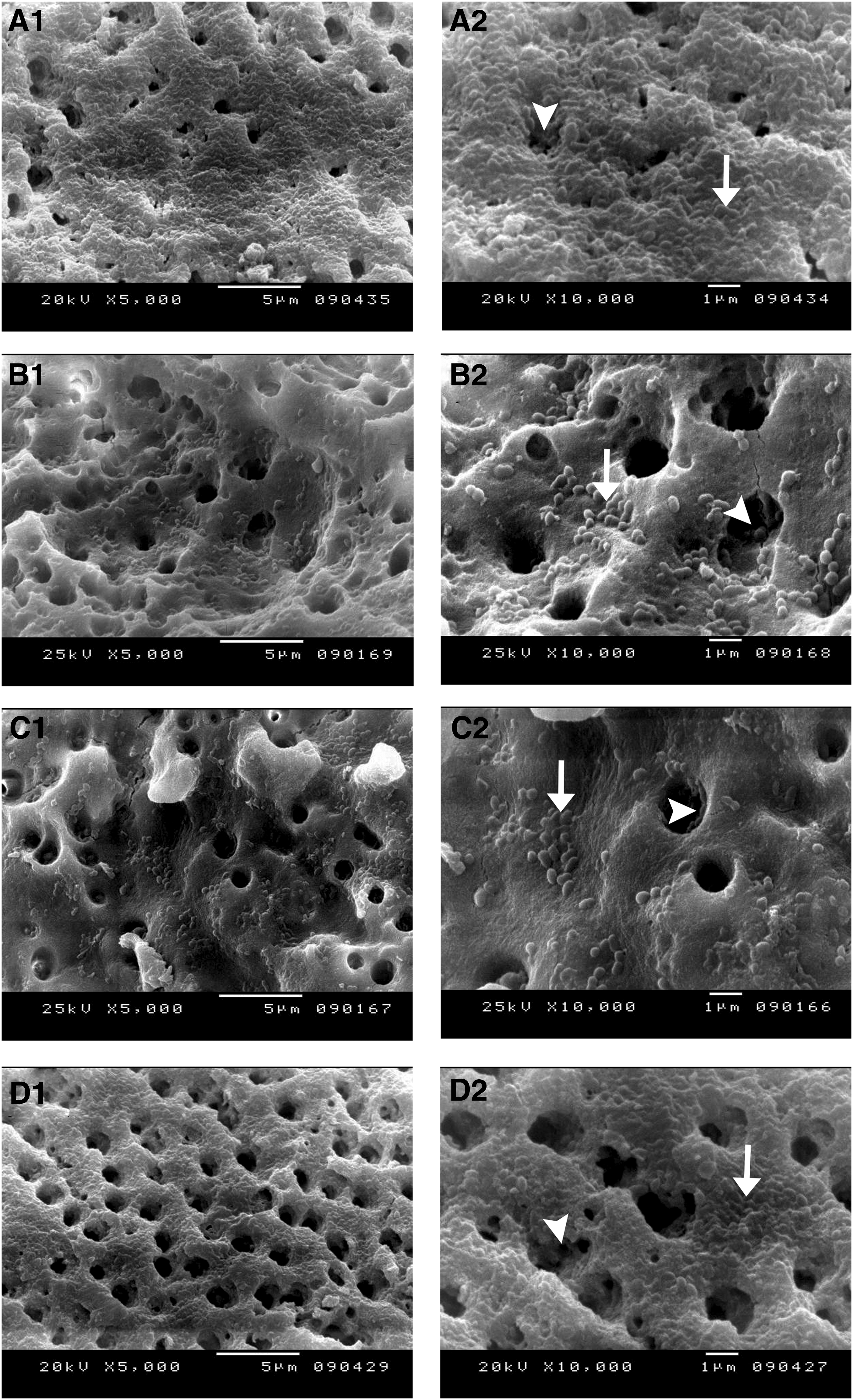
SEM images of representative Enterococcus faecalis biofilms after 24 hours of treatment.
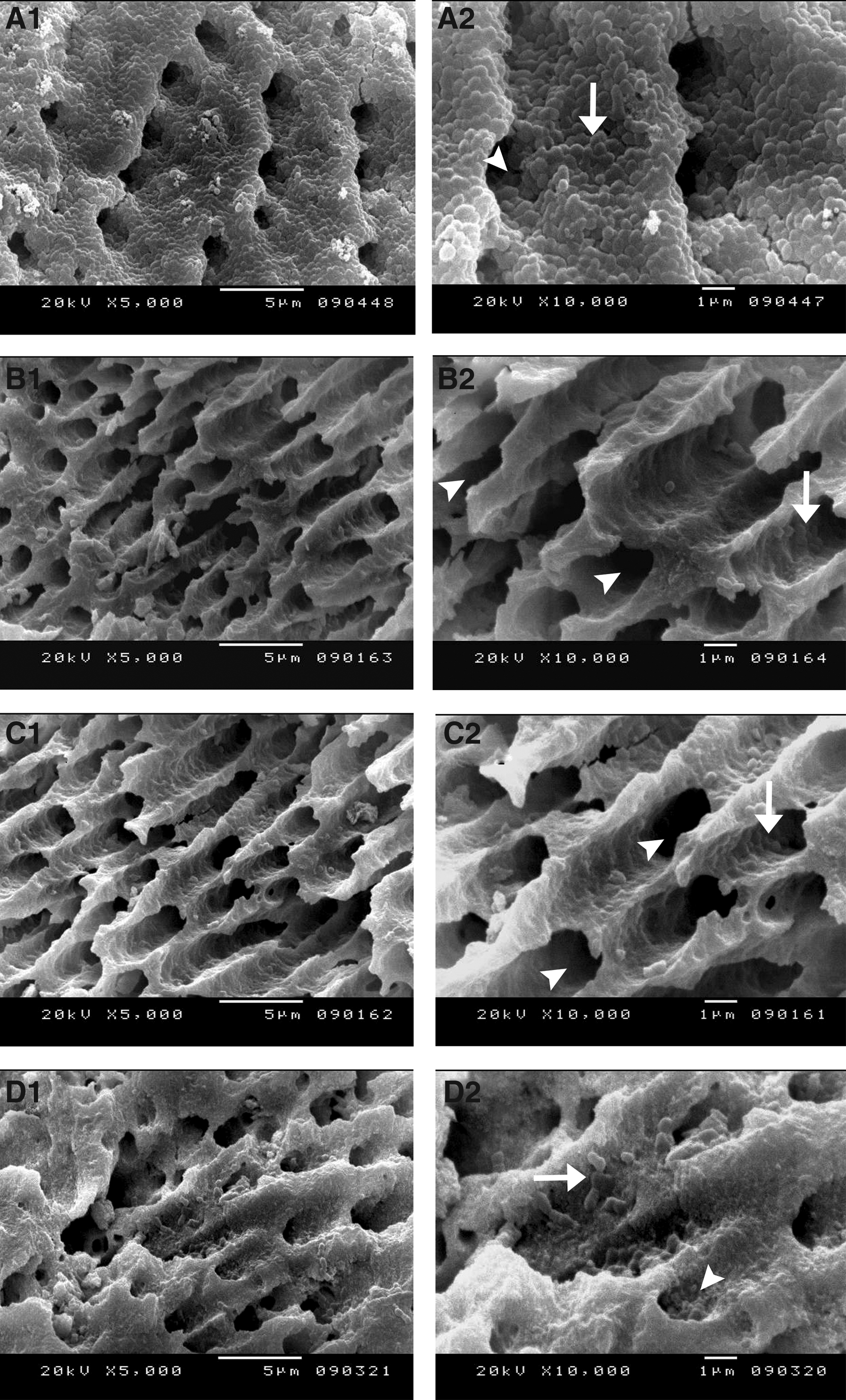
SEM images of representative Enterococcus faecalis biofilms after 7 days of treatment.
In the samples treated with TAMP-BG (100 mg/mL) for 24 hours, most areas were covered by E. faecalis biofilm colonizing root dentin surface and invading dentinal tubules (Fig. 6), but in the samples treated with it for 7 days, few areas were covered with bacterial cells (Fig. 7).
Discussion
Disinfection in regenerative endodontics (RE) is mainly based on chemical cleaning rather than mechanical shaping of the root canal system, especially in immature teeth with wide canals and thin dentinal walls, which limit mechanical instrumentation. Therefore, the antibacterial efficacy of the intracanal medicament used is very essential for successful RE treatment. 34 This study utilized AgNPs and TAMP-BG as antimicrobial agents for potential use in REPs and compared them with the more commonly used DAP combination. CFU test and SEM analysis were used to evaluate the effects of these antimicrobials on 3-week-old E. faecalis biofilms. E. faecalis was selected for the present study as this bacterial strain is dominant in the oral microbial ecosystem and in persistent/secondary endodontic infections. In addition, it is able to survive in case of low nutrients, high pH and temperature (>45°C), and it can maintain its viability in treated root canals for a long time. 35 In the present study, the biofilm was left to grow on the samples for 3 weeks to allow formation of a thick mat-like structure biofilm mostly covering the entire dentin surface and this was in accordance with Saber et al., 10 Wu et al., 12 and Tagelsir et al. 11 Moreover, the biofilm mode of bacterial growth offers many antimicrobial resisting mechanisms such as production of extracellular polymeric matrix that acts as a physical barrier and evokes phenotypical changes of the deeply located bacteria, leading to antimicrobial resistance enhancement. Moreover, the presence of persister cells that have very low metabolic activity allows them to enter a dormant state and survive with minimal nutrient requirements, which can, in turn, impede the effectiveness of many antimicrobials. 36
In the present study, the biofilm disruption assay was done followed by quantification of bacterial CFUs/mL, as it is considered one of the most valid methods to detect viable bacteria and was used in many recent studies.11,37,38 In addition, SEM imaging was performed to analyze the samples for the presence and quality of colonizing E. faecalis and their biofilm to qualitatively assess antimicrobial strategies on the targeted E. faecalis biofilms.
In this study, DAP rather than TAP was used as the antibiotic medicament because it was successfully used in clinical RE.39,40 In addition, its lower concentrations were suggested to have superior antibiofilm effects in comparison with the same concentration of TAP, besides minimizing many disadvantages of TAP.32,33 Indeed, in the present study, 1 mg/mL of DAP provided a significant antibiofilm effect at both time intervals (24 hours and 7 days), eliminated the majority of E. faecalis biofilm, and caused a 99.2% and 99.9% (Supplementary Fig. S1; Supplementary Data are available online at www.liebertpub.com/mdr) decrease in the viable bacteria, respectively. This finding agrees with a recent study that suggested a significant antibiofilm effect of 1 mg/mL of DAP. 11 Recent studies indicated no cytotoxic effect of 1 mg/mL of DAP against stem cells from apical papillae and dental pulp stem cells.41,42
However, another recent study has also shown that even lower concentrations of DAP may affect viability of dental stem cells. 32 Although there is a general consensus that 1 mg/mL of DAP may be used safely as an interappointment disinfectant during endodontic regeneration, there is a necessity to develop safer medicaments that will not affect the regenerated tissues. MC was used as drug delivery vehicle for DAP, AgNPs, and TAMP-BG. It is used as a common drug delivery vehicle for commercial intracanal medicaments as it is an inert polymer. Therefore, it was proposed to extend the therapeutic effect of medicament active ingredients 43 and may even induce dental pulp stem cell proliferation. 44
In this study, AgNP gel (in MC) had a significant antibacterial effect in both time phases of treatment as it caused a 99.4% and 99.9% (Supplementary Fig. S1) decrease in viable bacteria, respectively. This finding is in accordance with Kim et al. 45 who found that there was a significant reduction in bacterial count in both gram-positive and gram-negative bacteria after 24 hours of treatment with different concentrations of AgNPs. Moreover, Bo and Kayombo. 46 found that AgNP gel showed the ability to inhibit E. faecalis proliferation at different concentrations, even at much lower concentrations after 24 hours of treatment. In addition Wu et al. 12 stated that treatment with AgNP gel for 7 days destroyed E. faecalis biofilm and they attributed this finding to the nanoparticle ability to penetrate the dentinal tubules after this period of time (7 days). This suggested that the antibacterial efficacy of AgNPs depended on the concentration and duration of interaction. 47 In contrast, Mozayeni et al. 48 found that the AgNP gel was not effective enough against E. faecalis and they attributed that to two main reasons: the first reason was the different synthesis procedures of AgNPs and the second was the fact that the added gel may inhibit the release of silver ions. Moreover, according to recent studies, AgNPs minimize the liability of root canal reinfection as materials in nanoscale are more substantive 49 and infiltrative inside the dentinal tubules, lateral canals, and accessories than the materials in micro or larger scales. 50 Besides using AgNPs solely as an intracanal medicament, it can be used also in combination with calcium hydroxide. This mixture may enhance the antibacterial efficacy of calcium hydroxide. 38 Recent studies showed that it is not cytotoxic at low concentrations and with suitable particle size range (25.2 ± 6.5 nm). 51 This study differs from other previously published studies in that we tested AgNP as an intracanal medicament for 24 hours against a well-formed E. faecalis biofilm to evaluate the power of the onset of the antibacterial action, which may be suggesting its use for a shorter period of time (<7 days). The shorter time of application may minimize AgNP adverse effects either on stem cells 52 or radicular dentin. 53 The current study indicated that 100 mg/mL of TAMP-BG had a significant but limited antibiofilm effect when compared with DAP and AgNPs, particularly after 24 hours of treatment. The antibacterial behavior of TAMP-BG could be attributed to its reaction in the aqueous media (MC) as leaching of alkali ions (calcium and phosphorous) and dissolution of silicate network occurred. After the first 3 days from being suspended in the aqueous media, the leaching was reduced dramatically as the material surface was covered with the CaP precipitate. 54 According to a previous study, the pH of aqueous media containing (TAMP-BG) ranged from 7.3–7.4 during 1–7 days, 54 which suggested that the antibacterial effect of (TAMP-BG) was related to the effect of liberated ions or mineralization of bacterial cells rather than the pH effect, and this may explain also its delayed antibacterial effect.
The results of the present study regarding TAMP-BG's antibacterial effect agree with Krithikadatta et al. 55 who found that using BAG (S53P4) as an intracanal medicament caused a significant reduction in the number of E. faecalis colony counts after three different time points (1, 3, and 5 days). Also, Atila-Pektaş et al. 56 found that treatment with BAG for 7 days caused a significant reduction in the number of E. faecalis colony counts. They attributed that the antimicrobial effect of BAG might be due to its high pH, osmotic effects, or the Ca+2 concentration in the dentine environment. 28 Although BAGs have been widely studied in the literature as well as their antimicrobial properties,57–59 to our knowledge, this is the first study that evaluated the antimicrobial properties of 70S/30C TAMP-BG to be used as an antimicrobial intracanal medicament for REPs against E. faecalis biofilms.
In addition to being biocompatible, TAMP-BG has been previously shown to enhance hard tissue regeneration in a recently published clinical trial. 30 This fact coupled with the demonstrated antimicrobial properties of TAMP-BG may further urge this material to be used as a potential scaffold for dentin/pulp regeneration for REPs. 60 Also, this is the first study to use TAMP-BG suspended in water soluble vehicle (MC). This form was used to ease the delivery and removal of TAMP-BG rather than using it as a slurry paste and also to enhance TAMP-BG degradation and liberation of Ca, Si, and P ions. 56 Therefore, this formulation could participate in enhancement of its antibacterial effect.
One of the limitations of this study is that the evaluation of the antibacterial effects of the antibacterial agents used was done under in vitro conditions, which may be quite different from those of the clinical setting as clinically the presence of blood, exudates, infected pathological periapical tissue, and other strains of microorganisms may have an effect on the antibacterial behavior of the antibacterial agent used. 61 Also, it is worthy to note that the laboratory environment differs in the fact that all other strains rather than E. faecalis were eliminated by autoclaving and this may provide a better condition for growth and proliferation of the tested bacteria.
In addition, it is recommended to test the efficacy of DAP, AgNPs, and TAMP-BG in intermediate time points to evaluate the antibacterial behavior of each antibacterial agent more precisely from the starting time point (24 hours) to the end time point (7 days). Also, testing TAMP-BG in nanoscale may enhance its antibacterial effects as particle size has been shown to be related to the antimicrobial property of other bioglass formulations as it was shown that a higher dissolution rate of alkaline species thus elevated their antimicrobial efficacy. 62
Moreover, it is very crucial to test the cytotoxicity of the used antimicrobial concentrations on dental stem cells to confirm their suitability for use during REPs. This is particularly crucial since it has been shown that residual antimicrobial agents may be left on the root canal walls, which may later interfere with the attachment and proliferation of dental stem cells and hence retard the regenerative process. 63 Furthermore, in vivo studies using infected animal models are recommended to prove the efficacy of these agents in their current formulations.
Conclusion
DAP (1 mg/mL), 0.02% AgNPs, and TAMP-BG (100 mg/mL) can all serve as potent intracanal medicaments for REPs. However, complete elimination of E. faecalis biofilms was only possible with DAP and AgNPs at the proposed concentrations. Moreover, DAP and AgNPs are effective as early as 24 hours postapplication, but TAMP-BG (100 mg/mL) needs more time. Hence, these antimicrobials may serve as potential candidates for REPs.
Footnotes
Acknowledgments
The authors acknowledge the assistance of Noha Shazley for help during BAG preparation and the tissue engineering laboratories, Faculty of Dentistry, Alexandria University, for providing the facilities to do that.
Disclosure Statement
In the present study, no competing financial interests exist.
References
Supplementary Material
Please find the following supplemental material available below.
For Open Access articles published under a Creative Commons License, all supplemental material carries the same license as the article it is associated with.
For non-Open Access articles published, all supplemental material carries a non-exclusive license, and permission requests for re-use of supplemental material or any part of supplemental material shall be sent directly to the copyright owner as specified in the copyright notice associated with the article.