
Abstract
Yersinia enterocolitica biotype 1A strains are emerging pathogens, frequently isolated from clinical samples across the globe. Y. enterocolitica strains produce two chromosomal β-lactamases, BlaA and BlaB. BlaA is a constitutively expressed, Ambler class A, penicillinase, whereas BlaB is Ambler class C-type, inducible cephalosporinase. An earlier study from our laboratory indicated that instead of BlaB, Y. enterocolitica biotype 1A produced a “BlaB-like” enzyme. The objective of this work was to study the molecular characteristics of “Bla-B like” β-lactamases produced by biotype 1A strains to discern their similarity with AmpC-type β-lactamases and the basis of varied levels of minimum inhibitory concentration (MIC) to cefotaxime. Thus, the promoters and blaB genes were investigated in four strains of biotype 1A. Three-dimensional structures of the “BlaB-like” enzymes were modeled, and docked in silico with cefotaxime to understand how specific substitutions in gene sequences affect antibiotic susceptibilities. Our results indicated that all the reported key catalytic residues were present in variants of “Bla-B-like” enzymes, discerned in biotype 1A strains, but at different positions. Molecular docking of enzyme variants with cefotaxime revealed that lesser was the number of the H-binding residues with cefotaxime in a strain, lower was the MIC of cefotaxime in that strain. To the best of our knowledge, this is the first study in which the molecular characteristics and enzymatic interactions of “BlaB-like” cephalosporinases of Y. enterocolitica biotype 1A strains have been reported.
Introduction
Yersinia enterocolitica, an enteropathogenic bacterial species has been classified into more than 60 serotypes and 6 biotypes, with varied geographical distribution, ecological niche, and pathogenic potential. 1 Y. enterocolitica biotype 1B is highly pathogenic; biotypes 2–5 are moderately pathogenic, whereas the pathogenicity of biotype 1A has been controversial. 1 However, in the recent past biotype 1A strains have been regarded as emerging pathogens as these are being isolated frequently from clinical samples across the globe.2–5
Y. enterocolitica strains produce two types of chromosomal β-lactamases named β-lactamase A (BlaA) and β-lactamase B (BlaB). BlaA is a constitutively expressed, Ambler class A, Bush group III penicillinase, whereas BlaB is Ambler class C, Bush group I, AmpC-type inducible cephalosporinase. The expression of blaB in Y. enterocolitica is regulated by ampR and ampD. The ampR is transcriptional regulator of ampC and, AmpD is a cytoplasmic N-acetyl-anhydromuarmyl-
Several investigators have reported the minimum inhibitory concentration (MIC) and/or enzymatic data supporting the presence of AmpC-type β-lactamases in species of Yersinia.8–11 However, molecular data for AmpC-type β-lactamases, except for a strain of serotype O: 5b has not been reported for most of the bioserotypes of Y. enterocolitica.6,12 An earlier study from our laboratory indicated that BlaB of Y. enterocolitica biotype 1A was different from BlaB of other biotypes, as it revealed multiple bands of pIs 6.8 and 7.1 on isoelectric focusing. This suggested that Y. enterocolitica biotype 1A produced a “BlaB-like” enzyme. 13 The Y. enterocolitica biotype 1A strains harboring “Bla-B like” cephalosporinase also showed high MIC for cefoxitin (this study) and resistance to β-lactam/β-lactamase inhibitor combination, 14 indicating the possibility of “Bla-B like” produced by Y. enterocolitica biotype 1A strains to be actually an AmpC or AmpC-type enzyme. Thus, the objective of this work was to study the molecular characteristics of “Bla-B-like” β-lactamase produced by Y. enterocolitica biotype 1A strains and the basis of varied levels of MIC to cefotaxime to discern its similarity to AmpC-type β-lactamases. The promoter regions and the complete coding sequences (CCDS) of blaB genes in four strains of Y. enterocolitica biotype 1A were investigated. Three-dimensional (3D) structures of the “BlaB-like” enzymes were modeled, and docked in silico with cefotaxime to understand how specific substitutions in gene sequences affect antibiotic susceptibilities. To the best of our knowledge, this is the first study to investigate the molecular characteristics of “BlaB-like” enzyme of Y. enterocolitica biotype 1A.
Materials and Methods
Bacterial strains
Four strains of Y. enterocolitica biotype 1A maintained in our laboratory, (University of Delhi South Campus) at 4°C in trypticase soy agar, were examined. The details of the strains are given in Table 1. These were well-authenticated strains deposited with Yersinia National Reference Laboratory and WHO Collaborating Center, Pasteur Institute, Paris (France) and at the national repository at Microbial Type Culture Collection (MTCC) and Gene Bank located at Institute of Microbial Technology (IMTECH), Chandigarh (India).
Details of Yersinia enterocolitica Biotype 1A Strains and Their Antimicrobial Susceptibilities Determined by E-Test
The data on MIC for amoxicillin–clavulanic acid and cefotaxime has been retrieved from Singhal et al. 14
CLSI, 2010 guidelines for MIC breakpoints: amoxicillin–clavulanic acid, ≤8/4(S), 16/8(I), ≥32/16(R); cefotaxime, ≤1(S), 2(I), ≥4(R); cefoxitin—≤2(S), 10(I), ≥32(R).
Letters in parentheses indicate antibiotic susceptibility: I, intermediate; S, sensitive; R, resistant.
MIC, minimum inhibitory concentration.
Determination of antibiotic susceptibility for cefoxitin and phenotypic detection of AmpC production
Antibiotic susceptibility testing for cefoxitin—a second generation cephalosporin and phenotypic detection of AmpC production—was carried out with E-test strips (bioMérieux, Inc., MO) of cefoxitin and AmpC using methods described previously. 14 In brief, bacterial strains were grown on Muller–Hinton (MH) agar plates at 28°C, overnight. Single colonies were picked from agar plates and suspended in 1 mL of normal saline to reach a turbidity equivalent to 0.5 McFarland standards (∼1–2 × 10 8 colony-forming units/mL). A 100 μL of this cell suspension was spread on the surface of MH agar plates and E-test strips were placed over the surface. The MH agar plates were further incubated at 28°C and the MIC values were recorded. All strains were induced with imipenem for production of AmpC-type cephalosporinases using methods described previously. 9 The AmpC E-test strips contain cefotetan at one end and cefotetan+cloxacillin at the other end. A ratio of MIC of cefotetan and cefotetan+cloxacillin of ≥8 indicates production of AmpC. The MICs of the antibiotics were interpreted according to the guidelines of Clinical and Laboratory Standards Institute, 2010. 15
Isolation of genomic DNA, primer designing, and PCR amplification of promoter region and CCDS of ampC
DNA was isolated from bacterial pellet collected from 1 mL of the bacterial culture grown overnight at 28°C using DNeasy Tissue kit (Qiagen, Hilden, Germany) following manufacturer's instructions. Primes B11f (5′-CCTGACTTTTTCACGTATTAT-3′) and B12r (5′-GGGGATAGTGATAAAGGTAT-3′) were designed for amplification of promoters and partial gene regions of blaB genes, whereas B15 (5′-TGACGGAAAGCCGCAATTCT-3′) and B16 (5′-TCATAGAAGCGTCAACGCAA-3′) were designed for amplification of CCDS of blaB genes, using Primer-BLAST.a The primers were synthesized from Sigma-Aldrich. PCR amplification was performed in a My Cycler™ Thermal Cycler (Bio-Rad, CA). The PCR mixture comprised 1 × PCR buffer (10 mM Tris-HCl, 1.5 mM MgCl2, 1.5 mM KCl, and 0.1% Triton X-100), 200 μM of each of the 4 dNTPs (MBI Fermentas GmbH, Germany), 10 pmol each of forward and reverse primers (Microsynth GmbH), 2 U of Taq DNA polymerase (DyNAzyme™; Finnzymes, Finland), and 50–100 ng of genomic DNA in a total volume of 25 μL. The PCR conditions were the same as described previously 14 except annealing temperature which was 49°C and 54°C for primer pairs B11f, B12r and B15f, and B16r, respectively. The PCR products were analyzed by electrophoresis on 1% (w/v) agarose gel. The gels were stained with ethidium bromide (0.5 μg/mL) and visualized under UV transilluminator.
Sequencing of promoter regions and CCDS of blaB genes
PCR amplicons containing partial coding sequences of blaB genes including the promoter region and CCDS were purified using HiYield™ extraction kit (RBC Bioscience, New Taipei City, Taiwan) and sequenced following Sanger's method at a commercial facility (Invitrogen BioServices India Pvt. Ltd., Bangalore, India). The sequences obtained were analyzed further for similarity by homology search using NCBI-BLAST.b The nucleotide sequences amplified using primer pair B11f, B12r and B15f, and B16r were aligned together to construct the CCDS of blaB genes.
Analysis of “BlaB-like” proteins by conserved domain database, InterProScan, and multiple sequence alignment
The functional classification of “BlaB-like” proteins into families was carried out using the web tools—Conserved Domain Database and InterProScan. 16 Multiple sequence alignment (MSA) of promoters and translated CCDS of “BlaB-like” proteins of biotype 1A strains was carried out by Clustal Omega.c
Homology modeling of “BlaB-like” cephalosporinases: 3D structure prediction, modeling, and validation
MSA revealed that a few point substitutions were present in amino acid sequences of “BlaB-like” proteins; hence, protein structures of all four Y. enterocolitica strains were modeled in silico. The 3D structures of the proteins were predicted using MODELLER 9.10d (http://salilab.org/modeller). Of the predicted models, the best model was selected based on the lowest modeler objective function and validated using the programs–PROCHECK, 17 ERRAT, 18 and Verify 3D. 19 The protein models were visualized using PyMol.e
Molecular docking
Molecular docking is a method that is used to determine preferred binding orientations of two molecules to form a stable complex, which in turn predicts their binding affinity. The binding affinity of “BlaB-like” protein variants with cefotaxime, molecular docking of “BlaB-like” protein variants was performed using AutoDock 4.0. The PDB structure of cefotaxime (drug bank ID: DB00493) was downloaded from the database, DrugBank.f Protein structures were preprocessed for docking using AutoDockTools (ADT) version 1.5.6 by adding polar hydrogen atoms and Kollman charges, removing all nonprotein molecules, and converting the PDB file into PDBQT format. Ligand structures were also prepared for docking by adding hydrogen and charges and, converting it into PDBQT format. The docking search space was determined visually by centering the Grid Box at the known binding sites of the ligand and expanding the dimensions of the cubic search space so that it completely encompassed that site. Docking was performed at default parameters, other than the search space specifications mentioned previously. The maximum number of poses generated per ligand was set to 10. The final output was selected on the basis of binding global energy (kcal/mol) associated with each docked pose The hydrogen bonding and hydrophobic interactions in the enzyme–ligand complex were analyzed using LigPlot and PyMoL version 2.0 (Schrödinger, LLC).
Results and Discussion
It has been reported that bacteria which produce AmpC β-lactamases show reduced susceptibility to β-lactam/β-lactamase inhibitors like amoxicillin–clavulanic acid. 7 Thus, four strains of Y. enterocolitica biotype 1A that were resistant to amoxicillin–clavulanic acid were included in this study. 14 The production of BlaA was observed to be similar in these strains. 14 Resistance to cefoxitin - a cephamycin and second-generation cephalosporin - is considered as indicative of production of inducible cephalosporinases 7 ; hence, all Y. enterocolitica strains were also tested for susceptibility to second-generation cephalosporin - cefoxitin, and for AmpC production, using E-test strips of cefotetan/cefotetan+cloxacillin. E-test indicated that all the four biotype 1A strains showed intermediate susceptibility to second-generation cephalosporin-cefoxitin (Table 1). In addition, it was noted that E-test strips of cefotetan/cefotetan+cloxacillin were not suitable for phenotypic detection of AmpC production in Y. enterocolitica, as all strains tested negative for AmpC production, whereas enzymatic assays 9 and PCR amplification indicated that “BlaB-like” inducible cephalosporinases were present in Y. enterocolitica strains of biotype 1A.
Various studies have reported that Y. enterocolitica strains of the same biotype do not display similar antimicrobial susceptibilities.20,21 Similar findings were reported by us while studying susceptibilities of Y. enterocolitica biotype 1A strains for various cephalosporins. 14 In this study, we also tried to understand the molecular basis underlying differential interactions of “BlaB-like” β-lactamase of Y. enterocolitica biotype 1A strains with cefotaxime. Mutations and insertions in the promoter regions of β-lactamase genes have been associated with hyperproduction of β-lactamases.22,23 To understand if variations in promoter sequences or in amino acid sequences of “BlaB-like” proteins were responsible for differential susceptibilities of biotype 1A strains to cefotaxime, the corresponding gene sequences were analyzed. Primer pair B11f and B12r resulted in the expected amplicon of 1,076 bp in all strains. BLAST analysis confirmed that the amplicons encoded promoters and partial gene sequences of blaB. MSA of the promoter regions revealed that promoter sequences of blaB were identical in all strains (Fig. 1). Thus, these might not be responsible for differential susceptibilities of biotype 1A strains to cefotaxime.
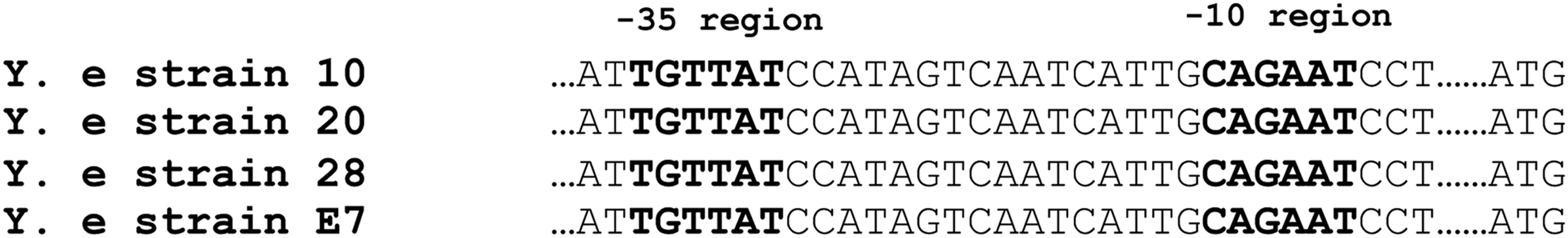
MSA of promoter region of blaB of Yersinia enterocolitica biotype 1A strains. MSA, multiple sequence alignment.
To investigate if variations in gene sequence were responsible for differential antibiotic susceptibilities of biotype 1A strains, genes of “BlaB-like” β-lactamase were PCR amplified, sequenced, and their CCDS were translated. The primer pair B15 and B16 resulted in the expected amplicons of 1,002 bp in all strains. Gene sequencing and BLAST analysis confirmed these to encode for “BlaB-like” β-lactamases of Y. enterocolitica. The key catalytic residues and sites, substitutions at which reportedly lower the enzymatic activity of AmpC enzymes are S64, K67, Y150, N152, K315, and A318. 24 Analysis of amino acid sequences of “BlaB-like” cephalosporinases of biotype 1A strains indicated that all the reported key catalytic residues were present in variants of “Bla-B-like” enzymes, but at different positions. The positions of the catalytic site residues in BlaB-like cephalosporinases were S-89, K-92, Y-175, N-177, K-342, and A-345. The two significant motifs conserved across the Ambler class C β-lactamase, that is, 151SXXK154 and 342KTG344 (where X represents any amino acid) were present in BlaB-like cephalosporinase of all biotype 1A strains. A 24 amino acid long signal peptide was present at the terminal end in all “BlaB-like” enzyme variants. As the signal peptide is cleaved before the mature enzyme is released in the periplasmic space, variations in it were excluded from comparative studies. MSA results indicated that although the amino acid sequences were similar, variations were present at a few places (Fig. 2). Variations like Q30L and K38N were observed in Y. enterocolitica strain 20; V56I, T74A, and C166W in Y. enterocolitica strains 20 and 10; E115K and I198M in Y. enterocolitica strain E7. The sites of variation in individual strains are given in Figure 2. Submission of “BlaB-like” enzyme sequences to Conserved Domain Database (CCD) and Interproscan retrieved no results.
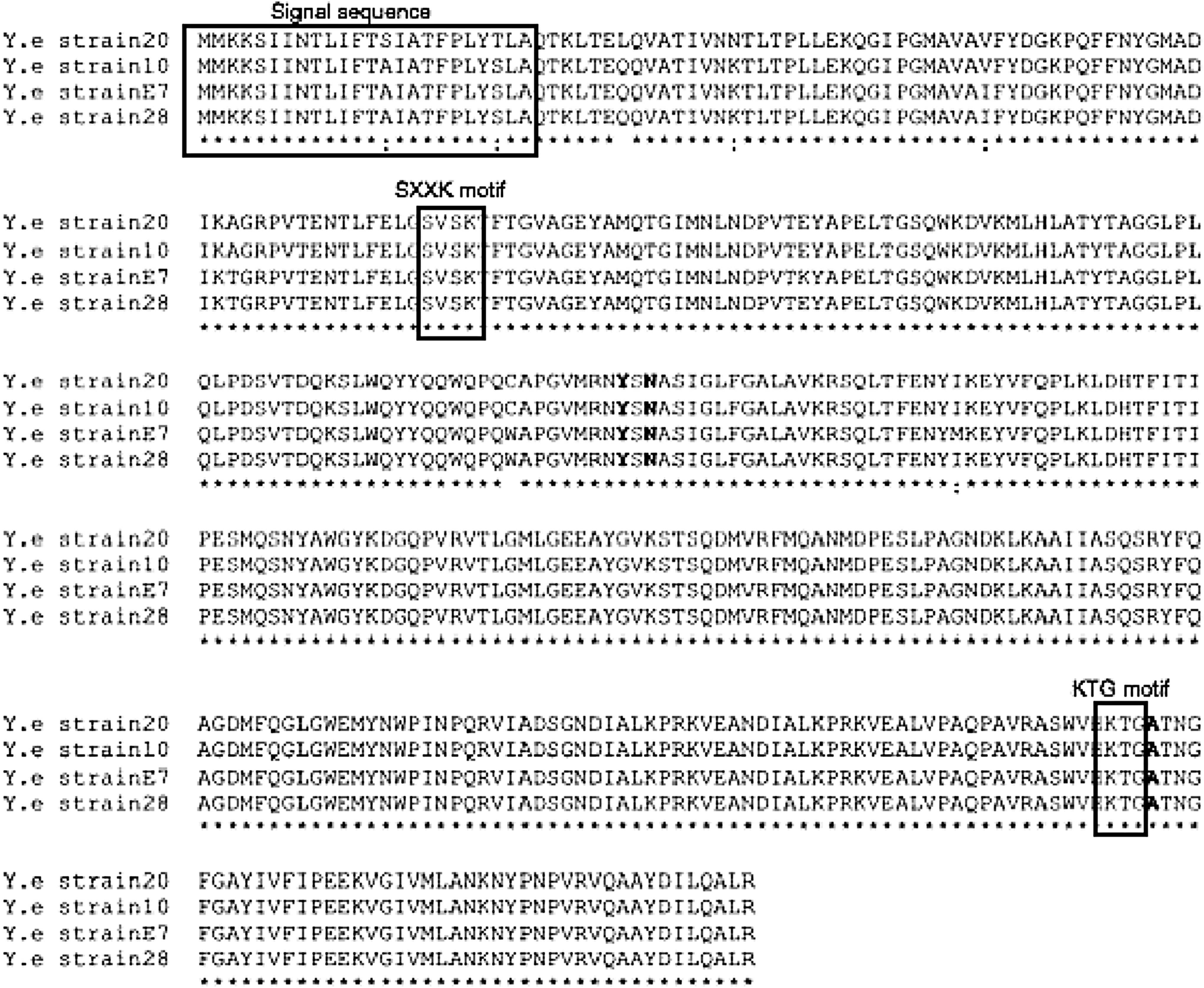
MSA of “BlaB-like” enzyme variants present in different strains of Y. enterocolitica biotype 1A. The key catalytic residues are given in bold. The active site residues 89 S and 175 Y were present at their respective positions.
The variants of “BlaB-like” enzymes discerned in biotype 1A strains were 3D modeled and docked with cefotaxime in silico. The top three templates (PDB ID: 5GGW_A, 1FSW_A, 2BLS_A) with a sequence identity >65% were chosen as templates to model the 3D structures. The predicted normalized Dope scores for the final selected models of “BlaB-like” enzyme variants in Y. enterocolitica strains 20, 10, 28, and E7 were −1.165, −1.198, −1.159, and −1.224, respectively. The selected protein models were validated through various structure assessment programs. Results of the PROCHECK program indicated that in “BlaB-like” enzyme variants of Y. enterocolitica strains 20, 10, 28, and E7, the residues present in the most favored regions of Ramachandran map were 93.4%, 92.8%, 91.6%, and 92.8%, respectively. Calculation of the nonbonded atomic interactions of the modeled protein variants by ERRAT program indicated an average ERRAT score of 83.24, 79.77, 76.71, and 84.53 for Y. enterocolitica strains 20, 10, 28, and E7, respectively. The Verify-3D scores of the protein models were 87.3%, 89.69%, 89.69%, and 81.70% for the protein models of Y. enterocolitica strains 8, 20, 10, 28, and E7, respectively. PROCHECK, ERRAT, and Verify-3D results confirmed that the predicted 3D structures were reliable and within the acceptable range.
Molecular docking analysis of the modeled protein structures of “BlaB-like” enzymes were carried out to identify their binding affinity for third-generation cephalosporin—cefotaxime. Amino acid residue variations at sites other than active sites of the enzyme might create local disturbances in the 3D structure of enzyme and increase/decrease its conformational flexibility, thereby affecting substrate binding. The global free energy, hydrogen bond (H bond), and hydrophobic interactions of cefotaxime with different “BlaB-like” enzyme variants were analyzed and the details are given in Table 2. In silico docking of the “BlaB-like” enzyme variants with cefotaxime revealed that in each enzyme variant, different amino acid residues were involved in H bonding and hydrophobic interactions (Fig. 3). It is well known that H bonds and hydrophobic interactions not only play a crucial role in molecular recognition and overall stability of the protein structure, these also stabilize the protein–ligand complexes. In a docked complex, the number of the residues involved in H-bond formation indicates the binding affinity between an enzyme and ligand. 25 This implies that lesser the number of H-binding residues in an enzyme–antibiotic complex, the lesser is the hydrolysis of the antibiotic by the enzyme and, lower is the MIC of the antibiotic. In this study it was observed that in Y. enterocolitica strain 20, the number of residues of “BlaB-like” enzyme involved in H bonding with cefotaxime was 2, and the MIC for cefotaxime in this strain was almost 10 times lesser than the MIC of cefotaxime observed for Y. enterocolitica strain 28, in which the number of residues involved in H-bond formation with cefotaxime was 6. A similar observation was made while comparing the H-bonding residues of other “BlaB-like” enzyme variants and their respective MICs for cefotaxime. Lesser the number of H-bonding residues with cefotaxime in a strain, lower was the MIC of cefotaxime in that strain (Table 1). Thus, in silico analysis explained the higher MIC for cefotaxime in strains 10, 28, and E7. However, further experiments are required to strengthen our findings that amino acid mutations affect the H-binding residues of “BlaB-like” enzymes and their interaction with cefotaxime. The foregoing data and discussion give further evidence that “BlaB-like” inducible cephalosporinase of Y. enterocolitica biotype 1A strains is an AmpC-type β-lactamase. To the best of our knowledge, this is the first study in which the molecular characteristics and enzymatic interactions of “BlaB-like” cephalosporinase of Y. enterocolitica biotype 1A strains have been reported.

Molecular docking analysis of cefotaxime with the four variants of “BlaB-like” enzymes discerned in Y. enterocolitica
Estimated Global Energy of Binding, H-Bond Interactions, and Hydrophobic Interactions of Cefotaxime with “BlaB-like” Enzyme Variants Discerned in Yersinia enterocolitica Biotype 1A
Footnotes
Acknowledgments
N.S. is supported by Science & Engineering Research Board (SERB) of Department of Science & Technology (SB/YS/LS-156/2014). D.P. is supported by fellowship from DST-INSPIRE (IF160262). The authors thank Rajesh Kumar Mahto for the technical help.
Disclosure Statement
No competing financial interests exist.