
Abstract
In the last 15 years, Acinetobacter baumannii has received special attention, mainly due to several resistance mechanisms and high rates of morbimortality. The ability to form biofilms contributes to the persistence of this microorganism in the hospital environment and facilitates the occurrence of nosocomial infections. Several studies have highlighted the pharmacological relevance of pyridines in the treatment and control of infectious diseases and others have related the anti-A. baumannii potential of hydrazine derivatives. Considering this scenario, we aimed to evaluate the antimicrobial and antibiofilm activity of 10 pyridinylhydrazone compounds against A. baumannii. The minimum inhibitory concentration of the compounds was determined by broth microdilution method and the antibiofilm activity was evaluated by inhibition and destruction biofilm assays. In addition, the cytotoxicity of the compounds in the J774A.1 cell line was also evaluated, and the selectivity index was calculated. Among the 10 pyridine compounds, the compounds B and D were able to inhibit the formation of biofilms and destroy bacterial biofilms even in a concentration of 12.5 μg/mL. Thus, the pyridine compounds evaluated can be a scaffold for the development of new substances with antimicrobial and antibiofilm activity.
Introduction
According to the American Society of Infectious Diseases, the “ESKAPE” pathogens (Enterococcus faecium, Staphylococcus aureus, Klebsiella pneumoniae, Acinetobacter baumannii, Pseudomonas aeruginosa, and Enterobacter spp.) are considered the six microorganisms most relevant in the world, being directly associated to multidrug resistance, nosocomial infections, morbimortality, and, consequently, increased cost of health care.1,2 Among these pathogens, A. baumannii has received special attention in the last 15 years, mainly due to its easy transmissibility, several resistance mechanisms, and high rates of morbimortality. 3
A. baumannii is naturally resistant to many antibiotics and the appropriate treatment of infections caused by this pathogen is hampered by this feature, which makes the therapeutic arsenal available limited. 4 In addition, the ability of A. baumannii to form biofilms on biotic and abiotic surfaces favors its survival in hostile environments and acts as a protective barrier, reducing the activity of antimicrobials and biocides. 5 The resistance mechanisms associated with this capacity contribute to its persistence in the hospital environment and facilitate the occurrence of nosocomial infections, such as urinary, respiratory, and cutaneous infections.6,7 Considering this set of characteristics, in 2017, A. baumannii was included in the list of priority pathogens for research, discovery, and development of new antibiotics. 8
However, as important as the development of new antibiotics is the discovery of inhibitory and/or biofilm-destroying substances, since this condition facilitates the stabilization and spread of microorganisms, including within the health care setting.9,10 Among several compounds that, besides showing antimicrobial, 11 antitumor, 12 and antifungal 13 activity, also show antibiofilm action are pyridine compounds.14,15
Pyridine is a heterocyclic compound and its chemical structure consists of a ring containing five carbon atoms and one nitrogen atom. Drugs such as isoniazid, nevirapine and phenazopyridine are derivatives of pyridines used to treat and control infectious diseases, with antimycobacterial, 16 antiviral, 17 and analgesic activity 18 reported, respectively. Therefore, considering the pharmacological potential of this class of compounds and the lack of studies related to the anti-A. baumannii potential of hydrazone derivatives,19,20 we evaluated the antimicrobial activity of 10 pyridinylhydrazone derivatives against clinical isolates of A. baumannii, and their ability to inhibit and destroy bacterial biofilms.
Materials and Methods
Compounds
The pyridinic compounds were prepared at the Institute of Drug Technology (FarManguinhos—FIOCRUZ-RJ). The synthesis of the pyridine-2-ylhydrazone derivatives, A-J, involved reactions of commercial pyridine-2-ylhydrazine X and arenecarbaldehydes in ethanol solution at room temperature (Fig. 1; Table 1). 21 The compounds were obtained in 44–81% yields (Table 1) and cLog P was calculated using Molinspiration software.

Synthesis of 2-pyridin-2-ylhydrazone derivatives
Pyridin-2-Ylhydrazone Derivatives A–J
Calculated using Molinspiration software.
For the assays, these compounds were solubilized in dimethyl sulfoxide. The antibiotics amikacin, ceftazidime, and ciprofloxacin were purchased and diluted according to the manufacturer's instructions. All compounds were stored at 10 mg/mL and maintained at 4°C until use.
Strains
A. baumannii strains were from the collection of Núcleo de Pesquisa em Microbiologia Médica of Federal University of Rio Grande—FURG. For antimicrobial activity assay, five clinical isolates were selected, which were from patients treated at University Hospital by Dr. Miguel Riet Correa Jr. in Rio Grande/RS, between August 2012 and July 2013, for urinary tract infection (UTI). In addition, the reference strain Acinetobacter baumannii ATCC 19606 was also included in all experiments. The identification of the strains was performed by biochemical (growth on MacConkey agar, motility, catalase, oxidase, citrate, and urease) and molecular tests (amplification of the blaOXA-51-like gene 22 ).
Antimicrobial activity assay
The minimum inhibitory concentration (MIC) of antibiotics (amikacin, ceftazidime, and ciprofloxacin) and pyridine compounds against planktonic A. baumannii was determined by microdilution method, according to Clinical Laboratory Standards Institute, 23 using resazurin as cell viability indicator. In brief, a serial (1:2) microdilution of 50 μL of the compound in 50 μL of Mueller–Hinton medium (MH) was performed in a 96-well microplate. The concentrations of the antibiotics ranged from 32 to 0.5 μg/mL and the pyridine compounds ranged from 200 to 6.25 μg/mL. The inoculum was prepared by diluting a bacterial suspension (adjusted to the 0.5 scale of McFarland) in MH in a ratio of 1:100, and 50 μL of this inoculum was added, to give a final concentration of 5 × 10⁵ colony-forming unit (CFU)/mL in each well. Bacterial growth (containing inoculum and medium) and sterility controls (medium) were also included in the plates. After 24 hours of incubation at 37°C, 15 μL of resazurin (0.02%) was added to each well and the result was determined after changing the positive control from blue to pink. MIC was considered as the lowest concentration of a compound capable of inhibiting bacterial growth. Pyridine compounds with MIC ≤200 μg/mL were considered active.
Cytotoxicity
The cytotoxicity of the compounds was evaluated on the J774A.1 cell line (ATCC TIB67), according to the methodology proposed by Pavan et al. 24 Cells were cultured in Dulbecco's modified Eagle's medium (DMEM) and, in a 96-well plate, was added 200 μL of a cell suspension in each well at the concentration of 1 × 10⁵ cells/mL. After 24 hours incubation at 37°C (5% CO2), the adhered cells were exposed to the same concentrations evaluated against A. baumannii (200–6.25 μg/mL), for 24 hours. Next, the contents of the test wells were removed, was added 30 μL of resazurin (0.01%) diluted in DMEM, and 6 hours later the fluorescence was measured (620 nm) for determination of IC50 (half maximal inhibitory concentration): concentration of the compound capable of maintaining the viability of 50% of the cells. The IC50 was calculated according to the following formula: (100 – (OD of exposed cells/OD of cells not exposed) × 100). In addition, the selectivity index (SI) of the compounds was also calculated, where SI = IC50/MIC. The higher the SI value, the less toxic the compound is for the cell.
Biofilms
The capacity of biofilm formation in vitro by strains of A. baumannii was evaluated using the methodology proposed by Gopal et al. 25 The strains were grown on MacConkey agar and incubated at 37°C overnight. From isolated colonies, a bacterial suspension was prepared in MH enriched with 0.2% of glucose, to give a final concentration of 1 × 10⁶ CFU/mL, and 100 μL of that suspension was transferred to a 96-well polystyrene flat bottom microplate. Plates were incubated at 37°C for 24 hours and then the contents of the wells were removed, washed three times with phosphate-buffered saline (PBS) 1 × (150 μL) and the adhered cells were fixed with methanol 100% (150 μL) and stained with violet crystal solution 0.4% (200 μL). Next, the wells were washed three times with distilled water (200 μL), and the dye was dissolved with 100% ethanol (200 μL). The optical density of the wells was measured at 620 nm using the Thermo Plate TP-Reader BioTek™ Elx800™; the results were interpreted mathematically according to Stepanović et al. 26 and then, the strains were classified as one of four categories: no biofilm producer, weak, moderate or strong biofilm producer. 27 For the next antibiofilm assays, just those strains that showed strong biofilm formation were selected.
Antibiofilm activity: biofilm inhibition
In this assay, the strains were exposed to different concentrations of the compounds before induction of biofilm formation, to determine if the compounds would be capable of inhibiting/avoiding bacterial biofilm formation. For this, from growth on MacConkey agar, a bacterial suspension of each A. baumannii strain was prepared in MH enriched with 0.2% of glucose, with a final concentration of 1 × 10⁶ CFU/mL. One hundred microliters of suspension was transferred to a 96-well polystyrene flat bottom microplate and was exposed to the following concentrations of the active compounds, previously determined in the antimicrobial activity assay: 2 × MIC, MIC, and 1/2 MIC. The working solutions were prepared so that the volume of compound required for the assay in each well did not exceed 10 μL. Controls were included: medium containing the solvent used for solubilization of the compounds, to confirm cell viability; controls of biofilm formation; and sterility controls, containing only culture medium. The plates were incubated for 24 hours at 37°C. Next, the steps of washing, fixing, and staining of biofilm were carried out and quantification of the biofilm formation was performed by measuring the optical density at 620 nm, according to the methodology previously described in “Biofilms” topic. Those compounds with MIC values ≤200 μg/mL against planktonic A. baumannii were selected for the biofilm inhibition assay.
Antibiofilm activity: biofilm destruction
In this essay, the step of bacterial biofilm formation was performed previously and the adhered cells were exposed to different concentrations of the pyridine compounds to evaluate their ability to destroy the bacterial biofilm. Then, from growth on MacConkey agar, a bacterial suspension of each A. baumannii strain was prepared in MH enriched with 0.2% of glucose, with a final concentration of 1 × 10⁶ CFU/mL. One hundred microliters of suspension was transferred to 96-well polystyrene flat bottom microplates and, next, the plates were incubated for 24 hours at 37°C, for the formation of biofilms. After the incubation period, the contents of the wells were removed, and washing was carried out with PBS 1 × to eliminate unadhered cells. Next, 100 μL of MH enriched with 0.2% of glucose containing the following concentrations of the active compounds were added: 2 × MIC, MIC, and 1/2 MIC. The plates were reincubated for more 24 hours at 37°C and, the steps of washing, fixing, and staining of biofilm were carried out and quantification of the biofilm formation were performed by measuring the optical density at 620 nm, according to the methodology previously described in “Biofilms” topic. Those compounds with MIC values ≤25 μg/mL against planktonic A. baumannii strains were selected for the biofilm inhibition assay.
Processing of data
The experiments were performed in triplicate, and the data were processed according to mean values and standard deviation. The final result was expressed as a percentage, using the formula: 100 × (1 – (ODtreatment – ODnegative control)/(ODpositive control – ODnegative control)). Where ODtreatment is the OD of bacterial biofilm exposed to different concentration of compounds; ODnegative control is the OD of wells containing only medium; and ODpositive control is the OD of wells containing the biofilm. The concentration of the compounds capable of inhibiting 50% of biofilm formation (BIC50) and eradicating 50% of mature biofilms (BEC50) was also calculated. For statistical analysis, one-way ANOVA was used, followed by Bonferroni's Multiple Comparison Test. Values of p < 0.05 were considered statistically significant. The graphics were constructed using the software GraphPad Prism 5.00.
Results
From 10 pyridine compounds, B and I were active against all strains evaluated, with MIC between 25 and 200 μg/mL; four compounds (A, C, G, and H) were not active for any of the six strains of A. baumannii; and compounds D, E, F, and J showed antimicrobial activity against at least one of the strains evaluated. Regarding the antimicrobial activity of the antibiotics, A. baumannii strains showed different levels of susceptibility to amikacin, ceftazidime, and ciprofloxacin (Table 2). 28
Minimum Inhibitory Concentration of the Compounds and Antibiotics Amikacin, Ceftazidime, and Ciprofloxacin Against Six Strains of Acinetobacter baumannii
Cytotoxicity values on J774A.1 lineage and SI values. Breakpoints of antibiotics: AMK: S: ≤16, I: 32, R: ≥64; CTZ: S: ≤8, I: 16, R: ≥32; CIP: S: ≤1, I: 2, R: ≥4. (S): susceptible; (I): intermediate; (R): resistant. 30
The calculation of SI was performed considering the MIC values for the reference strain A. baumannii ATCC 19606.
AMK, amikacin; CIP, ciprofloxacin; CTZ, ceftazidime; MIC, minimum inhibitory concentration; NE, not evaluated; SI, selectivity index.
Although compound A was not active for any of the A. baumannii isolates evaluated, the addition of a nitrogen dioxide at the meta position of the phenolic ring (compound B) was able to increase antimicrobial activity by at least fourfold, being it active against all isolates. Another structural change that may have been fundamental for the antimicrobial activity, comparing with compound A, was the addition of a hydroxyl in the phenolic ring (compound D), also in the meta position and, in relationship to pharmacological safety, the compound D showed the higher SI value and was considered the less toxic compound.
Comparing the compounds, I and J, the structural difference between these is the position of the nitrogen in one of the two pyridinic rings. According to the results shown in Table 2, the change of nitrogen from the meta position (I) to the para position (J) results in maintenance or reduction of the antimicrobial activity of the compound (Table 2).
Moreover, all the six A. baumannii strains were able to form biofilm in vitro. Four strains showed weak (Strain 2) or moderate formation of biofilm (Strains 1, 3, and 5), while Strain 4 and ATCC 19606 showed strong biofilm formation and were those selected to biofilm inhibition and destruction assays.
Regarding the inhibition assay, all the active compounds for planktonic form of the strains ATCC 19606 and Strain 4 had the capacity to inhibit the formation of A. baumannii biofilms (Fig. 2A, B) and the concentrations of the compounds that inhibit 50% of biofilm formation were calculated and are represented in Fig. 3. Moreover, to perform the biofilm destruction assays and to determine the concentration capable of eradicating 50% of the bacterial biofilm (BEC50), only those compounds with MIC = 25μg/mL against ATCC 19606 and Strain 4 were selected (B and D).
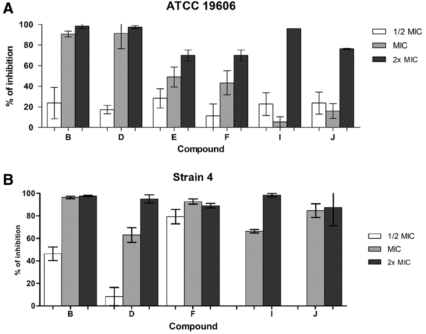
Representation of the biofilm inhibitory capacity of the active pyridine compounds (MIC ≤200 μg/mL) for Acinetobacter baumannii strains ATCC 19606

Values of BIC50: the concentration of the compounds that inhibit 50% of biofilm formation.
Compounds B and D showed antibiofilm potential, with inhibitory and destructive action for both strains and, in relationship to the capacity of disruption of A. baumannii biofilms, showed more promising results for Strain 4, with a statistically significant difference when compared to the ATCC 19606 strain (Fig. 4). Moreover, for each strain, there was no statistical difference between the action of these compounds, except for the concentration of 1/2 MIC (12.5 μg/mL) for Strain 4 (Fig. 4). In addition, for ATCC 19606 strain, the BEC50 of the compounds B and D were 12.8 and 28.2 μg/mL, respectively.

Representation of the biofilm destruction capacity of compounds B and D for strains of Acinetobacter baumannii ATCC 19606 and Strain 4. Values of p < 0.05 were considered significant. Differentiation between uppercase and lowercase letters indicates statistically significant difference between the percentage of biofilm destruction in the concentrations of the compounds B and D evaluated for the strains, indicating more promising results for Strain 4 in comparison to the ATCC 19606. The asterisk indicates that for strain 4, compound F had a higher biofilm destruction capacity at 12.5 μg/mL compared to the same concentration of compound D, with a difference statistically significant.
Discussion
The biofilm formation can be a source of nosocomial infections and a barrier in the treatment of several diseases. Therefore, it is important to highlight the relevance of the development of substances that present not only antimicrobial activity but also act against pathogenic biofilms. In our study, we evaluated the antimicrobial and antibiofilm activity of 10 pyridine derivatives against six strains of A. baumannii. Six compounds were considered active, with MIC values ≤200 μg/mL, for at least one of the strains in the planktonic form. In addition, these compounds were also able to inhibit the formation of A. baumannii biofilms. Among them, the compounds B and D showed the capacity to destroy mature biofilms of the two A. baumannii strains classified as strong in vitro biofilm-forming, even at the concentration of 12.5 μg/mL.
In general, the formation of bacterial biofilm occurs basically in three phases. First, adhesion involves mainly the expression of genes related to structures such as pili, flagella, and fimbriae, which help in the interaction between the microorganism and the surface to be colonized. 29 In a second step, the bacterial density and the induction of the expression of genes related to the quorum sensing increase, resulting in the maturation of the biofilm, and the production of an extracellular matrix composed of polysaccharides, proteins, and DNA. These substances coat the adhered cells, providing protection and, as a result, normally biofilm cells are more resistant to antimicrobial agents such as antibiotics and biocides. 30 And, finally, the dispersion of cells of the biofilm occurs mainly due to the induction of enzymes and surfactants substances capable of degrading the extracellular matrix.31,32
The capacity of biofilm formation by A. baumannii, specifically, is directly related to the induction of the expression of genes, which code for a pili that assists in the adhesion of this pathogen to abiotic surfaces.30,33,34 Considering the results related to the inhibition of A. baumannii biofilms, it can be inferred that the evaluated compounds that are capable of inhibiting biofilm formation probably can act on some of these mechanisms associated with the first step of surface adhesion and/or on inhibition of signaling mechanisms (Fig. 5A, B). In addition, our results indicate that probably the compounds evaluated in the inhibition tests do not act directly in Step 3, since these compounds could reduce the capacity of biofilm formation in 24 hours, in at least one of the assessed concentrations (Fig. 2). The cetylpyridinium chloride, for example, is a nitrogenous compound which has a pyridine in its chemical composition and shows antimicrobial activity, 35 including against bacterial biofilms. 36 As related by Pandit et al., 37 this substance is able to inhibit dental biofilms, being more effective in the early stages of the biofilm formation process.

In relationship to the biofilm destruction assay, the compounds B and D were able to disrupt the attached cells, with more promising results for Strain 4. All evaluated concentrations of the two compounds were able to eradicate at least 65% of the biofilm formed by this isolate. In addition, it should be noted that the subinhibitory concentrations for planktonic cells presented destructive activity in biofilms, indicating that these pyridine compounds, unlike those with inhibitory action, probably act in another phase of the metabolism of A. baumannii or even in specific structures present in the mature biofilm, as in the extracellular matrix (Fig. 5B). This ability may be an advantage aiming the development of new compounds that can be administered in combination with commonly used antimicrobials, acting as adjuvant and facilitating the action of drugs; and/or in the development of biocides for external application, as in surfaces and various hospital materials.
It should be noted that the emergence of resistant pathogens, mainly the biofilm formers, has been an increasing concern, since it represents a challenge for the treatment of several infections, especially in the hospital environment, besides being a source of nosocomial infections. 38 A. baumannii can adhere and form biofilms on abiotic surfaces, being considered a uropathogen intimately related to UTI associated with catheter use, mainly due to its capacity to form biofilm on the surface of this material.39,40
In the treatment of UTI, some compounds are used concomitantly with drugs, such as phenazopyridine—a pyridine derivative, which presents analgesic and anesthetic effects on the urinary tract, reducing the clinical symptoms of the infection. 41 Considering that five A. baumannii strains evaluated in this study were isolated from patients with UTI, and our pyridine compounds showed antimicrobial and antibiofilm activity against them, we once again highlight the potential of these compounds, especially B and D, as a basis for the development of new substances that can be used as adjuvants, including in the treatment of infections such as UTI.
Conclusions
The pyridine compounds B and D showed antimicrobial activity against at least five of the six strains of A. baumannii evaluated in our study in planktonic form and, besides being able to inhibit the formation of biofilms, were also able to destroy bacterial biofilms in a concentration of 12.5 μg/mL. Thus, our results reinforce the importance of these pyridine compounds as scaffolds for the development of new substances such as new drugs or adjuvants for the treatment of A. baumannii infections, and/or as biocides that could be used for external application in hospital devices and surfaces, with the aim of both destroying formed biofilms and preventing bacterial adhesion.
Footnotes
Ethical Conduct of Research Statement
This research followed the precepts of the Resolution No. 466/12 of Conselho Nacional de Saúde, which regulates research involving humans and was approved by the Ethics Committee of FURG (Comitê de Ética em Pesquisa na Área da Saúde—CEPAS—no. 85/2013).
Disclosure Statement
This research was conducted in the absence of any commercial or financial relationships that could be construed as a potential conflict of interest.
Funding Information
This work was supported by National Council for Technological and Scientific Development (CNPq) and Coordination for the Improvement of Higher Education Personnel (CAPES).