
Abstract
Background:
Metabolic syndrome is associated with cardiovascular disease and oxidative stress. The aim of this study was to investigate the differences of novel oxidative stress parameters and lipid profiles in men and women with metabolic syndrome.
Methods:
The study population included 88 patients with metabolic syndrome, consisting of 48 postmenauposal women (group I) and 40 men (group II). Premenauposal women were excluded. Plasma levels of total antioxidant status (TAS) and total oxidative status (TOS) were determined by using the Erel automated measurement method, and oxidative stress index (OSI) was calculated. To perform the calculation, the resulting unit of TAS, mmol Trolox equivalent/L, was converted to μmol equivalent/L and the OSI value was calculated as: OSI = [(TOS, μmol/L)/(TAS, mmol Trolox equivalent/L) × 100]. The Student t-test, Mann–Whitney-U test, and chi-squared test were used for statistical analysis; the Pearson correlation coefficient and Spearman rank test were used for correlation analysis. P ≤ 0.05 was considered to be statistically significant.
Results:
Both women and men had similar properties regarding demographic characteristics and biochemical work up. Group II had significantly lower levels of antioxidant levels of TAS and lower levels of TOS and OSI compared with group I (P = 0.0001, P = 0.0035, and P = 0,0001). Apolipoprotein A (ApoA) levels were significantly higher in group I compared with group II.
Conclusions:
Our findings indicate that women with metabolic syndrome have a better antioxidant status and higher ApoA levels compared with men. Our findings suggest the existence of a higher oxidative stress index in men with metabolic syndrome. Considering the higher risk of atherosclerosis associated with men, these novel oxidative stress parameters may be valuable in the evaluation of patients with metabolic sydrome.
Introduction
M
In 2005, The International Diabetes Federation (IDF) provided a definition of metabolic syndrome that was accepted worldwide.3 According to the IDF definition, a participant was defined as having metabolic syndrome if he or she had central obesity (waist circumference ≥80 cm in women, ≥94 cm in men) plus two or more of following criteria: (1) raised triglyceride levels of 1.69 mmol/L (150 mg /dL) or higher or specific treatment of this lipid abnormality; (2) reduced high-density lipoprotein cholesterol (HDL-C) levels of less than 1.04 mmol/L (40 mg/dL) in men, less than 1.29 mmol/L(50 mg/dL) in women, or specific treatment of this lipid abnormality; (3) raised systolic or diastolic blood pressure of 130/85 mmHg or higher or previously diagnosed hypertension; and (4) raised fasting plasma glucose level of 5.6 mmol/L(100 mg/dL) or higher or previously diagnosed type 2 diabetes mellitus.
Whether the risk of cardiovascular disease is greater than the risk attributable to the components of metabolic syndrome is debatable, it is known that oxidative stress is elevated in metabolic syndrome as well as cardiovascular disease. Gender differences in relation to oxidative stress in patients with metabolic syndrome, which is usually considered as a “precursor” of cardiovascular diseases, is an important issue. Therefore, in this study, we aimed to compare the oxidative/antioxidative status in relation to lipid profile in men and women presenting with metabolic syndrome.
Materials and Methods
A total of 88 patients (mean age 56.1 ± 10.9 years), who were diagnosed as metabolic syndrome defined by IDF criteria3 were included. All of these patients had central obesity as defined by IDF criteria (waist circumference ≥80 cm in women, ≥94 cm in men).
Our study group consisted of 48 postmenauposal women (group I), who had a mean age of 55.9 ≥ 9.9 years, and 40 men (group II), who had a mean age of 56.4 ≥ 12.0 years. Patients with acute corınary syndromes, obvious renal dysfunction (creatinine >2 mg/dL), tumors, or severe peripheral vascular disease and premenauposal women and smokers were not included.
None of our patients were taking statins, β-blockers, or hormone replacement therapy. In both groups, about 23 (48%) of women were using thiazides and 19 (47%) of the men were taking thiazides; all of the patients were taking angiotensin-converting enzyme (ACE) inhibitors. In all, 42 (47%) of our patients were diabetic; 22 (46% of group I) of them were women and 20 (50% of group II) of them were men. All of the diabetic patients were taking metformin.
Demographic and clinical characteristics, body mass index (BMI), and waist circumference were recorded. Additional biochemical factors such as fasting glucose, glycosylated hemoglobin (HbA1c), lipid markers, serum apolipoprotein A (ApoA), high-sensitivity C-reactive protein (hsCRP), uric acid, as well as fibrinogen and plasma levels of total antioxidant status (TAS) and total oxidative status (TOS) were analyzed. For blood sampling and laboratory measurements, blood samples were drawn after 12 h of fasting at rest between 8 and 10 mL with venous occlusion from the antecubital vein into 10-cc Vacutainer tubes and ethylenediaminetetraacetic acid (EDTA) tubes. Venous blood sample tubes were centrifuged for 10 min, and serum was stored at −70°C until analysis. Lipid parameters were measured in fresh serum by the use of commercial kits (Thermo Diagnostic) and Opera (Technicon RA System, Bayer) analyzer. Serum tryglicerides, total cholesterol, glucose, HDL-C, and, low-density lipoprotein cholesterol (LDL-C) were measured by using the standard methods. hsCRP and ApoA concentrations were determined by a quantitative nephelometric method using serum in a PROSPEC analyzer (Dade-Behring). HbA1c was measured quantitatively by high-performance liquid chromatography
Plasma TAS levels were determined using a novel automated measurement method, developed by Erel.4 In this method, the hydroxyl radical, which is the most potent radical, is produced via a Fenton reaction. In the classical Fenton reaction, the hydroxyl radical is produced by mixing a ferrous ion solution and a hydrogen peroxide solution. In the most recently developed assay by Erel, the same reaction is used. In the assay, the ferrous ion solution, which is present in reagent 1 was prepared by dissolving 114 mg of xylenol orange (Merck 8677) and 8.18 grams of NaCl (Riedel-de Haën 13423) in 900 mL of H2SO4 solution (Merck 713), 25 mM. The pH value of the reagent was 1.75. It was mixed with hydrogen peroxide (Merck 8597), which is present in the reagent 2. Reagent 2 was ferrous ion 5 mM and 10 mM o-dianisidine (Sigma D-3252) in a 25 mM H2SO4 solution. These methods were applied to an automated analyzer, Opera (Technicon RA System, Bayer) mean ± standard deviation (SD), and based on a linear type of calibration. The sequentially produced radicals, such as the brown-colored dianisidinyl radical cation, produced by the hydroxyl radical, are also potent radicals. In this assay, the antioxidative effect of the sample against the potent free-radical reactions, which are initiated by the produced hydroxyl radical, was measured. The assay has excellent precision values, which are lower than 3%. The results are expressed as mmol Trolox equiv/L.5
Measurement of total oxidant status
Plasma TOS levels were determined using a novel automated measurement method, developed by Erel.6 In this method, oxidants present in the sample oxidized the ferrous ion–odianisidine complex to ferric ion. The oxidation reaction was enhanced by glycerol molecules (Carlo Erba 346165), which are abundantly present in the reaction medium. The ferric ion makes a colored complex with xylenol orange in an acidic medium. The color intensity, which can be measured spectrophotometrically, is related to the total amount of oxidant molecules present in the sample. The assay was calibrated with hydrogen peroxide, and the results were expressed in terms of micromolar hydrogen peroxide equivalent per liter (μmol H2O2 equiv/L). This method was applied to an automated analyzer, Opera (Technicon RA System, Bayer).
Oxidative stress index
The oxidative stress index (OSI) was calculated as: OSI = (TOS/TAS) × 100.The percent ratio of the TOS to the TAS gave the OSI, an indicator of the degree of the oxidative stres. To perform the calculation, the result unit of TAS, mmol Trolox equivalent/L, was converted to μmol equivalent/L and the OSI value was calculated as below formula; OSI = [(TOS, μmol)/(TAS, mmol Trolox equivalent/L) × 100].4 –6
Statistical analysis was done using the SPSS for Windows 16.0 package. Continuous variables were presented as the mean followed by SD, and categorical variables were presented as frequency and percent. Data were tested for normality of distribution by the Kolmogorow–Smirnow test. Continuous variables between two groups were compared using the Student t-test for normally distributed data and the Mann–Whitney-U test for data not normally distributed. Categoric parameters were evaluated by the chi-squared test. Relations between variables of interest were assessed using the Pearson correlation coefficient or Spearman rank order correlation for variables not normally distributed. For all statistical evaluations, a two-tailed P value of 0.05 or less was considered to be statistically significant with a 95% confidence interval (CI).
Results
Baseline characteristics of both groups are given in Table 1. Group I and group II did not have any significant differences for BMI, type II diabetes mellitus, waist circumference, and systolic and diastolic pressure. hsCRP values were relativly high but did not differ between two groups (4.8 ± 2.5 mg/L versus 6.3 ± 1.7 mg/L). A comparison of the biochemical parameters TAS, TOS, and OSI is shown in Table 2. Group I had significantly higher ApoA and TAS levels compared with group II (2.0 ± 1.4 vs. 1.4 ± 0.2; P = 0.011 and 0.9 ± 0.2 vs. 0.7 ± 0.2; P = 0.0001, respectively). On the other hand, group II had significantly higher TOS and OSI levels compared with group I (10.9 ± 1.7 vs. 9.8 ± 1.4, P = 0.035; and 16.3 ± 5.2 vs. 12.2 ± 3.2, P = 0.0001).
B
Abbreviations: NS, not significant; BMI, body mass index ; DM-II, type II diabetes mellitus
B
Abbreviations: HDL, high-density lipoprotein; LDL, low-density lipoprotein; HbA1c, glycosylated hemoglobin; hsCRP, high-sensitivity C-reactive protein; ApoA, apolipoprotein A; H2O2, hydrogen peroxide.
One-way analysis of variance (ANOVA) analysis was done to correlate with BMI, waist circumference, HbA1C, HDL, triglycerides, and ApoA in the total cohort and in men and women to see which factors contributed to the TAS or TOS. TOS was strongly associated with triglycerides (Fig. 1) in women.
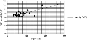
Total oxidative status (TOS) and triglyceride association in women.
Discussion
Metabolic syndrome patients were grouped according to gender in our study. We found no differences between men and postmenauposal women in terms of clinical and lipid profiles except that there were significantly higher levels of HDL and its main component ApoA in women. Our findings that ApoA was significantly higher in women in concordance with a better antioxidant profile is reflected by higher TAS levels in this group. This marker was significantly higher in women compared with men. This finding is in accordance with the data that men tend to have more cardiovascular events than women. 7Both of our groups had high CRP levels, classifying them into high-risk groups for future vascular events. We believe that gender imbalance in oxidant status becomes even more important in this setting.
Triglycerides were significantly correlated with TOS in women. Although women may have higher HDL and ApoA levels, rises in triglycerides may be associated with higher TOS levels within this group. An interesting similar finding was observed in hemodialysis patients who had HDL-C, and ApoA was higher in the polysulfone and hemophan than in the cuprophane groups whereas triglyceride was lower.8
Oxidative stress arises as a result of an imbalance in the human oxidative and antioxidative status.9 Oxidant molecules are produced endogenously in organisms, and they are also taken from the outer environment. The electron transport chain and a range of oxidase enzymes, including xanthine oxidase, glycolate, and monoamine oxidases, make major endogenous reactive oxygen species (ROS) sources. Oxidative stress has been implicated in aging, atherosclerosis, and various diseases.10 , 11 There is evidence that increases in oxidative stress contributes to complex functional and structural changes that ocur in the vessel wall.12
Oxidative stress and lipid peroxidation have been implicated in the pathogenesis of atherosclerosis. The oxidative modification of LDL is mediated by reactive ROS. Plasma antioxidants levels and prevalence of atherosclerosis have a strong inverse association, i.e., patients with low antioxidant capacity levels may be at increased risk for coronary atherosclerosis. 7High activity of ROS levels during oxidative stress contributes to the complex functional and structural changes that occur in the blood vessel wall. There is recent evidence that gender hormones many influence vascular oxidative stress. There is now strong evidence that levels of vascular ROS are lower in female animals compared with male animals under healthy, hypertensive, and atherosclerotic conditions. 13Metabolic syndrome has been associated with a higher fraction of oxidized LDL and thus with higher levels of circulating oxidized LDL.14 Dyslipidemia and insulin resistance in obese LDL receptor-deficient mice were associated with increased oxidative stress and impaired HDL-associated antioxidant defense, resulting in accelerated atherosclerosis due to increased macrophage in filtration and accumulation of oxidized LDL in the aorta.15
Considering these data, oxidative stress induces modification of proteins. ApoA is the main component of HDL, representing 65% of the content. 16Both ApoA, the major HDL apolipoprotein, and sphingosine-1-phosphate, a minor bioactive lipid, have been proposed to contribute significantly to HDL-mediated cytoprotection. Altered conformation of ApoA-1 in small HDL particles in metabolic syndrome may result in their deficient biological acitivity, thereby weakening the defense of HDL antiapoprotic activity from density and ApoA-I content. High levels of oxidative stress, low-grade inflammation, and hyperglycemia constitute important characteristics of metabolic syndrome.17 Oxidative stress is increased in metabolic syndrome and may contribute to the pathogenesis of early atherosclerosis. 18Obesity, dyslipidemia, hyperglycemia, and hyperinsulinemia are characteristics of most rodent models that mimic the metabolic syndrome.19 The metabolic syndrome, which often accompanies obesity, has also been independently linked with increased oxidative stress and inflammatory burden. 20
There are many methods for assessment of oxidative status. We have used the Erel method for this purpose.4 This novel assay is rapid, reliable, sensitive, inexpensive, and fully automated. The developed method has high linearity and the results are highly reproducible. The reagents are easy to prepare and their lifetimes are long. The prevalance of coronary heart disease is higher in men compared with women (6.9% vs. 5.4 %).7 Identification of changes in oxidative stress by gender may be important in this setting. HDL is the most antiatherogenic and antioxidant of all lipoproteins. Ridker et al. has found standard lipid measures as valuable as Apo A-I/B100 in women. 20 This may be due to high ApoA content in HDL. Some hormones, such as estrogens and progestin, may also influence the degree of vascular oxidative stress by modulating the expression and function of antioxidant enzymes within the blood vessel wall. 13 Gender steroids have an antioxidant capacity; their reduced levels in female with menopause and in males with lower testosterone may contribute to the accelerating development of atherosclerosis in this section of the population.7
On the other hand, TOS and OSI are markers of oxidative damage. These markers were significantly higher in men compared with women. In a recent study, Cavalca et al. have found that glutathione levels were lower in men, and in coronary artery disease (CAD) and elderly patients. 21 Total antioxidant capacity was previously associated with sex hormones. 22Just a lower antioxidant capacity may not be the only reason for higher TOS in men. There could be an increase in the production of ROS that may be responsible for the higher TOS in men. In animal studies, ROS generated in response to angiotensin II (AngII) are lower in female versus male mice.23 Karali et al. 24 suggest that there may be differing regulating mechanisms through gender differences; they have found that other oxidative stress parameters such as advanced oxidation protein products, nonprotein thiol, Fe2+, levels and the redox index were significantly higher in male rats compared with female rats. These findings support our hypothesis that an increase in production of ROS may be a contributing factor for higher TOS in men. There is a causal relationship according to gender between different tumors and type 2 diabetes/metabolic syndrome as well. 24Supposed pathomechanisms are obesity, cytokines secreted excessively in adipose tissue, permanent and postprandial hyperglycemia, hyperinsulinism, and insulin resistance, and other growth factors, like proinsulin, insulin like growth factor-1, ROS, angiogenesis, inflammation, and the multiple effects of inflammatory cytokines.
Inflammation plays a role in all phases of atherosclerosis. The acute-phase reactant CRP, as a marker of inflammation, has been regarded as a major cardiovascular risk factor. 25hsCRP assessment adds prognostic information25 , 26 in metabolic syndrome. In conclusion, women with metabolic syndrome have a better antioxidant status and lipid profile compared with men. Our findings suggest the existence of a higher oxidative stress index in men with metabolic syndrome. The complex mechanisms between lipid profile and oxidative stress needs further investigation. Considering the higher risk of atherosclerosis associated with men, these novel oxidative stress parameters may be valuable in the evaluation of patients with metabolic sydrome
Limitations
The major limitation of our study is that these measurements of oxidation status were done once and time-related changes in the course of the disease process were unavailable. Also our population size was relatively small.