
Abstract
A promising method that offers both time- and site-specific delivery of macromolecules is photochemical internalization technology (PCI). Here, we have characterized various polyamidoamine (PAMAM) carriers [generation (G) 0–7], for light-directed delivery of nucleic acids in vitro by the use of PCI technology. A number of parameters for optimal delivery of nucleic acids into human cancer cells, that is, various light-doses, carrier-doses, and small interfering RNA (siRNA)/messenger RNA (mRNA) doses were investigated for either up- or down-regulation of enhanced green fluorescent protein (EGFP) gene expression. In summary, our results showed in an osteosarcoma cell line (OHS) [EGFP] model system the possibility for efficient light-directed siRNA silencing (>80% silencing) when using PAMAM G3 to G7 as carriers. Surprisingly, no EGFP mRNA up-regulation was detected either with or without PCI after EGFP mRNA/PAMAM (G0–G7) transfection in standard OHS cells. We have here identified properties for PAMAM formulations enabling light-directed siRNA delivery with the aim of developing a site-specific strategy for delivery of nucleic acids in vivo.
Introduction
Besides chemical modification of carrier or cargo, photochemical internalization (PCI) technology is an alternative method for specific delivery of macromolecules entrapped in the endocytic pathway (Berg et al., 1999). In PCI, a prelocalized photosensitizer (PS) in the endosomal membrane is triggered by illumination and generates reactive oxygen species. This event causes endosomal rupture and subsequent release of the entrapped material to the cytosol (Hogset et et al., 2004). Of importance, a novel PS, the disulfonated tetraphenyl chlorin (TPCS2a), or Amphinex, has recently been developed for clinical utilization of PCI (Berg et al., 2011). A number of carriers/siRNA complexes have earlier been combined with PCI treatment in vitro, such as jetSI-ENDO (Boe et al., 2007), Lipofectamine 2000 (Oliveira et al., 2007), various branched polyethylenimine (PEI) formulations (Boe et al., 2008), dextran nanogels (Raemdonck et al., 2010), and beta-cyclodextrin (Boe et al., 2010b). However, the majority of these carriers are less well suited for in vivo applications.
The cationic polyamidoamine (PAMAM) dendrimer, possesses several advantages compared to other carriers (e.g., PEI) previously tested for PCI-induced delivery. PAMAM dendrimers are a group of highly branched and distinct polymers that have the exclusive benefit of a controllable nanoscale size, high monodispersity, and controlled number of surface groups (Tomalia et al., 1985). The cationic primary amine groups on the spherical surface of full-generation PAMAMs are able to form electrostatic interactions with anionic charged phosphate groups in the nucleic acid and form stable and uniform dendriplexes (Labieniec and Watala, 2009). PAMAM dendrimers also possess numerous tertiary amines in the interior that are protonated in the acidic endosomes, resulting in endosomal disruption followed by release of nucleic acids from PAMAM/nucleic acid dendriplexes (Sonawane et al., 2003). Of particular interest, Zhou and colleagues recently demonstrated that PAMAM generation 5 (G5) dendrimer-mediated siRNA delivery can avoid interferon responses and toxicity in vivo after systemic administration in mice (Zhou et al., 2011). At present, no PAMAM formulation has yet been evaluated for PCI-directed delivery of siRNA or mRNA molecules. PAMAM dendrimers have previously been used in combination with PCI in vitro to deliver doxorubicin to Ca9-22 cells (human gingival cancer) (Lai et al., 2007), saporin to Ca9-22 and KJ-1 cells (human nasopharyngeal carcinoma) (Lai et al., 2008), and plasmid DNA to HeLa cells (human cervical epithelioid carcinoma) (Shieh et al., 2008). The intentions in the former studies were to enhance the toxicity of doxorubicin/saporin by triggering a synergetic cell killing effect, mediated by the photosensitizer (AlPcS2a).
In this study, we have evaluated the possibility of using unmodified PAMAM dendrimers, generation 0 to 7 (G0–G7) for light-directed gene modulation. After optimizing various parameters, we show that PAMAM formulations have the potential for effective light-directed siRNA delivery, resulting in potent silencing of the gene of interest compared to non-PCI treated samples. Surprisingly, our results also show that PAMAM dendrimers are less well suited for mRNA delivery.
Materials and Methods
Cell line and culture conditions
The osteosarcoma cell line, OHS, was established from a bone tumor biopsy (Fodstad et al., 1986). In addition, we used an OHS cell line stably transfected with enhanced green fluorescent protein (EGFP). Both the regular OHS cells and the OHS cells expressing EGFP were cultured and maintained in RPMI 1640 medium (Bio Whittaker), supplemented with 10% fetal calf serum (PAA Laboratories) and GlutaMAX (Invitrogen). The cells were maintained at 37°C in a humidified atmosphere containing 5% CO2 and were routinely tested for mycoplasma infections (VenorGeM, Minerva Biolabs).
Nucleic acids
A prevalidated siRNA targeting EGFP and a negative siRNA control were ordered from Ambion. EGFP mRNA was produced as described previously (Saeboe-Larssen et al., 2002).
Transfection
We evaluated various PAMAM formulations for PCI-induced delivery of nucleic acids (Table 1). All PAMAM carriers were ordered from Sigma-Aldrich. The carriers were vacuum centrifuged by a GeneVac EZ-2 (Genevac) to remove methanol waste from the stock solution, and diluted in sterile water prior to transfection. Dendriplex formations were carried out in RPMI 1640 medium under serum-free conditions. The dendriplex solution was added to serum-containing RPMI 1640 medium after 30 minutes. The transfection solution, either with or without 0.5 (μg/mL of the photosensitizer TPPS2a (Porphyrin Products), was added to either OHS or OHS EGFP cells in 12-well plates. After 18 hours, cells were washed 3 times with fresh RPMI 1640 medium and reincubated for 4 hours prior to light treatment. Cell plates were exposed to blue light (5.1 mW/cm2) by a LumiSource prototype (PCI Biotech AS) and reincubated before mRNA harvesting or EGFP signal measurement 48 hours post PCI treatment. Due to the half-life of EGFP (>24 hours) (Barrow et al., 2005), we re-incubated the cells for 96 hours post PCI for detection of EGFP by fluorescence microscopy. Cells were light protected using aluminum foil during the experiments.
PAMAM, polyamidoamine.
Detection system
Total cellular RNA was isolated with the GenElute Mammalian Total RNA Miniprep Kit (Sigma-Aldrich), and the iScript cDNA synthesis kit (BioRad) was used for reverse transcription. We evaluated the gene expression of EGFP by quantitative reverse transcription polymerase chain reaction (qRT-PCR) with SYBR Green 1 real-time detection (BioRad). The EGFP primer set used amplifies a 71-bp segment (forward primer 5′-GTCCAGGAGCGCACCATCT-3′ and reverse primer 5′-CCCTCGAACTTCACCTCGG-3′). Samples were normalized to untreated control relative to the 2 housekeeping genes, the TATA box-binding protein (with forward primer 5′-GCCCGAAACGCCGAATAT-3′ and reverse primer 5′-CGTGGCTCTCTTATCCTCATGA-3′) and the human acidic ribosomal phosphoprotein P0 (with forward primer 5′-CGCTGCTGAACATGCTCAAC-3′ and reverse primer 5′-TCGAACACCTGCTGGATGAC-3′). These housekeeping genes were chosen because they were unaffected by the different treatment modalities in this study. Calculations were based on the delta-delta cycle threshold (ΔΔCT) method (Vandesompele et al., 2002). For evaluation of cell viability, the MTS solution [3-(4,5-dimethylthiazol-2-yl)-5-(3-carboxymethoxyphenyl)-2-(4-sulfophenyl)-2H-tetrazolium] (Promega) was added to the cells 48 hours after light treatment. The absorbance was measured at 490 nm in wells containing RPMI 1640 (with serum and GlutaMAX) diluted with MTS to a final volume of 1200 μL/well (1:6 dilution). EGFP mRNA transfected cells were evaluated for transfection efficiency at 488 nm by a Wallac Victor2 1420 Multilabel counter (PerkinElmer) and samples were calculated as percentage of untreated control. For dendriplex size measurements, 0.7 μg EGFP siRNA and 0.35–0.7 μg PAMAM G0–G7 were prepared in serum-free RPMI medium and measured at 25°C with a scattering angle of 173° on a Zetasizer Nano ZS (Malvern Instruments Ltd.), and determined using the Z-average value with approved instrumental expert advice criteria for measurements. Fluorescence images of EGFP siRNA transfected cells were visually examined on a Zeiss inverted microscope, Axiovert 200 equipped with filter for fluorescein isothiocyanate (450- to 490-nm bandpass (BP) excitation filter, a 510-nm farbteiler (FT) beam splitter, and a 515- to 565-nm longpass (LP) emission filter). Images were composed by the use of Carl Zeiss Axiocam HR, Version 5.05.10 and Axiovision 3.1.2.1 software. Pictures were prepared with Adobe Photoshop 7.0 (Adobe) and Zeiss LSM Image Browser (Version 3).
Results and Discussion
Light-directed down-regulation of gene expression using EGFP siRNA molecules
We evaluated the possibility for light-directed down-regulation of gene expression when combining PAMAM G0–G7 with siRNA and PCI. When using 0.7 μg PAMAM G0–G7 in combination with 0.7 μg EGFP siRNA (50 nM), ∼90% silencing was measured in G4 to G7 samples, compared with untreated cells and negative siRNA control, using a light-dose of 153 mJ/cm2 (Fig. 1c). In contrast, PAMAM G0 showed no significant silencing, compared to untreated cells or negative siRNA control, while the silencing effect in G1, G2, and G3 samples were measured to ∼20%, ∼30%, and ∼80% in PCI-treated samples, respectively. Of note, the silencing effect of siRNA increased by increasing molecular weights (MW) of the PAMAM dendrimer (G7>G0), both with and without irradiation. Furthermore, the optimal targeting effect (difference between PCI and non-PCI treated samples) was measured in G4 (89% silencing with PCI vs. 19% without PCI). We next explored the cell viability, and a minor reduction of less than 20% was detected in G3 to G7 PCI-treated samples, compared to untreated control levels (Fig. 1d). No significant reduction in cell viability was detected in the G0 or G1 samples, neither with nor without PCI, while in the G2 samples, ∼10% reduction in cell viability was observed after PCI treatment. Finally, less than 10% reduction in cell viability was measured in G3 to G7 without PCI treatment. Our negative siRNA control sample when transfected with PAMAM G7 resulted in a similar reduction in cell viability as the EGFP siRNA/G7 sample. We next investigated the effect of light-directed siRNA gene silencing when using PAMAM concentrations above (1.4 μg) or below (0.35 μg) the previously tested 0.7 μg dose presented in Fig. 1c. Of note, we kept the siRNA dose (50 nM EGFP siRNA) and light dose (153 mJ/cm2) constant. Our results demonstrated efficient silencing down to ∼10% of remaining EGFP mRNA levels when using 1.4 μg G4 to G7, compared to untreated control levels (Fig. 1e). With this PAMAM concentration, cell viability was however reduced with ∼45% in G4–G7 samples treated with PCI (Fig. 1f). The highest targeting effect when using 1.4 μg PAMAM was measured in G3 (84% silencing with PCI vs. 26% without PCI). When lowering the PAMAM concentration to 0.35 μg, silencing above 90% was detected only in the G7 samples after PCI treatment (Fig. 1a). The targeting effect was also highest in the G7 samples (93% silencing with PCI vs. 21% without PCI). The 21% silencing effect measured in samples not exposed to PCI is most likely a result of a minor “proton sponge” activity from the PAMAM carrier under the given parameters. With the low PAMAM concentration, reduction in cell viability was less than 5% in both PCI-treated and non PCI-treated G0-G7 samples (Fig. 1b). To demonstrate that EGFP was silenced also at the protein level, PAMAM G6 was utilized with the same concentrations and light dose as in Fig. 1c. The gene-silencing effect of PCI in combination with PAMAM G6 and EGFP siRNA was hereby confirmed (Fig. 1g).
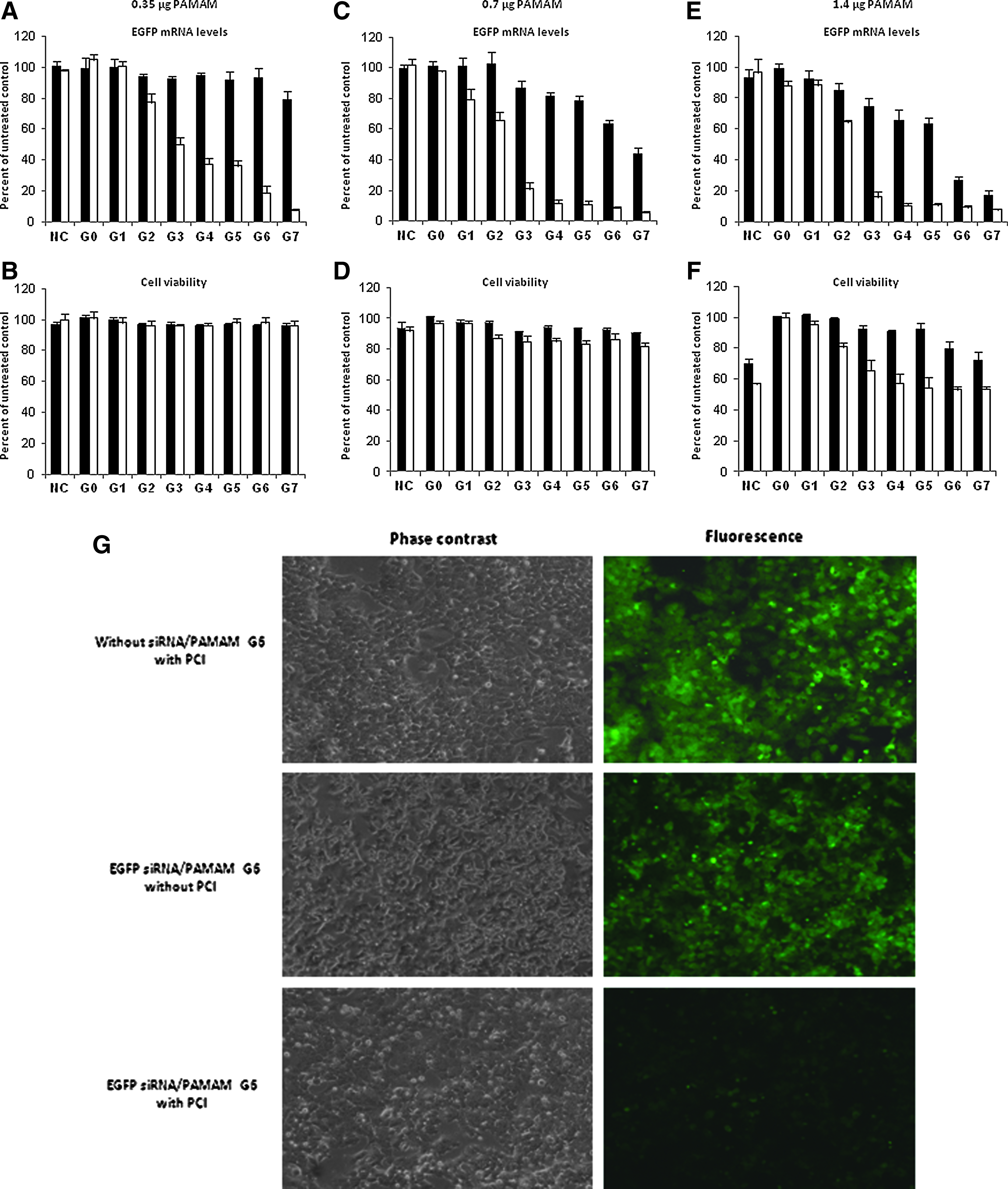
Light-directed down-regulation of gene expression in vitro in the osteosarcoma cell line (OHS) [enhanced green fluorescent protein (EGFP)] cell line using EGFP small interfering RNA (siRNA) molecules and a light-dose of of 153 mJ/cm2.
Light-directed down-regulation of EGFP gene expression under conditions with lower doses of light and siRNA
Because we are focusing on a transfection protocol with in vivo potential, it is beneficial to use a minimum light dose to avoid phototoxic cell death, and also to use a low siRNA concentration to decrease off-target effects. Using a lower light dose of 76.5 mJ/cm2, under identical PAMAM and siRNA concentrations as Fig.1c, resulted in reduced silencing (Fig. 2a). Only G7 resulted in ∼90% gene silencing compared to untreated control levels. The highest targeting effect with the low light dose was measured in G5 (77% silencing with PCI vs. 22% without PCI). Under these conditions, we measured less than 10% reduction in cell viability in G0-G7 samples, both with and without PCI (Figure 2b). Finally, we evaluated the effect of lowering the siRNA-dose on siRNA gene silencing using 0.7 μg PAMAM G0–G7 and 10 nM (0.14 μg) EGFP siRNA with a light dose of 153 mJ/cm2. Our real-time qRT-PCR results showed optimal silencing down to ∼15% of remaining EGFP mRNA levels using G7, compared with untreated control levels (Fig. 2c). As in the previous experiments, the silencing effect was also here increased by increasing the MW of the PAMAM dendrimer (G7>G0), both with and without irradiation. The highest targeting effect was measured in G7 (86% silencing with PCI vs. 26% without PCI). With the low siRNA doses, cell viabilities were reduced with less than 10% in G0–G7 both with and without PCI-treatment (Fig. 2d).
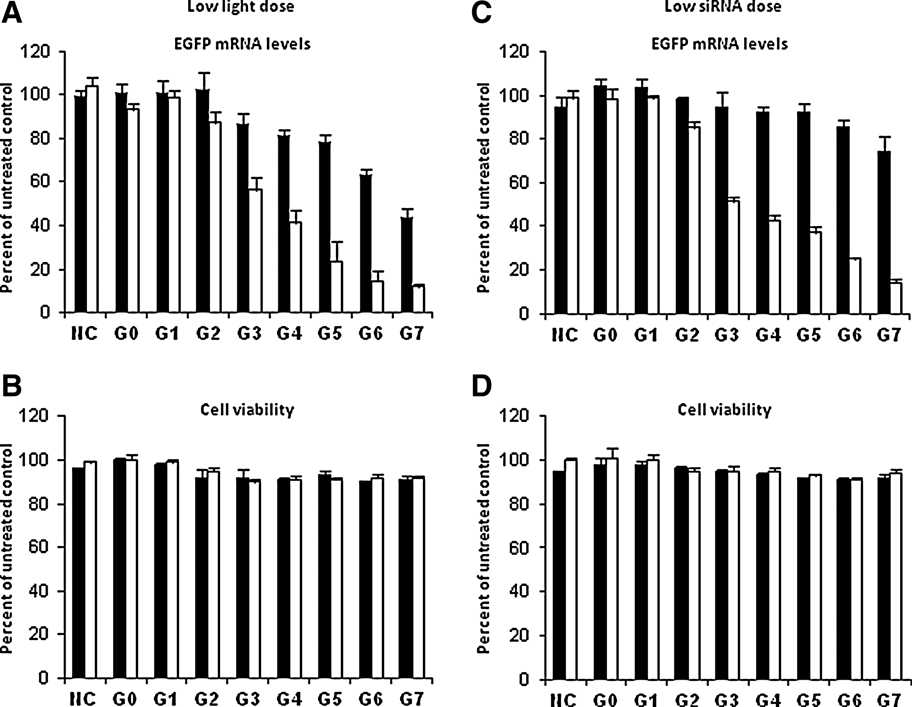
Light-directed down-regulation of EGFP gene expression under conditions with lower doses of light and siRNA. PAMAM formulations G0–G7 were investigated for their gene delivery properties and cell viabilities in vitro in the OHS [EGFP] cell line using a low light dose of 76.5 mJ/cm2, 0.7 μg PAMAM, and 50 nm EGFP/negative control siRNA
When comparing the PAMAM data with our previous reports, we find that PAMAM shows improved PCI-mediated gene silencing in vitro compared to beta-cyclodextrin (Boe et al., 2010b) and approximately similar silencing effect as was observed with jetSI (Boe et al., 2007) and 25 kDa B-PEI (Boe et al., 2008). The targeting effect of PCI (measured by the difference in gene silencing between PCI vs. non-PCI treated samples) was approximately similar to what we have detected previously with beta-cyclodextrin and 25 kDa B-PEI (>70% PCI targeting), but improved compared to jetSI/-ENDO (50-60% PCI targeting). Interestingly, PAMAM G7 demonstrated gene silencing efficacy above 85% even under conditions such as low siRNA concentration (10 nM), or with a very low light-dose (76.5 mJ/cm2), or with a low PAMAM dose (0.35 μg) when combined with PCI. The features of the PAMAM G7 to work under a low siRNA dose, under a low PAMAM dose, or under a low light dose are all very useful for further in vivo investigations. Because our focus is limited to the delivery of nucleic acids with PCI, and not cell killing, the low doses of PAMAM and light required for gene silencing suit an in vivo situation where the aim exclusively is delivery. Moreover, a low siRNA dose reduces the possibility for off-target effects. To our knowledge, no polymeric carrier system for PCI mediated siRNA delivery has shown the same gene silencing efficacy as presented here, when using as low as 10 nM siRNA concentration or a light dose of only 76.5 mJ/cm2. Another advantage using PAMAM formulations is the distinct surface of PAMAM dendrimers, which ensures the same MW from batch to batch, and in this way is beneficial regarding reproducibility of experiments. This is in contrast to other polymeric carrier systems.
To investigate a possible correlation between dendriplex size and gene silencing effect, we evaluated dendriplex sizes of siRNA with various PAMAM carriers under the described conditions (Fig. 3). Our results showed only minor differences in dendriplex sizes measured with 0.35, 0.7, and 1.4 μg PAMAM formulations when the siRNA concentration was kept constant at 0.7 μg. For G6–G7, samples were larger at 0.35 μg than at 0.7 μg, and smallest at 1.4 μg. The G4 and G5 formulations decreased in size from 0.35 to 0.7 μg, and increased in size from 0.7 to 1.4 μg. Of note, all dendriplexes measured ranged from ∼400 nm to ∼600 nm. These results are in accordance with Jensen and colleagues who reported that siRNA dendriplexes with G4 and G7 decreased in size when the dendrimer concentration was elevated (Jensen et al., 2011). In the same study, they concluded that G1 was unable to form dendriplex aggregates with siRNA, which is partly due to the large size of siRNA molecules (18 kDa) compared to G1 (1.43 kDa). In our study, dendriplex measurements of G0–G3 and dendriplexes with 0.14 μg siRNA consistently appeared to indicate polydisperse spheres, to a level that did not pass the quality criteria from the Zetasizer Nano ZS instrument.
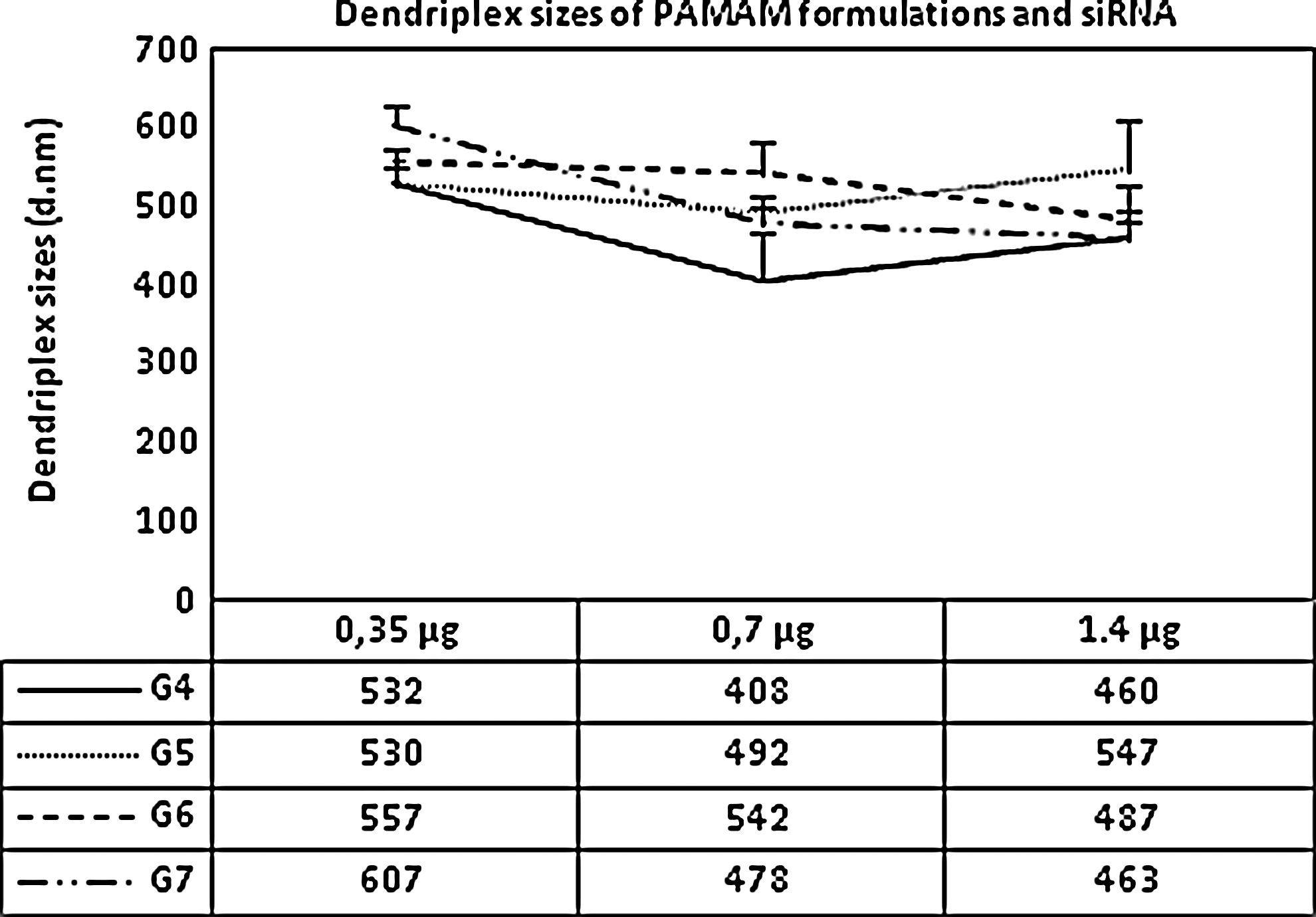
Dendriplex sizes of PAMAM G4–G7 and siRNA. The concentration of PAMAM formulations ranged from 0.35–1.4 μg, while the siRNA concentration was kept constant at 0.7 μg. Error bars show standard error of the mean±SEM.
We were also interested in whether PAMAM formulations G0–G7 could be used to deliver EGFP mRNA molecules for light-directed up-regulation of gene expression. Our results showed that none of the PAMAM formulations were effective for up-regulation of EGFP signal when testing 1, 2, and 4 μg mRNA in combination with 0.35, 0.7, or 1.4 μg G0–G7, neither with nor without PCI treatment (data not shown). Thus, PAMAM formulations were not effective for mRNA delivery, in contrast to B-PEI, which showed PCI-induced mRNA transfection efficacy up to 90% (Boe et al., 2010a). Differences between PEI and PAMAM formulations with respect to complex formation/packing (due to size and structure variation of the given carriers) are likely to play a critical role. The EGFP mRNA molecule we utilized for transfection has a length of approximately 800 bases, which is substantially longer than the EGFP siRNA used for transfection. Dendriplexes of PAMAM formulations and mRNA/siRNA are most likely packed in different ways, and this appears to have a great impact on transfection.
In conclusion, we have evaluated and optimized the parameters for light-directed gene siRNA silencing when using various PAMAM formulations. We find that PAMAM compares favorably, with a high light-directed targeting effect and low toxicity after optimization. We foresee that the protocol described here can inspire development of future in vivo gene therapy applications, using siRNA molecules combined with various PAMAM- based carriers for light-directed delivery.
Footnotes
Acknowledgments
The authors are grateful for the PhD grant allocated by the Norwegian Gene Therapy Programme, the Norwegian Radium Hospital.
Authors Disclosure Statement
No competing financial interests exist.