
Abstract
We report the toxicological and pharmacokinetic properties of the synthetic, small interfering RNA (siRNA), QPI-1007, following intravitreal administration. QPI-1007 is a chemically modified siRNA designed to act via the RNA interference (RNAi) pathway to temporarily inhibit expression of the caspase 2 protein and is being developed as a neuroprotectant for the treatment of nonarteritic anterior ischemic optic neuropathy and other optic neuropathies such as glaucoma that result in the death of retinal ganglion cells. The half-life of QPI-1007 in the vitreous and retina/choroid in the Dutch Belted rabbit was about 2 days, and there was no sign of accumulation after repeated administrations at either 2- or 4-week dosing intervals in the rabbit. QPI-1007 was well tolerated in Dutch Belted rabbits following single or repeated intravitreal administrations of up to 11 doses over 9 months. Test-article–related effects were limited to the eyes, with minimal to mild vitreal cellular infiltration being the major finding, which was reversible. In repeated-dose studies, a modest reduction in B-wave amplitude obtained by electroretinography was observed in animals treated with the highest dose level tested (3 mg, which is equivalent to a 12 mg/eye human dose) that was not considered to be clinically meaningful. Administration in the rat of either a single bolus intravenous (i.v.) injection of 100 mg/kg or daily bolus i.v. injections of 75 mg/kg/day for 28 days failed to elicit any macroscopic or microscopic changes, suggesting a low risk for systemic toxicity. QPI-1007 was negative in three genetic toxicity studies. Overall, the nonclinical studies support the further development of QPI-1007.
Introduction
QPI-1007
Death of retinal ganglion cells in NAION occurs primarily through apoptosis (Kaja et al., 2003; Brao-Osuna et al., 2007; LEVIN, 2007). The caspases play a major role in apoptosis, and caspase 2 has been shown to be activated specifically in retinal ganglion cells in rat models of retinal ischemic insult Ahmed et al., 2011. Since they are unable to divide, loss of retinal ganglion cells results in irreversible loss of vision. Therefore, preventing apoptosis by inhibiting caspase 2 expression could protect retinal ganglion cells and thereby preserve vision.
Preclinical data on QPI-1007 were reported previously by Ahmed et al. (2011). These authors demonstrated that the chemical modifications incorporated into QPI-1007 siRNA were effective in abrogating RNAi-mediated activity arising from either the passenger strand or the seed region of the guide strand, thereby improving the specificity and reducing the potential for unwanted off-target effects. QPI-1007 was readily taken up by retinal ganglion cells following intravitreal injection in the rat, and RNAi-mediated cleavage of caspase 2 mRNA could be detected in rat retina as soon as 4 hours after injection, demonstrating that this 19-bp siRNA can be delivered to the retina in the absence of complex delivery formulations. Ahmed et al. further showed that QPI-1007 did not activate recombinant murine or human recombinant Toll-like receptor 3, 7, 8, or 9, nor did it induce either cytokine release or activation of the interferon pathway in human peripheral blood mononuclear cells, indicating a low risk of immunostimulatory activity. Finally, the authors demonstrated that siRNAs targeting caspase 2 mRNA afforded protection of retinal ganglion cells in two models of acute optic nerve damage. Here we report the results from nonclinical pharmacokinetic and toxicity studies used to support single- and repeated-dose clinical trials.
Materials and Methods
QPI-1007 siRNA
The molecular sequence of QPI-1007 is:
Passenger strand: 5′ ia-GCC AGA AUG UGG AAC UCY U 3′
Guide strand: 5′ AgG aGu UcC AcA uUc UgG c 3′
where uppercase letters represent unmodified RNA nucleosides, lowercase letters represent 2′-O-methyl sugar modified RNA nucleosides, “Y” represents
QPI-1007 was manufactured by Agilent Technologies (Boulder, CO). Except where noted, QPI-1007 was provided as bulk powder and formulated on the day of dosing in commercially available phosphate-buffered saline (PBS) to the nominal concentrations indicated using a factor to correct for purity and moisture content. The formulation accuracy and homogeneity of dosing solutions were verified by Agilent Technologies.
Quantification of QPI-1007 in plasma and tissues
A sandwich hybridization assay was developed by Charles River Laboratories Preclinical Services, Montreal Inc., to specifically quantify the Guide Strand of QPI-1007, which is considered to be directly proportional to the concentration of duplex QPI-1007 on a molar basis, since both strands of QPI-1007 are present in equivalent amounts in the hybridized duplex. The method involves hybridization of the 3′ end of the antisense strand of QPI-1007 to a 10-nucleotide (nt) “capture probe” tethered to the surface of a 96-well plate, and subsequent hybridization of the 5′ end of the antisense strand to a biotinylated 9-nt “detection probe.” A horseradish peroxidase–streptavidin conjugate is then used for detection in combination with the colorimetric substrate 3,3′,5,5′ tetramethylbenzidine.
The method was developed and fully validated in human plasma (K2EDTA, potassium ethylenediaminetetraacetic acid) and partially validated in rat plasma (K2EDTA), rabbit plasma (K2EDTA), and rabbit vitreous diluted with rabbit plasma. In addition, the method was also fully validated in rabbit whole eye for the determination of QPI-1007 in rabbit ocular tissues.
Single-dose rabbit pharmacokinetic/distribution study
This study was performed at Charles River Laboratories Preclinical Services, Arkansas. Dutch Belted rabbits (Myrtle's Rabbitry, Thompsons Station, Tennessee) received a single administration of QPI-1007 (0.3 mg/eye or 3 mg/eye) via bilateral intravitreal injection. At dosing, animals were 18 weeks of age. The males weighed 1.9 to 2.0 kg and the females weighed 1.9 to 2.1 kg. There were 16 animals per sex per group. Parameters evaluated during this study were clinical observations, body weights, ophthalmic examinations, toxicokinetic analysis, and macroscopic evaluation of the tissues. Blood (plasma) samples, vitreous humor and retina choroid tissue from both eyes, as well as samples of liver and kidney, were collected from two animals per sex per group per time point at 0.5 (blood samples collected only), 1, 4, 8, 24, 48, 96, 168, and 336 hours postdose for evaluation of QPI-1007 concentrations.
Toxicity studies
A list of the studies performed is provided in Table 1. The rat single-dose intravenous (i.v.) toxicity study, and the rabbit single-dose intravitreal pharmacokinetic (PK) and toxicity studies were performed by Charles River Laboratories, Preclinical Services, Arkansas. The rabbit repeated-dose intravitreal toxicity study was performed at Covance Laboratories, Madison, Wisconsin. The rat repeated-dose i.v. study was performed at Charles River Laboratories, Preclinical Services, Ohio. In vitro genetic toxicity assays (bacterial reverse mutation and chromosomal aberration) were performed by Charles River Laboratories Preclinical Services, Montreal. Each facility that conducted animal work (for either the toxicity studies or the PK/distribution study), had an Institutional Animal Care and Use Committee (IACUC) which reviewed the individual study protocols and conducted evaluations of the institution's animal care and use. The IACUC approvals for each study are filed with study documentation at each respective facility.
i.v., Intravenous; PBMCs, peripheral blood mononuclear cells; PBS phosphate-buffered saline; S9, supernatant fraction obtained by centrifugation, from rat liver homogenate; sac., sacrificed.
Single-dose intravitreal toxicity and toxicokinetic study in the rabbit
Dutch Belted rabbits (Myrtle's Rabbitry) received PBS (vehicle control) or QPI-1007 (0.3, 1, or 3 mg/eye) via bilateral intravitreal injection on study day 1. The animals were 18 weeks old with body weights ranging from 1.8 to 2.0 kg for the males and 1.8 to 2.2 kg for females. There were three animals per sex per group for the main study and all animals were sacrificed on study day 4. Additional recovery animals (two per sex) were included in the PBS and QPI-1007 high-dose groups and were sacrificed on study day 29.
The following parameters were evaluated: clinical signs (daily during the treatment/recovery phase), body weight (days 1, 3, or 4 for main study animals and days 1, 8, 22, 28, and 29 for recovery animals), food consumption (daily), ophthalmology examinations (predose and prior to and after the dose on day 1, day 3 or 4, day 11 or 12, and within 3 days of the scheduled day 29 necropsy), intraocular pressures (pretest, day 3 or 4, and day 29), electroretinography (predose, day 3 or 4, and day 29), hematology (predose, and on days 4 and 29), serum chemistry (predose, and on days 4 and 29), coagulation (predose and on days 4 and 29), toxicokinetics (plasma collected at 1, 4, 8, 24, 48, and 72 hours postdose and on day 7), macroscopic observations at necropsy, organ weights, and histopathology.
Repeated-dose intravitreal toxicity and toxicokinetic study in the rabbit
Dutch Belted rabbits (Covance Research Products Inc., Denver, Pennsylvania) received PBS or QPI-1007 (0.3, 1, or 3 mg/eye/dose) via bilateral intravitreal injection for a total of either three administrations every 2 weeks, or a total of five injections (first three administrations every 2 weeks followed by administrations every 4 weeks), or a total of 11 injections (first three administrations every 2 weeks followed by administrations every 4 weeks). At initiation of dosing for each respective cohort, the animals were 15 to 40 weeks old and their body weights ranged from 1.6 to 2.1 kg for males and 1.5 to 2.2 kg for females. There were two animals per sex per group in the cohorts receiving three administrations and three animals per sex per group for animals receiving either 5 or 11 injections. These main-study animals were sacrificed 6 days following the last dose of QPI-1007. Additional recovery animals (two per sex) were included in the PBS and QPI-1007 high-dose groups in cohorts receiving either 5 or 11 injections and were sacrificed 29 days following the last dose of QPI-1007.
The following parameters were evaluated: clinical signs (daily during the treatment/recovery phase), body weight (predose and weekly during dosing and recovery phases), food consumption (daily), ophthalmology examinations (all animals pretest and prior to each administration; for animals receiving three administrations, 2 days after the first and third dosing; for animals receiving five administrations, 2 days after the first, third, and fifth dosing; for animals receiving 11 administrations, 2 days after the first, fifth, seventh, and eleventh administrations; and for all recovery animals, 22 or 23 days following the last administration), intraocular pressures (generally in conjunction with ophthalmic exams), electroretinography (all animals predose; for animals receiving three administrations, 3 days after the third dose; for animals receiving five administrations, 2–3 days after the third and fifth doses; and for recovery animals, 23 days after the fifth dose; for animals receiving 11 administrations, 2–3 days after the fifth and eleventh doses; and for recovery animals: 24 days after the eleventh dose), hematology/serum chemistry/coagulation (all animals predose; for animals receiving three administrations, 2 days after the third dose; for animals receiving five administrations, 2 days after the third and fifth dose and for recovery animals on the day of sacrifice; for animals receiving 11 administrations, 2 days after the fifth and eleventh dose and for recovery animals on the day of sacrifice), toxicokinetics (plasma collected prior to the final dose for each cohort, and at 0.5, 3, 8, 24, 72, 144, and 336 hours postdose for the following intervals: for animals receiving three administrations, after the first and third doses; for animals receiving five administrations, after the first, third, and fifth doses; for animals receiving 11 administrations, after the first, fifth, and eleventh doses), organ weights and histopathology. Tissues from each animal were preserved in 10% neutral-buffered formalin, with the exception of the eyes, optic nerves, and testes, which were preserved in modified Davidson's fixative. The following ocular tissues were processed and examined from all animals in all groups at all sacrifices: eye with bulbar conjunctiva, upper and lower eyelids with palpebral conjunctivae, harderian gland, lacrimal gland, nictitating membrane, and optic nerve. The following additional tissues were processed and examined: the full set of nonocular tissues from all animals in the control and high-dose groups sacrificed at the 1-month and 3-month interim dosing phase sacrifices, the dosing phase final sacrifice, and all animals that died or were sacrificed at an unscheduled interval, and macroscopic lesions from all animals in the low- and mid-dose groups were also processed and examined microscopically.
Single-dose i.v. toxicity and toxicokinetic study in the rat
Rats (Sprague Dawley from Charles River Laboratories, Raleigh, North Carolina) were administered PBS or QPI-1007 (4, 20, or 100 mg/kg) by intravenous injection via a tail vein on day 1. At the start of treatment, the animals were 8 weeks of age and ranged in weight as follows: males, 248 to 299 g; females, 183 to 216 g. There were 10 animals per sex per group. Five per sex per group of these animals were sacrificed on day 2 of the study, and five per sex per group were designated as recovery animals. Recovery animals were maintained untreated for a 14-day recovery period following dose administration. An additional six per sex were included in the QPI-1007 low- and high-dose groups for toxicokinetic sampling.
The following parameters were evaluated: clinical signs (daily during the treatment/recovery phase), body weight [pretest, and on study days 1, 2 (main study animals only), 8, 14, and 15], food consumption (weekly), hematology (days 2 and 15), serum chemistry (days 2 and 15), coagulation (days 2 and 15), toxicokinetics (plasma drug levels; samples collected from dedicated animals), macroscopic observations at necropsy, organ weights, and histopathology.
Repeated-dose i.v. toxicity and toxicokinetic study in the rat
Rats (Sprague Dawley from Charles River Laboratories, Portage, Michigan) were administered PBS or QPI-1007 (10 or 75 mg/kg/day) by intravenous bolus injection via a tail vein once daily from days 1 to 28. At the start of treatment, animals were 8 weeks of age and with body weights ranging from 221 to 253 g for the males and 172 to 206 g for the females. There were 10 animals per sex per group for the main study. All of these animals were sacrificed on day 29 of the study. Additional recovery animals (five per sex) were included in the PBS group and the high-dose (75 mg/kg/day) QPI-1007 group. An additional four per sex were included in the QPI-1007 dose groups for toxicokinetic sampling. Recovery animals were maintained untreated for a 15-day recovery period following the last dose.
The following parameters were evaluated: clinical signs (daily during the treatment/recovery phase), body weight (twice pretreatment and on days 1, 8, 15, 22, 28, 35, and 42), food consumption (days 1 to 8, 8 to 15, 15 to 22, 22 to 28, 28 to 35, and 35 to 42), ophthalmology (pretreatment and on day 27, by a board-certified veterinary ophthalmologist), hematology (days 29 and 43), serum chemistry (days 29 and 43), coagulation (days 29 and 43), toxicokinetics (plasma drug levels; samples collected from dedicated animals), macroscopic observations at necropsy, organ weights and histopathology. In addition, bone marrow was collected from the main study animals and evaluated for micronuclei, as an indicator of potential genotoxicity of QPI-1007 (see below for details on the micronucleus evaluation).
Bacterial mutation test
Salmonella typhimurium strains (TA1535, TA1537, TA98, TA100) and Escherichia coli strain WP2 uvrA were treated with QPI-1007 at a range of concentrations up to 5,000 μg per plate (the standard limit dose for this assay) in the presence and absence of a supplemented liver fraction (S9 mix), using the plate incorporation and pre incubation versions of the bacterial mutation test. Bacteria were incubated with standard positive control agents, and the response of the various bacterial strains to these agents confirmed the sensitivity of the test system and the activity of the S9 mix.
Chromosome aberration test
Human peripheral blood lymphocytes were stimulated to divide in culture, then treated with the test article at a range of concentrations up to the standard limit of 5 mg/mL. Cultures were treated for 4 hours in the absence and presence of rat S9 mix and for 21 hours in the absence of S9; appropriate concurrent vehicle and positive controls were included for each treatment regimen. In the absence of toxicity, metaphases from cultures treated with the three highest dose levels of test article (together with appropriate vehicle and selected positive control cultures) were subjected to detailed examination for the presence of chromosomal aberrations using light microscopy.
Micronucleus evaluation
An in vivo micronucleus evaluation was conducted as part of the repeated-dose toxicity and toxicokinetic study in the rat (see below for details on study design). Five animals per sex per group were designated for micronucleus assessment at 18–24 hours following the final dose administration. Bone marrow was obtained from one femur (i.e., the one that was not collected for histopathological evaluation) from each animal. Smears were prepared from bone marrow cells, and the smears were fixed in methanol and stained by mounting with the fluorescent metachromatic dye acridine orange. Three positive control slides were also included and the slides were randomized and encoded to minimize potential operator bias. Slides were examined by fluorescence microscopy using a blue excitation filter and a yellow barrier filter. A total of 2,000 immature (polychromatic) erythrocytes per animal were examined for the presence of micronuclei. A negative result indicated that individual and group mean values fell within (or close to) the historical control range.
Results
Pharmacokinetic profile of QPI-1007
The PK, ocular distribution, and systemic distribution profiles of QPI-1007 were evaluated in a dedicated pharmacokinetic study/ocular distribution study in the rabbit, and in toxicity studies in the rat and rabbit.
Absorption and distribution following intravitreal administration in the rabbit
Bilateral intravitreal injections of 0.3 or 3 mg per eye QPI-1007 in Dutch Belted rabbits resulted in prolonged exposure in the vitreous and retina/choroid (Fig. 1A). Terminal half-lives were long and ranged from 42.1 to 45.0 hours in the vitreous and 37.6 to 53.3 hours in the retina/choroid. Both maximum concentration (Cmax) and area under the concentration-time curve (AUCinf) were generally dose proportional (data not shown). It should be noted that terminal half-lives were calculated for retina/choroid while there were still significant concentrations of QPI-1007 in the vitreous. Therefore, these values may reflect the elimination of QPI-1007 from the vitreous rather than the intrinsic elimination from retina/choroid. The dose-normalized AUCinf_obs was used to compare the relative QPI-1007 exposure of vitreous and retina/choroid or plasma. The relative QPI-1007 exposure in retina/choroid after intravitreal administration was approximately 20% to 27% of that in vitreous (based on g/mL). There was very limited systemic exposure in plasma after bilateral intravitreal administration, with plasma AUCinf_obs values only 0.002% to 0.003% of those observed in the vitreous. This likely reflects slow transfer of QPI-1007 from ocular tissues as well as more rapid clearance from plasma (709 to 759 mL/hour, see also below) compared with vitreous (0.015 to 0.023 mL/hour).
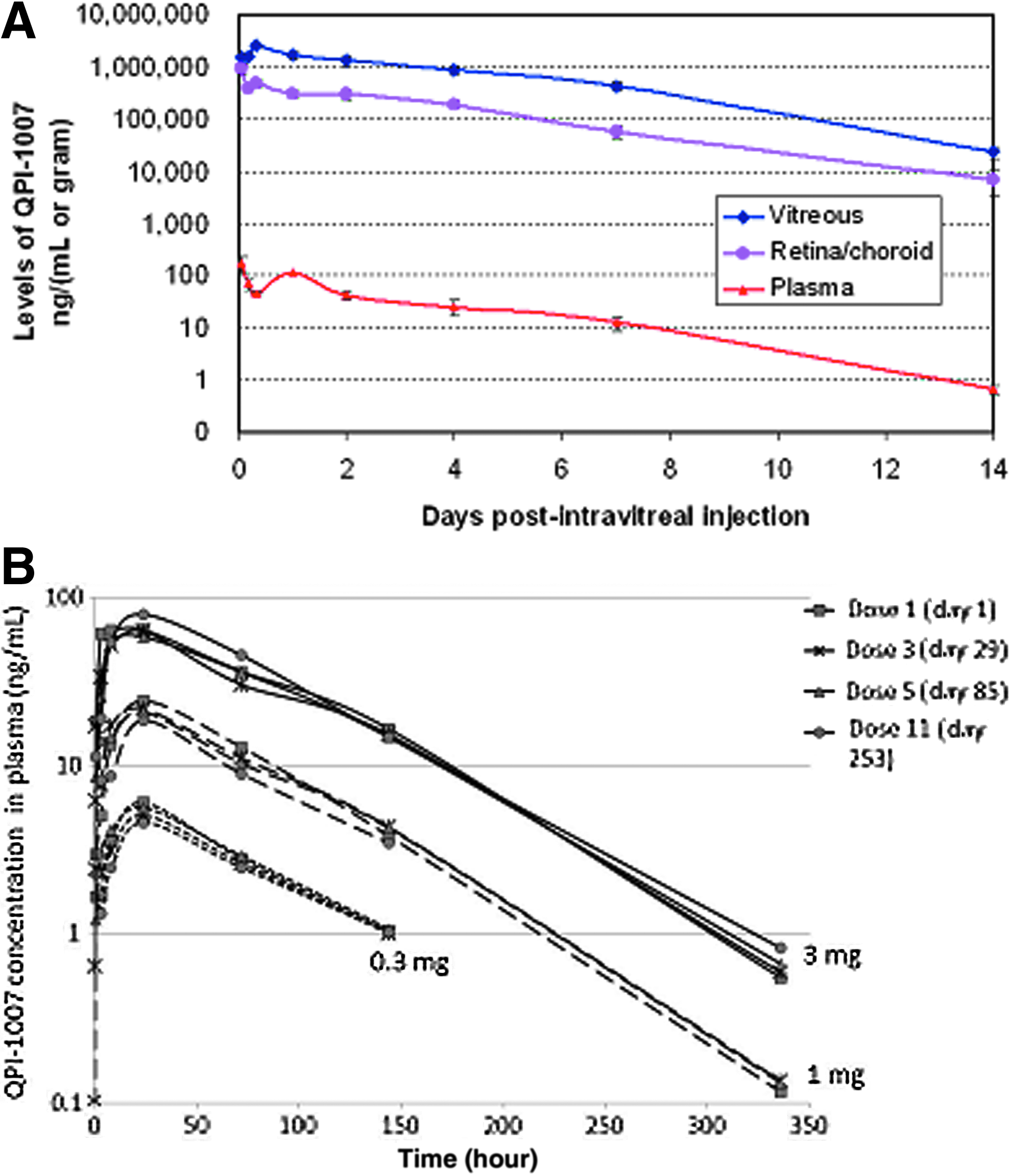
Pharmacokinetic profile of synthetic, small interfering RNA QPI-1007 following either a single or repeated intravitreal administration in the rabbit.
QPI-1007 was quantified in plasma in a repeated-dose intravitreal toxicity study in rabbits receiving either 0.3, 1, or 3 mg per eye per dose for a total of three administrations over 4 weeks, five administrations over 3 months, or eleven administrations over 9 months. In general, mean plasma concentrations of QPI-1007 were maximal at 24 hours after intravitreal injection and declined slowly thereafter (Fig 1B). The peak mean plasma levels were dose related and roughly linearly dose proportional, and this was generally true for other time points. The plasma levels were very similar following intravitreal injection on each collection day (i.e., following doses 1, 3, 5, and 11) indicating a lack of accumulation in plasma with successive injections at either 2- or 4-week intervals.
Plasma clearance following i.v. administration in the rat
The clearance of QPI-1007 from plasma was characterized in single-dose and repeated-dose i.v. toxicity studies in Sprague-Dawley rats that were intended to assess possible systemic toxicity of QPI-1007. When QPI-1007 was administered as a single bolus tail vein injection in rats at doses of 4 and 100 mg/kg, QPI-1007 was found to clear rapidly from plasma (Fig. 2) with an elimination half-life of less than an hour and mean residence time of less than 20 minutes. This is in good agreement with published information on other synthetic siRNAs like QPI-1007 that demonstrated rapid clearance from the blood compartment via renal filtration following i.v. administration (van de Water et al., 2006; Thompson et al., 2012). In the repeated-dose i.v. study in the rat, there was no accumulation of QPI-1007 in the plasma following daily injections of 75 mg/kg/day for 28 days (data not shown).

QPI-1007 plasma levels in rats following a single intravenous (i.v.) administration. Sprague-Dawley rats received i.v. bolus injections of 4 or 100 mg/kg (dosing volume: 5 mL/kg) via the tail vein.
Toxicology studies
QPI-1007 was assessed in a series of nonclinical studies using i.v. and intravitreal routes of administration. The program included single-dose and repeated-dose intravitreal studies in Dutch Belted rabbits, single-dose and repeated-dose i.v. studies in Sprague-Dawley rats, two in vitro genetic toxicity studies, one in vivo genetic toxicity study in conjunction with the i.v. rat toxicity studies.
Intravitreal toxicity studies
A single-dose intravitreal study was conducted in Dutch Belted rabbits that received either 0 (vehicle), 0.3, 1, or 3 mg per eye QPI-1007. There were no clinical observations seen during this study that were considered to be related to administration of QPI-1007. No differences in body weight, body weight changes, feed consumption, clinical pathology parameters, or organ weights occurred that were considered to be related to QPI-1007. There were no gross necropsy observations that were considered to be related to QPI-1007. There were no findings from the ophthalmic examinations that were considered QPI-1007 related. In addition, electroretinography and intraocular pressure measurements did not reveal any effects of QPI-1007. QPI-1007–related effects were limited to microscopic changes in the eyes. Intravitreal injection of QPI-1007 at a dosage of 1 or 3 mg per eye was occasionally associated with granular basophilia of the vitreous and basophilic staining of the posterior lens capsule and the retinal surface, which was presumed to simply reflect deposition of QPI-1007 in these tissues and was not considered adverse. Such changes were not evident in QPI-1007–treated animals terminated on study day 29, suggesting that QPI-1007 was cleared from the eye within the 4-week recovery period. QPI-1007-related effects in animals dosed at 3 mg per eye (human equivalent dose of 12 mg/eye) were limited to reversible mild vitreal inflammation in 4 of 12 eyes.
A repeated-dose study was conducted in Dutch Belted rabbits, with administration of QPI-1007 at dose levels of 0 (vehicle), 0.3, 1.0, and 3.0 mg per eye for a total of either three doses over 1 month, a total of five doses over 3 months, or a total of eleven doses over 9 months, with an approximate 4-week recovery phase after administration of either five or eleven doses. No QPI-1007-related deaths or moribundity occurred. No QPI-1007-related clinical signs or effects on body weight or body weight change, hematology, coagulation, or clinical chemistry test results were noted, and no QPI-1007–related macroscopic observations were noted at necropsy. Ophthalmic examinations showed an increased incidence of a generally mild degree of cellular infiltrates in the vitreous in the high-dose (3.0 mg/eye/dose) animals. This mainly occurred during the period of every-other-week dosing in the first month of the study and was not observed during the recovery phase. The presence of these cells was reversible and had no apparent detrimental effect on other ocular structures. Hence, these observations were not considered adverse, especially after the dosing interval was increased to every 4 weeks. A modest reduction in B-wave amplitude in the 3.0 mg per dose per eye group was observed in electroretinograms. The changes in electroretinography (ERG) values were generally moderate and not severe, and there was large variation in the control group values. In addition, differences between the means of treated groups from control values were generally less than the 50% reduction that would indicate significant clinical impact. Further, no animals exhibited an extinguished or “flat” ERG. Therefore, the ERG changes observed at the highest dose level (3.0 mg/eye/dose) of QPI-1007 were not considered clinically meaningful. Microscopic examination showed no alterations in any nonocular tissues of high-dose (3.0 mg/eye/dose) animals, and QPI-1007-related microscopic changes were limited to minimal to slight basophilia of the lens capsule and the retina, as well as basophilic granules in the cytoplasm of retinal ganglion cells, both of which were reversible. The presence of oligonucleotides in tissues is known to confer basophilic staining with standard hematoxylin and eosin slide preparations, and these findings were interpreted as simply reflecting deposition of the QPI-1007 siRNA in the affected ocular tissues similar to that observed in the single-dose study described above. Given the lack of significant toxicological findings in this study, the no-observable-adverse-effect-level in rabbits receiving 11 intravitreal injections of QPI-1007 over 9 months was considered to be 3 mg per eye, which represents a 12 mg per eye human equivalent dose.
Intravenous toxicity studies
A single-dose i.v. toxicity study was conducted in Sprague-Dawley rats with administration of QPI-1007 at 0, 4, and 100 mg/kg by bolus tail vein injection. There were no early deaths in this study. There were no clinical observations or differences in body weight, feed consumption, clinical pathology parameters, or organ weights that were considered to be related to QPI-1007. There were no gross macroscopic or microscopic findings that were considered to be related to QPI-1007. In summary, no QPI-1007–related findings were noted in this study. Hence, the no-observed-effect-level (NOEL) in the rat following a single i.v. administration of QPI-1007 was the highest dose level tested, 100 mg/kg.
A repeated-dose study was conducted in Sprague-Dawley rats with administration of QPI-1007 at 0, 10, and 75 mg/kg/day by intravenous bolus injection for 28 days. In this study, there was no QPI-1007-related mortality, and no QPI-1007–related clinical signs or ocular findings were observed. There were no toxicologically meaningful differences in body weight, body weight changes, food consumption, hematology parameters, coagulation parameters, clinical chemistry parameters, or absolute or relative organ weights in QPI-1007-treated males or females compared to vehicle controls. There were no QPI-1007–related gross necropsy findings or microscopic changes noted in the evaluated tissues, and there was no evidence of clastogenicity based on the micronucleus evaluation performed as part of this study. Based on these results, the NOEL was considered to be at least 75 mg/kg/day (the highest dose level tested).
Genotoxicity
QPI-1007 was not genotoxic in the bacterial mutation assay, the chromosome aberration assay, or the in vivo micronucleus evaluation included in the rat repeated-dose i.v. toxicity study. These data are in good agreement with other synthetic oligonucleotides that have consistently been negative in these test systems (Henry et al., 2002; Sazani et al., 2010; Ponstein-Simarro et al., 2013; Oligonucleotide Safety Working Group, personal communuication).
Discussion
QPI-1007 is a double-stranded (19-bp) chemically modified, synthetic small interfering RNA (siRNA) targeting caspase 2 mRNA and is designed to temporarily inhibit the expression of caspase 2 via the RNAi pathway. QPI-1007 is being developed as a neuroprotectant for the treatment of NAION and other optic neuropathies, such as glaucoma, that result in the death of retinal ganglion cells. siRNAs targeting caspase 2 mRNA have been shown to protect retinal ganglion cells in the two models of optic nerve damage in the rat (Ahmed et al., 2011).
Synthetic siRNAs have been reported to activate the innate immune system (Judge and MacLachlan, 2008; Kleinman et al., 2008); however, QPI-1007 failed to activate recombinant murine and human Toll-like receptors (TLRs) 3, 7, 8 and 9, and also failed to activate cytokine production or the interferon pathway in human peripheral blood mononuclear cells (PBMCs). TLRs 3, 7, 8, and 9 comprise part of the mammalian innate immune system designed to identify the presence of foreign nucleic acids and are primarily localized in endosomes, although TLR3 has also been reported to be present on the surface of some endothelial cells (Kleinman et al., 2008). RIG-I and MDA-5 are cytoplasmic helicases that also recognize foreign nucleic acids, thereby initiating a signaling cascade leading to the secretion of pro-inflammatory cytokines and chemokines (Medzhitov et al., 1997; Chaudhary et al., 1998; Chuang and Ulevitch, 2000). Immunostimulatory activity of synthetic siRNAs can be abrogated by careful selection of the siRNA sequence, limiting the length of the siRNA, and/or by the incorporation of chemical modifications into the siRNA duplex (Judge and MacLachlan, 2008). Particularly effective is the introduction of 2′-O-methyl-modified nucleotides into the siRNA (Kariko et al., 2004; Judge et al., 2006; Zimmermann et al., 2006; Cekaite et al., 2007). Finally, Ahmed et al. (2011), reported that the research version of QPI-1007 siRNA that contained similar chemical modifications as QPI-1007 was not immunostimulatory in in vitro studies in human peripheral blood mononuclear cells and failed to induce expression of interferon-responsive genes in rat retinas following intravitreal injection in rats.
Synthetic siRNAs can also exhibit off-target activity, here defined as RNAi-mediated activity against RNAs other than caspase 2 mRNA. Off-target activity can derive from either the seed region (nucleotides 2–7) or the full-length passenger strand, the seed region (nucleotides 2–7) of the guide strand, or RNAs containing sequences that are substantially similar to the target region of caspase 2 mRNA. The target region in caspase 2 was carefully chosen to insure that the human transcriptome lacks sequences exhibiting significant similarity to the QPI-1007 siRNA. Regarding potential off-target activity from the seed regions or the passenger strand, chemical modifications can be incorporated into synthetic siRNAs to mitigate such off-target activity (Chiu and Rana, 2002; Birmingham et al., 2006; Jackson et al., 2006). The potential of QPI-1007 to elicit off-target activity was evaluated previously in cell culture using a Renilla luciferase reporter gene construct in which either the full-length target (one copy of the 19 nucleotide target) or the corresponding seed region target (three tandem copies of targets for nucleotides 1–8) of either the guide and passenger strand were cloned into the 3′ untranslated region of the reporter gene (Ahmed et al., 2011). QPI-1007 was active against the full guide strand target that is identical to the target site in caspase 2 mRNA; in contrast, no silencing activity was detected against the corresponding seed region, indicating that the guide strand retains slicer activity against the caspase 2 target sequence, but potential seed region-mediated off-target activity was significantly attenuated. QPI-1007 was inactive against either the full target site or the seed region of the passenger strand, indicating that RNAi activity of the passenger strand was abolished. These studies demonstrated that the chemical modifications incorporated into QPI-1007 were effective in improving the specificity of the siRNA.
PK/ADME profile of QPI-1007
The ability of retinal ganglion cells (the first cell layer in the mammalian retina that is in direct contact with the vitreous humor) to take up siRNAs after intravitreal administration was previously evaluated in the rat (Ahmed et al., 2011). Four hours after intravitreal injection, siRNA was readily detected in retinal ganglion cells and to a lesser extent in other layers in the retina by both confocal microscopy and in situ hybridization. RNAi-mediated cleavage of caspase 2 mRNA in the rat retina was confirmed by rapid amplification of cDNA ends Ahmed et al., 2011. These studies demonstrated that retinal ganglion cells efficiently take up siRNAs like QPI-1007 following intravitreal administration without the need for complex formulations.
No formal metabolism studies have been conducted with QPI-1007. However, the stability of QPI-1007 in rabbit vitreous humor was evaluated in vitro by incubating siRNA with rabbit vitreous at 37°C and assessing integrity of the siRNA. QPI-1007 was demonstrated to be stable in this matrix for at least 24 hours with little degradation (Ahmed et al., 2011).
The half-life of QPI-1007 in the vitreous and retina/choroid in the rabbit following intravitreal administration was about 2 days. QPI-1007 was quantifiable in plasma of rabbits at least 14 days (the last sampled time point) after single or repeated intravitreal injection(s). This persistence of detectable QPI-1007 in plasma likely reflects continued slow egress of the test article from the eye, as the decline in plasma levels in the rabbit mirrored the decline in the vitreous. This is supported by the toxicokinetic results from the i.v. toxicity study conducted in rats, in which the calculated elimination half-life was less than an hour, and the mean residence time was less than 20 minutes. There was no evidence of accumulation following repeated intravitreal administration every 2 or 4 weeks for up to 11 doses (9 months). These results are in good agreement with intravitreal studies done with other synthetic siRNAs (Dejneka et al., 2008).
Synthetic siRNAs like QPI-1007 are known to clear rapidly via renal filtration from the plasma compartment following i.v. administration (Lee et al., 2000; van de Water et al., 2006; Molitoris et al., 2009; Thompson et al., 2012), and QPI-1007 was indeed cleared rapidly from the blood compartment of rats following i.v. administration. Elimination of QPI-1007 from the vitreous after intravitreal administration is much slower and is likely the rate-limiting step in the pharmacokinetic profile of QPI-1007 in the retina/choroid and plasma after intravitreal administration.
Overall, the data indicate that QPI-1007 is chemically stable in the vitreous and that the elimination pathway for QPI-1007 following intravitreal injection is slow egress from the vitreous humor with limited metabolism followed by rapid clearance from the blood via renal filtration.
Toxicological properties of QPI-1007
There was no indication of systemic toxicity following single or repeated i.v. administrations of QPI-1007. In addition, there were no effects outside of the eye associated with the intended use of QPI-1007 (i.e., when given by single or repeated intravitreal injections). There was no indication of genetic toxicity from either the two in vitro systems (bacterial mutation assay and the chromosome aberration assay) or the in vivo micronucleus evaluation included in the rat i.v. toxicity study. Therefore, the focus of attention regarding the safety profile of QPI-1007 was on the local ocular effects. The only significant finding in the rabbit single- and repeated-dose intravitreal studies that was considered related to QPI-1007 was inflammatory cell infiltration in the vitreous. Although a modest reduction in B-wave amplitude of the electroretinogram was observed at the 3.0 mg per eye per dose group in the repeated-dose study, this was not considered to be clinically meaningful.
Overall, the results of the nonclinical toxicology program for QPI-1007 indicate that there are no significant systemic or ocular safety issues that would preclude further development of QPI-1007.
Footnotes
Author Disclosure Statement
No competing financial interests exist.