
Abstract
The synthetic cytosine–phosphate–guanosine oligodeoxynucleotide 107 (CpG ODN107) is a novel radiosensitizer for glioma treatment. However, the information related to its pharmacokinetics and toxicity remains unclear. Therefore, the plasma pharmacokinetics, distribution, elimination, and acute toxicity of CpG ODN107 in mice were investigated in the present experiments. The results from the liquid chromatography–tandem mass spectrometry (LC-MS/MS) assay showed that the plasma elimination half-life (t1/2β) of CpG ODN107 in BALB/c mice varied slightly with the dose, and it was 0.65, 0.49, and 0.50 h at the intravenous doses of 2.5, 5, and 10 mg/kg, respectively. CpG ODN107 rapidly and widely distributed in organs/tissues, except the brain and testes. The highest concentrations were found in the liver (28.6% of the administered dose after 0.5 h) and the kidneys (5.7% of the administered dose after 1 h). CpG ODN107 (0.3, 3, and 30 μg/mL) could highly bind to human and mouse plasma proteins in vitro. CpG ODN107 in the forms of prototype was excreted in urine (1.79%) and feces (0.91%), and its shortened metabolites were excreted in urine (2.1%) and feces (2.2%) within the first 24 h. The mice in vivo optical image showed CpG ODN107 labeled with Alexa Fluor 680 fluorochrome (AF680) accumulated in the brain after orthotopic injection, eliminated very slowly, and excreted in urine compared with poly T labeled with AF680. The median lethal dose (LD50) of CpG ODN107 was 75.7 mg/kg for mice; this dose only could produce apparent spleen and liver damage, in line with the distribution features of CpG ODN. In conclusion, our present pharmacokinetic and toxicity investigation will provide helpful information to further pharmacodynamic and pharmacokinetic research of CpG ODN107 and other oligodeoxynucleotide drugs in the future.
Introduction
T
There are several different classes of oligonucleotides being developed as therapeutic agents and their pharmacokinetic properties, which are defined by classes, are primarily based on their backbone chemistry, the relationship between the backbone chemistry and the biological stability, and plasma protein-binding properties [13,14]. The certain pharmacokinetics and toxicities of various phosphorothioate (PS)-ODNs have been described after intraparenchymal [15], subcutaneous [16], intrapulmonary [17], intradermal [18], intraperitoneal, and intravenous injection [19,20], as well as oral administration in rodents, primates, and humans [21–23].
CpG ODN107, containing only 12 deoxynucleotides (5′-TGGCGCGGGGCG-3′), is a type of ODN that was previously designed and screened from 21 of the sequences in our laboratory. It is made with a fully PS backbone to render it more nuclease resistant. In our previous study, CpG ODN107 was presented to possess a radiosensitizing effect in vitro and in vivo through TLR9-mediated NF-κB activation, NO production, and cell cycle arrest at the G1 phase. In subcutaneous and orthotopic implantation tumor models, tumors of mice treated with peritumoral injection of CpG ODN107 in combination with local radiotherapy were less angiogenic and hemorrhagic than those in mice with other treatments [24,25]. In the orthotopic implantation model of nude mice, the survival rate of mice significantly increased after treatment with CpG ODN107 (0.083 mg/kg) in combination with radiotherapy (10 Gy) compared with local radiotherapy alone. CpG ODN107 in combination with radiotherapy significantly decreased microvessel density, vascular endothelial growth factor (VEGF) level, and HIF-1α expression in orthotopic implantation glioma, suggesting that the radiosensitizing effect of CpG ODN107 is tightly related to its antiangiogenic activity through suppression of HIF-1α/VEGF pathway [24,25].
Following these positive findings, CpG ODN107 has been investigated as a potential radiosensitizer for glioma treatment. As well known, the physiological disposition of drug is tightly related to its activities. Plasma pharmacokinetics, tissue distribution, elimination, and protein-binding data in animal models can be extrapolated to humans to understand and predict drug behavior in the clinical trials. Therefore, investigation on pharmacokinetics and toxicity has become increasingly important.
Previously, a liquid chromatography–tandem mass spectrometry (LC-MS/MS) quantitative method with soft electrospray ionization (ESI) for the investigation of CpG ODN107 and its metabolites in plasma was carried out in our laboratory [26]. Based on the above results, in the present experiments, LC-MS/MS was continued for the quantitative analysis of CpG ODN107 and its metabolites in urine, feces, tissues, and organs following intravenous administration in mice to bring about new insight with regard to the pharmacokinetics, distribution, and excretion of CpG ODN107. In addition, the mice in vivo optical image was used for distribution investigation after CpG ODN107 labeled with Alexa Fluor 680 fluorochrome (AF680) was given through orthotopic administration.
Materials and Methods
Chemicals and reagents
The CpG ODN107 sequence is 5′-TGGCGCGGGGCG-3′ [12-mer, Mw 3917.4 Da]. The sequences of its metabolites (5′N-1, 3′N-1, 3′N-2, and 3′N-3) are 5′-GGCGCGGGGCG-3′ (Mw, 3599.4 Da), 5′-TGGCGCGGGGC-3′ (Mw, 3575.6 Da), 5′-TGGCGCGGGG-3′ (Mw, 3270.6 Da), and 5′-TGGCGCGGG-3′ (Mw, 2922.6 Da), respectively. The internal standard (IS) used in this work was an oligodeoxynucleotide with 15 nucleotides (5′-TTTTTTTTTTTTTTT-3′; Mw, 4725.0 Da) (Poly T). All of the above oligodeoxynucleotides with a PS backbone were synthesized by Invitrogen Biotechnology. Human serum albumin (HSA, batch number 975444, >96% pure) was purchased from Bering GmbH. Human plasma was gifted from Southwest Hospital and collected from healthy male volunteers (Chongqing, China). High Performance Liquid Chromatography-grade (HPLC-grade) acetonitrile and methanol were purchased from Merck. All other chemicals were of analytical grade. Water for HPLC was deionized and further purified with a Milli-Q system (Millipore). Oasis Hydrophile-Lipophile Balance (HLB) cartridges (1 cm3, 10 mg) that were used in solid-phase extraction (SPE) were purchased from Waters Corporation.
Animals
Specific pathogen-free male BALB/c mice (18±2 g) and nude mice (athymus with deficient immune system), 5–6 weeks old and weighing 19.3±0.2 g, were obtained from the Experimental Animal Center of Third Military Medical University (Chongqing, China). During the test period, animals were fed a standard laboratory diet and kept under controlled conditions. The animals were given a commercial diet and water ad libitum for 1 week before the study. All animals were weighed immediately before dosing. All animal experiments were performed in accordance with the National Guidelines for the Care and Use of Laboratory Animals and in accordance with the National Guidelines for Animal Care and Use. At the end of the experiment, the ether-inhaled anesthetic was used for euthanasia of the animals.
Preparation of plasma, urine, feces, and tissue samples
CpG ODN107 was extracted from plasma (100 μL) by liquid–liquid extraction (LLE) and SPE according to previously established methods [26]. CpG ODN107 and its metabolites were extracted from urine (100 μL) or feces (100 mg) using the following protocols. Urine samples were centrifuged and passed through a 0.22-μm nylon membrane filter before extraction. The resulting urine sample (pooled from five mice) was mixed with 5.0 μL of IS working solution (11.0 μg/mL), and then 0.1 mL of phenol/trichloromethane (1:1, v/v) was added. This mixture was vortex mixed for 20–30 s in a 2-mL tube and centrifuged for 5 min at 12,000 g. The aqueous solution was transferred to a clean tube, and then 0.5 mL of the triethanolamine/acetate (TEAA) buffer [200 mM triethanolamine (TEA), pH 10.0] was added and mixed before SPE. Feces samples were ground with 2 mL of TEAA buffer (200 mM TEA, pH 10.0). Typically, 5.0 μL of IS working solution (11.0 μg/mL) was added to the samples before the extraction. This mixture was shaken for 15 min at 25°C and centrifuged for 5 min at 12,000 g. The aqueous solution was transferred to a clean tube, and then 0.5 mL of TEAA buffer (200 mM TEA, pH 10.0) was added and mixed before SPE.
Tissue samples were prepared as follows. Samples were removed from the associated protein and endogenic nucleotide through a DNA adsorption column (TIANamp Genomic DNA Kit; Tiangen Biotech). Tissue samples (30 mg or 10 mg for spleen tissue) were ground with 0.5 mL of pure water on ice. Subsequently, buffer solution GA (200 μL; TIANamp Genomic DNA Kit) was added to the mixture. After vortex mixing, 20 μL of proteinase K was added and mixed. The mixture was placed in a water bath (56°C) for 1 h to facilitate sufficient digestion. Buffer solution GA (200 μL) was then added, and the mixture was shaken before incubation at 70°C in a water bath for 10 min. Ice-cold ethanol (200 μL) was added to the sample and vortex mixed for 15 s. The resulting mixture was transferred to an adsorption column (CB3) and centrifuged at 3,000 g for 3 min. The supernatant was transferred to a clean tube, and then 0.5 mL of 5% ammonia solution was added. This mixture was shaken for 15 min at 25°C and centrifuged for 5 min at 11,000 g. The supernatant was transferred to a clean tube, and then 0.5 mL of TEAA buffer (200 mM TEA, pH 10.0) was added. The analytes were extracted on an Oasis HLB SPE cartridge.
The SPE cartridge was first conditioned with 1 mL of methanol followed by 1 mL of TEAA buffer (200 mM TEA, pH 10.0) and 1 mL of deionized water. The mixture from LLE was then loaded onto the SPE cartridge. The cartridge was washed with 1 mL of TEAA buffer (200 mM TEA, pH 10.0), followed by 1 mL of 15% methanol. The analyte was eluted with 1 mL of 70% methanol, and the eluant was collected using a glass tube. The eluant was evaporated to dryness under a stream of nitrogen at 35°C. The evaporated sample was reconstituted with 300 μL of the mobile phase and dialyzed on a 0.22-μm nylon membrane for LC-MS/MS analysis.
LC-MS/MS analysis of plasma, urine, feces, and tissue samples
An Agilent 1260 HPLC system (Agilent Technologies) was equipped with a G1322A vacuum degasser, a G1312B binary pump, a G1316B column oven, and a G1367D autosampler. The chromatographic separation was performed on an Extend-C18 column (150×2.1 mm, i.d. 3.5 μm), which was protected by a flex capillary C18-A column (10×1.2 mm) (Agilent Technologies), and eluted at a rate of 0.3 mL/min with a mobile phase of 0.05% acetonitrile and aqueous NH3 (20:80, v/v). Before use, the mobile phase was degassed by vacuum filtration through a 0.22-μm nylon membrane filter. The column temperature was maintained at 35°C, and the injection volume was 20 μL. The total analysis time was 2.0 min per sample. The detection was performed on an Agilent 6410 Series Triple Quadrupole Mass Spectrometer (Agilent Technologies) with an ESI source. Multiple reaction-monitoring (MRM) modes were used for the determination of CpG ODN107 and its metabolites. The MRM transitions for CpG ODN107 were m/z 558.7→94.7 and m/z 558.7→343.9; the transitions for 5′N-1, 3′N-1, 3′N-2, and 3′N-3 were m/z 513.2→94.8 and 513.2→149.8, m/z 509.8→94.9 and m/z 509.8→343.9, m/z 544.1→94.7 and m/z 544.1→343.9, and m/z 486.1→94.9 and m/z 486.1→343.9, respectively. The MRM transitions for the IS were m/z 589.7→318.9 and m/z 589.7→125.0. Each transition was assigned a dwell time of 30 ms. The optimized collision energy was from −15 eV to −25 eV for the analytes. The nebulizer was set at 45 psi. The nitrogen drying gas temperature and flow rate were maintained at 350°C and 12 L/min, respectively. The mass spectrometer and the data acquisition were fully controlled using Agilent MS Workstation MassHunter 1.0 software. The specificity of the method was evaluated using blank mouse samples from five different mice.
Three-dose pharmacokinetic study
One hundred sixty-five BALB/c mice were randomly divided into three groups, each of which was administered a different dose regimen. Each group received a single intravenous bolus dose (2.5, 5, or 10 mg/kg) of CpG ODN107 through the tail vein to investigate the plasma pharmacokinetics. Blood samples (∼0.5 mL) were anticoagulated with EDTA-2K and collected through the fossa orbitalis at 0 min (predose), 2, 5, and 15 min and 0.5, 1, 2, 3, 6, 9, and 12 h, respectively. After the blood samples were centrifuged at 4000 g for 10 min, plasma samples were collected and stored at −70°C until analysis.
Distribution study
Thirty mice were divided into six groups. One single intravenous bolus dose (5 mg/kg) was given to investigate the distribution of CpG ODN107 in mice. Selected tissues or organs (heart, lungs, intestines, spleen, testes, liver, kidneys, muscle, and brain) were removed from euthanized animals (ether-inhaled anesthetic followed by exsanguination through the fossa orbitalis) at 0 min (predose) and 0.5, 1, 2, 4, 10, and 24 h. Following removal, tissues/organs were trimmed of extraneous fat or connective tissue, emptied and cleaned of all contents, blotted on filter paper, individually weighed before homogenization, and then stored at −70°C until processed for analysis. The results of the tissue distribution are expressed as μg/g tissue, which was calculated through the following equation: CT(μg/gtissue)=CSVS/P, where CT is the tissue concentration (μg/g), CS is the supernatant concentration, VS is the supernatant volume, and P is the weight of the tissue sample.
In addition, a single orthotopic injection (0.083 mg/kg/0.021 nmol/kg) was administered to investigate the distribution of CpG ODN107 labeled with Alexa Fluor 680 fluorochrome (AF680) in nude mice by living imaging. Nude mice were randomly divided into four groups (two mice/group): group 1 (spontaneous fluorescence control group) only accepted orthotopic injection of saline; group 2 (markers control group) accepted orthotopic injection of AF680 (0.021 nmol/kg); group 3 accepted orthotopic injection of CpG ODN107 labeled with AF680 (0.083 mg/kg/0.021 nmol/kg); group 4 (non-CpG control group) accepted orthotopic injection of Poly T labeled with AF680 (0.021 nmol/kg). Nude mice were anesthetized with pentobarbital sodium (35 mg/kg weight) through intraperitoneal injection. After fixing the mouse's head in the stereotaxic apparatus (Rewardst), a 5–6 mm midsagittal incision was made directly down the midline, and coronal suture was exposed. The periosteum was stripped and a 2 mm burr hole was made with a dental broach at 2.5 mm to the right of the midline and 1 mm anterior to the coronal suture. To limit the back flow along the needle track, the needle tip was initially sunk to a depth of 3.5 mm and then withdrawn 1 mm to a final depth of 2.5 mm. Each agent (AF680, CpG ODN107 labeled with AF680, and Poly T labeled with AF680) was dissolved in 0.5 μL of normal saline, and the injection time was set at 20 min. The needle was subsequently left for another 2 min, and then it was slowly withdrawn. Bone wax was applied over the injected site, and then skin was closed with surgical stitches. Fluorescence was investigated by an IVIS Spectrum (Interactive Video Information System, Perkin Elmer) at 0.5, 4, and 24 h. Excitation and absorption wavelength was 679 and 702 nm, respectively. The environment temperature was 37°C.
The plasma protein binding of CpG ODN107 was estimated using an ultrafiltration method. The absorbability of the ultrafiltration membrane, sample incubation time, and temperature were investigated in a preliminary experiment. Human plasma, 4% HSA, and mouse plasma (600 μL) were mixed with 50 μL of 4.0, 40, and 400 μg/mL CpG ODN107, giving a final concentration of ∼0.3, 3, and 30 μg/mL, respectively. The samples were vortex mixed completely and then incubated at 37°C for 60 min. Ultrafiltration was performed in tubes with a filter cut off of 10 kDa (Millipore Ultrafree®-MC; Millipore). A sample from each incubation mixture was poured into a precooled filter cup and centrifuged at 7000 g for 10 min at 4°C. Aliquots (four replicates) were pipetted from the filtrate and the initial incubation mixture. The sample concentrations of the in vitro plasma samples and their respective ultrafiltrates were analyzed by the LC-MS/MS method.
Plasma protein-binding ratio (%)=(ρ2–ρ1)/ρ2 × 100%, where ρ2 is the total CpG ODN107 in plasma (the concentration of CpG ODN107 in the initial incubation mixture) and ρ1 is the unbound fraction of CpG ODN107 in plasma (the concentration of CpG ODN107 in the filtrate).
Single-dose elimination study
Six male BALB/c mice were intravenously injected with a single-dose of CpG ODN107 (5 mg/kg). Each mouse was placed in a metabolism cage and fed a commercial diet and water ad libitum to investigate urinary and feculent excretion. Urine and feces were collected at 24 h before dosing and at intervals of 0–2, 2–4, 4–6, 6–8, 8–10, 10–12, and 12–24 h following CpG ODN107 injection. Total voided urine was collected, and each metabolism cage was washed following collection. Total voided feces were collected from each animal at various time points, and feces samples were dried by open-air drying before weighing. The volume of urine and the weight of feces were measured, and samples were then frozen at −70°C until LC-MS/MS analysis. Elimination percentage (%)=
Single-dose toxicity study
The median lethal dose (LD50) of CpG ODN107 was primarily evaluated within 24 h in mice following intravenous injection. Healthy 50 BALB/c mice were randomly divided into five groups with equal number and sex. Each animal was intravenously injected with a single dose of CpG ODN107 through the tail vein, and the injection volume was 10 mL/kg. Mice in all the groups were intravenously administrated CpG ODN107 once at the dose of 180.0, 126.0, 88.2, 61.7, and 43.0 mg/kg in 2 or 3 min, respectively. After administration, the acute toxic response caused by CpG ODN107 was observed and the number of deaths in each group was counted. If death happened, the mouse was dissected immediately to observe the possible death reasons.
According to the above results, 14-day toxicity was investigated based on the dose range from effective dose to the LD50 dose. Thirty male BALB/c mice were randomly divided into three groups and intravenously injected with CpG ODN107 (10.0, 30.0, and 75.0 mg/kg) once. The survived mice were retained for a 14-day recovery period to estimate the reversibility persistence and delayed occurrence of toxic effects. The following parameters were evaluated (predose and on day 1, 4, 7, and 14): body weights, hematology, macroscopic observations at necropsy, and histopathology.
Data analysis
The compartmental and/or noncompartmental analysis methods were used for pharmacokinetic characterization of the plasma and tissue concentration. The pharmacokinetic parameters were calculated using Data Analysis System software (DAS, version 3.0.4) supplied by the Pharmacological Society of China (Beijing, China). The area under the plasma concentration–time curve from the beginning of the experiment to the time of the last measurable concentration (AUC0–t) was calculated using the linear trapezoidal method. The elimination rate constant (Ke) was calculated according to the linear regression of the terminal semilog plot of the plasma concentration versus time. Dose proportionality after a single intravenous (i.v.) injection of different dosages was determined by comparison of the dose-normalized Cmax and AUC0–t across dosage levels using an analysis of variance and linear regression analysis. The protein-binding ratios at the three concentration levels or the different genera were compared by Student's t-test. The LD50 was calculated using SPSS software by the Bliss method. Mean±standard deviation (SD) values were used for the comparison, and P>0.05 was taken as the criteria for no significant difference between the means.
Results
Validation of analytical methods
SPE or a DNA column of the adsorbent removed the endogenous constituents of the biological samples, and endogenous signal interference from compounds was not observed in the MRM. A complete method validation for plasma has been previously reported in detail by our laboratory [26]. Therefore, method validations for urine, feces, and organ tissues were partial and as follows: the calibration curves for urine and feces were linear within the ranges of 20.0–4000.0 μg/L (R2=0.9958) and 0.02–4.00 μg/g (R2=0.9992), respectively. To quantify CpG ODN107 in the tissue samples, nine calibrated curves were prepared—one for each tissue analyzed—using the respective tissue homogenates as the sample matrix. The calibration curve of each tissue was linear within the range of 0.1–40.0 μg/g (R2>0.9998). CpG ODN107 metabolites in urine and feces were found in the range of 10.0–2500.0 μg/L (R2>0.9998). The relative SDs of accuracies for urine (15 replicate samples) were 2.5%, 1.2%, and 3.8% at 50, 500, and 4000 μg/L, respectively. The relative SDs of accuracies for feces (15 replicate samples) were 3.6%, 6.2%, and 2.6% at 0.05, 0.50, and 4.00 μg/g, respectively. The precisions determined for same- and between-day samples were all within 100%±15% of the actual values in urine and feces. In the present study, CpG ODN107 was stable if the urine samples were kept at an ambient temperature for <4 h, at 4°C for 24 h, or at −20°C for 5 days; fecal samples were kept at ambient temperatures for <4 h or at −20°C for 5 days. However, CpG ODN107 was unstable when the feces sample was stored at 4°C for 24 h, and the RE [RE%=(Observed concentration/Nominal concentration-1)×100%] was −18.9%. The LC-MS/MS method described in the Methods section has previously been proved to be sensitive, selective, and rapid for the determination of CpG ODN107 in mouse plasma, urine, feces, and organ tissues. The optimized method was validated to guarantee accuracy.
Plasma pharmacokinetics
After intravenous administration of CpG ODN107 to mice at three doses (2.5, 5, and 10 mg/kg), CpG ODN107 plasma concentrations were found to decrease rapidly within the first hour and then declined more slowly with a long terminal phase. At 6 h and with a dose of 2.5 mg/kg, CpG ODN107 could not be detected. A representative semilogarithmic plot of the fitted curve was attenuated and the blood kinetics of intravenously administered CpG ODN107 was found to be dose dependent in mice (Fig. 1). Plasma concentration–time data for CpG ODN107 were fitted to a two-compartment open model with first-order elimination from the central compartment. The distribution half-life times (t1/2α) for the three doses of CpG ODN107 were ∼2 min, while the elimination half-life times (t1/2β) were estimated to be <40 min. In addition, noncompartmental analysis was used for the pharmacokinetic characterization of the plasma concentration. The mean residence times (MRT0-t) for the three doses of CpG ODN107 were similar and rapid, with typical MRTs of 0.94, 0.97, and 0.90 h for 2.5, 5, and 10 mg/kg, respectively. The total clearance (Cl) was 0.74, 0.68, and 0.75 L/h/kg for 2.5, 5, and 10 mg/kg, respectively. The volume of distribution (Vd) of 0.19 L/kg (or 0.13, 0.19 L/kg) was higher than the total blood volume (about 0.078 L/kg) of the mice (Table 1). The pharmacokinetic parameters, AUC0-t and Cmax, of three CpG ODN107 doses were fitted to a linear model.
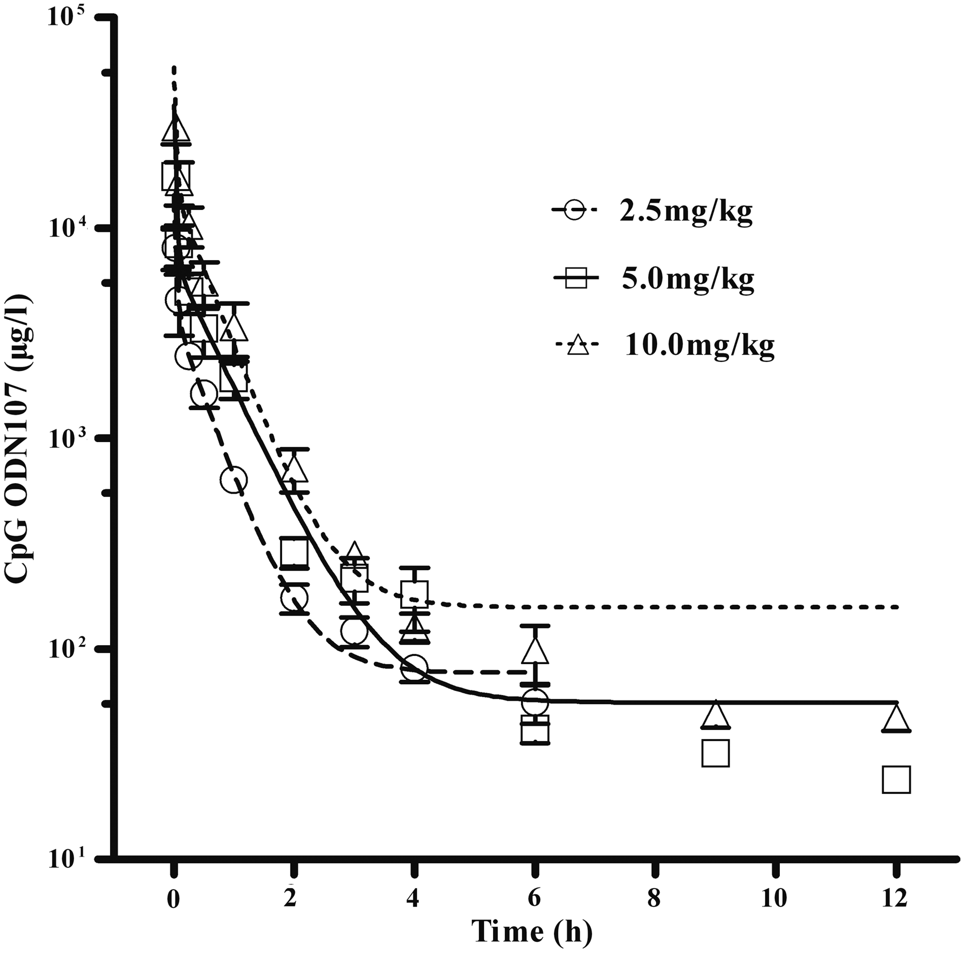
Plasma semilogarithmic concentration–time profiles of cytosine–phosphate–guanosine oligodeoxynucleotide 107 (CpG ODN107) following a single intravenous bolus injection (2.5, 5, and 10 mg/kg) to male BALB/c mice (n=6). Symbols represent the observed data and lines represent the fitted curve. Data are presented as mean±standard deviation.
Data are presented as mean±standard deviation (SD) derived at each time point from six male BALB/c mice.
CpG ODN107, cytosine–phosphate–guanosine oligodeoxynucleotide 107; SD, standard deviation; Vd, volume of distribution.
Tissue distribution of CpG ODN107
After intravenous administration of 5 mg/kg CpG ODN107 to mice, the concentration of CpG ODN107 in the tissues was determined using LC-MS/MS. The results showed that CpG ODN107 extensively and rapidly distributed throughout the body between 0.5 and 24 h (Table 2). However, CpG ODN107 was not detected in the brain. At all time points except 1 h, the highest concentration of CpG ODN107 was found in the liver and decreased from a peak concentration of 21.67 μg/g after 0.5 h to 3.99 μg/g after 24 h. At 1 h, the concentration of CpG ODN107 in the kidneys reached a maximum, with a concentration of 20.25 μg/g; however, it was subsequently cleared from the kidneys at a faster rate compared to the liver. After 24 h, CpG ODN107 had been almost completely eliminated from most tissues and organs, including the muscle, kidneys, heart, lungs, testes, and intestines. AUC0–24h in the tissues was used to signify the relative amounts of CpG ODN107. The accumulation of CpG ODN107 followed the order: AUC(Liver)>AUC(Kidneys)>AUC(Spleen)>AUC(Muscle)>AUC(Heart)>AUC(Lungs)>AUC(Testes)>AUC(Intestines)>AUC(Brain) (Fig. 2A). MRT0–24h in the tissues was used to signify the relative residence time of CpG ODN107 in the tissue or organs and followed the order: MRT(Lungs)>MRT(Spleen)>MRT(Heart)>MRT(Muscle)>MRT(Intestines)>MRT(Testes)>MRT(Liver)>MRT(Kidneys) (Fig. 2B). The distribution ratios (tissue vs. injected dose) of CpG ODN107 in important tissues, such as the liver, spleen, heart, and kidney, were compared. The highest distribution ratio was found in the liver at all time points, followed by the kidneys, the spleen, and then the heart. The highest distribution ratio for CpG ODN107 was found in the liver at 0.5 h (23.86%) (Fig. 3).

Mean AUC0–24h and mean MRT0–24h of CpG ODN107 in mouse tissues and organs. Mean AUC0–24h

Tissue and organ distribution ratios of CpG ODN107 at different time points following a single intravenous bolus injection of 5 mg/kg to male BALB/c mice. Data are presented as the mean derived from five male BALB/c mice. ID on figure is the abbreviation of injection dose.
“−” indicates CpG ODN107 was not detected. Data are presented as the mean±SD derived at each time point from five male BALB/c mice.
A mass of red fluorescent was observed in the brain tissue after orthotopic injection of AF680, CpG ODN107 labeled with AF680, and Poly T labeled with AF680, respectively (Fig. 4). In the nude mouse injected with AF680, red fluorescence was detected only in the thorax and bladder at 30 min following orthotopic injection, while red fluorescence intensity increased at 4 h and significantly reduced at 24 h. In the nude mouse with Poly T labeled with AF680, a significant amount of red fluorescence could be detected at the injection site at 30 min, and just a little in the neck and bladder. However, in the nude mice with CpG ODN107 labeled with AF680, a significant amount of red fluorescence could be observed at the injection site at 30 min, neck and bladder, and little in the thorax. Then, the red fluorescence in the neck apparently decreased at 4 h and it was very few at 24 h. Comparing the red fluorescence distribution of CpG ODN107 and Poly T labeled with AF680, CpG ODN107 was more widely distributed than Poly T in the neck and bladder at 0.5 and 4 h, and less distributed than Poly T in the thorax at 4 h. However, the other tissues and organs could not be detected because of the method limitation.
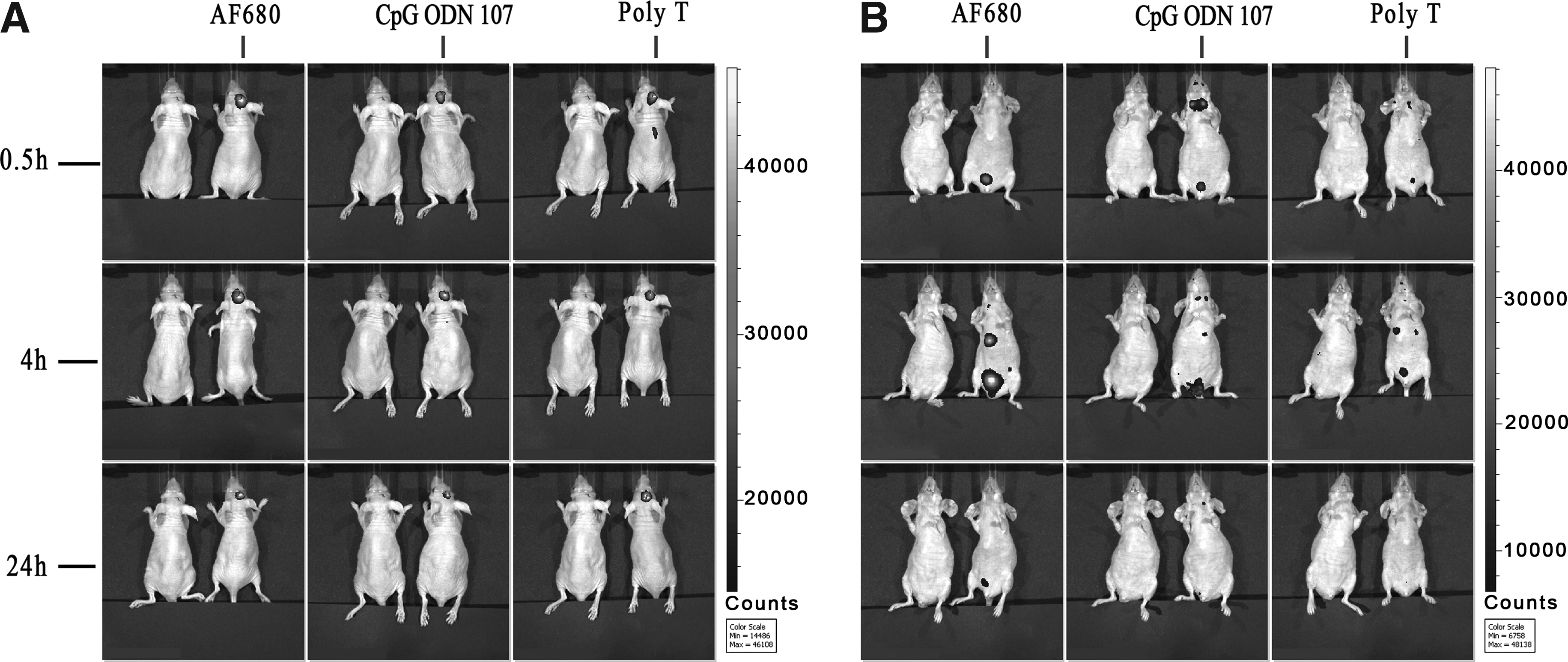
In vivo image of CpG ODN107 in nude mice following a single orthotopic injection. In each picture, left, with saline to rule out the spontaneous fluorescence; right, mouse was orthotopic and injected with same concentration (0.021 nmol/kg) of AF680, CpG ODN107 labeled with AF680 (abbreviated as CpG ODN107 on figure), and Poly T with AF680 (abbreviated as Poly T on figure), respectively. Fluorescence intensity of mice in creep position
The plasma protein binding of CpG ODN107 was determined based on the plasma concentrations from mice experiments. To assess the whole plasma protein binding of CpG ODN107 in vitro, samples from both mice and humans were studied and then compared. Moreover, the protein-binding characteristics of CpG ODN107 were evaluated with physiologic concentrations of HSA (4%). In mouse and human models, CpG ODN107 was highly bound (>96%) to whole plasma proteins within the concentration range of 0.3–30 μg/mL. At a plasma concentration of 30 μg/mL, CpG ODN107 was 90% bound to HSA in the human plasma (Table 3).
Data are presented as the mean±SD derived from five samples.
Significance value of aP<0.01 is shown compared among groups comparison.
Significance value of bP<0.05 is shown compared to group human.
Significance value of cP<0.01 is shown compared to 3.0 μg/ml and 0.3 μg/ml group in homogenic plasma.
HAS, human serum albumin.
Excretion study
Results from the urine and feces collected over a 24-h time period following intravenous administration of CpG ODN107 indicated that traces of the oligonucleotide can be found in urine and feces (Fig. 5). The excretion of CpG ODN107 increased rapidly between 0 and 2 h following intravenous administration, and then slowly increased from 2 to 24 h. Fecal excretion of CpG ODN107 accounted for ∼0.91% of the administered dose. Approximately, 1.79% of CpG ODN107 was recovered in urine. We additionally assessed the levels of CpG ODN107 metabolites in excreted urine and feces, which followed a similar trend to the parent drug. Specifically, 2.1% of the administered dose was associated with four CpG ODN107 metabolites found in the urine (Fig. 6A), while ∼2.3% of the administered dose was observed in the feces (Fig. 6B).
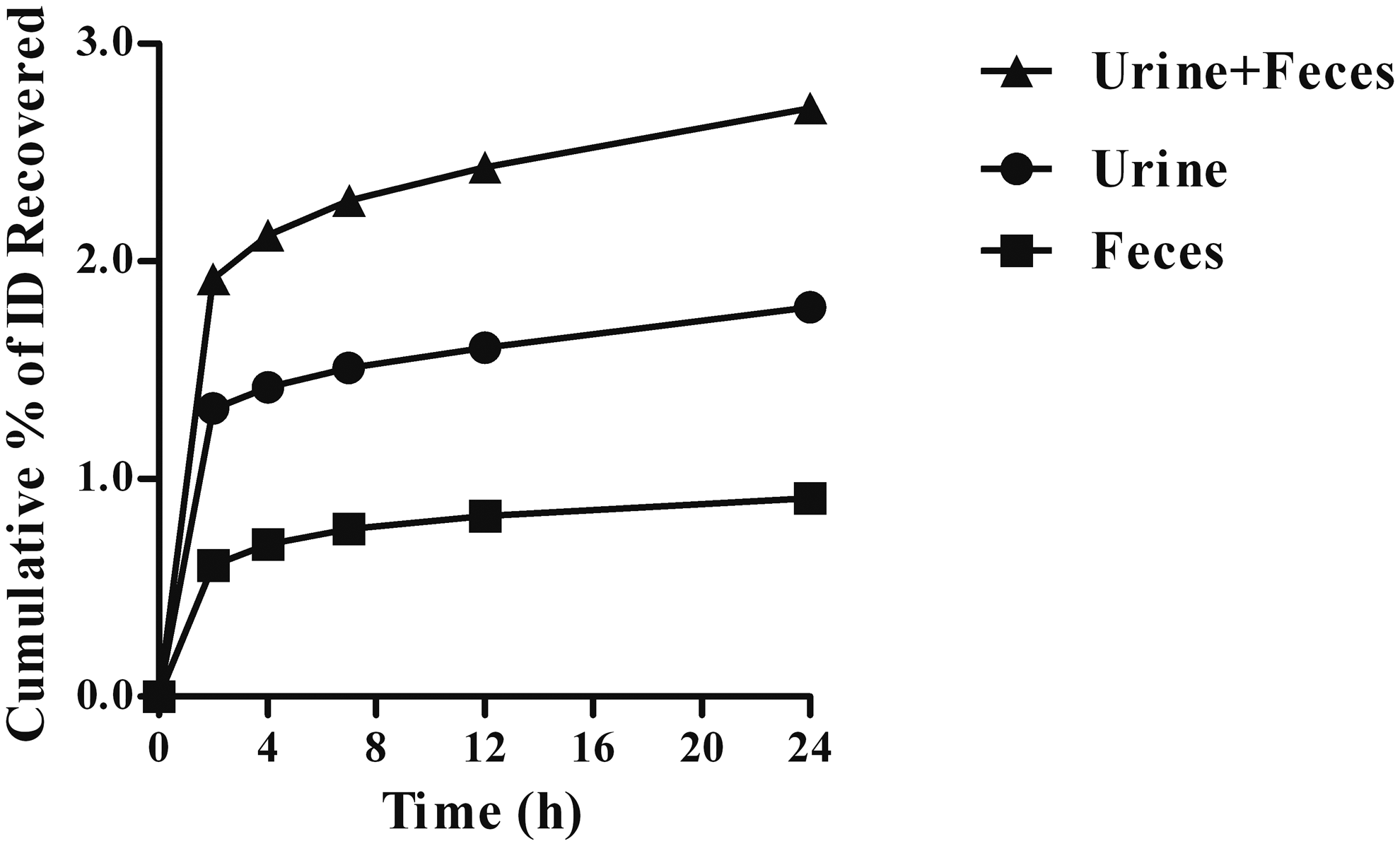
Cumulative total CpG ODN107 from urine (including the cage rinse) and feces during the period examined (24 h postdose), following a single intravenous injection of CpG ODN107 (5 mg/kg). Data are presented as the mean derived from six male BALB/c mice. ID on figure is the abbreviation of injection dose.

Cumulative total of CpG ODN107 metabolites found in urine
Toxicology studies
Acute toxicity of CpG ODN107 was primarily evaluated within 24 h in mice. After intravenous administration, diaphragmatic breathing and motor activity decrease were observed. Loose stools were observed at 1 h and death was only found between 30 min and 4 h. Symptoms before death displayed busy, clonicity, scrunch, gasping, and mouth-rrhage. On the brink of death, mice displayed restlessness, appetite decrease, and so on. The body weight of the survived mice increased stably during the following recovery periods. The dose level decreased to 43 mg/kg in additional mice, with no adverse affects observed. LD50 was 75.7 mg/kg, and 95% confidence interval was found between 61.5 and 92.0 mg/kg (Table 4). The PROBIT Model was P=−10.823+5.759x. Fitting was good, and fitting test results showed that the P was 0.957.
In 14-day toxicity studies, no differences of body weight, feed consumption, hematology parameters, or organ weights were found to be related to CpG ODN107 with one single bolus dose of 10.0, 30.0, and 75.0 mg/kg. Microscopic examination showed that no changes could be observed in organs and tissues, except the spleen and liver of mice treated with 75.0 mg/kg of CpG ODN107. Edema could be observed in the liver on D1, D4, D7, and D14. Liver cell cytoplasm loose and edema were more serious on D1 and D4, which were reversible on D7 and D14. However, there was extensive hyperemia in the spleen on D1 and D4. In addition, tissue necrosis and friability of spleen were observed on D7 and D14 (Fig. 7).
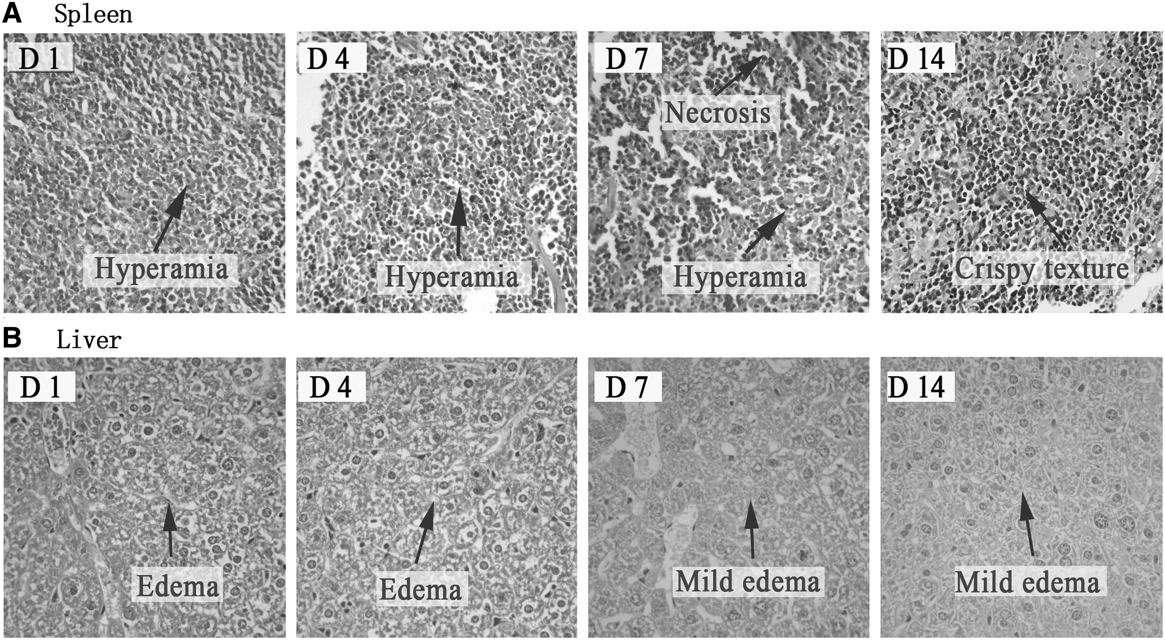
Histological analysis of tissue in mice after intravenous injection of CpG ODN107 (75 mg/kg) through the tail vein.
Discussion
CpG ODN107 is a potential candidate as a radiosensitizer for human glioma by means of NO production through CpG ODN/TLR9-mediated NF-κB activation and less angiogenesis through HIF-1α/VEGF pathway [24,25]. Herein, the pharmacokinetic and toxicological properties of CpG ODN107 were investigated. The results demonstrated CpG ODN107 highly bound to human and mouse plasma proteins, and predominantly distributed in the liver, kidneys, and spleen, but very little distributed in other tissues or organs after intravenous administration and mainly accumulated in the brain after orthotopic injection. This agent very rapidly cleared from the plasma and excreted into the urine and feces in the form of prototype and metabolites in mice. LD50 was 75.7 mg/kg for mice, and this dosage could elicit microscopic changes in the spleen and liver.
The LC-MS/MS method was used for detecting other oligonucleotide drugs, such as PF-ODN for the treatment of lung cancer [19,27,28]. This method was also used for CpG ODN107 in our laboratory [26] and further optimized in our present experiments. Commonly, poly-T oligonucleotides (20-mer −27-mer) were widely used as ISs to determine the ODN in the early studies [19,27–29]. For PF-ODN assay, a 20-mer poly-T oligonucleotide was used as an IS. Based on the selection principle that the IS should have similar physicochemical properties (such as molecular weight, chemical structure, chromatographic properties, and ionization state) with the analyte [30]. Herein, a 15-mer poly-T oligonucleotide was selected as an IS for all samples, including plasma, urine, feces, organs, and tissues, due to its similar but different molecular weight and ionization state [26].
Previously, pharmacokinetic profiles of many PS oligodeoxynucleotides were widely investigated in rats. Intravenous administration of [3H]CGP 69846A, a 20-mer PS oligodeoxynucleotide against the 3′-untranslated region of human c-raf.1 kinase mRNA, presented a biphasic profile for the disappearance of radiolabel from blood in rats; a rapid distribution phase (t1/2α=0.32 h/19 min) was followed by a prolonged elimination phase (t1/2β=12.9 h/775 min) [19]. Pharmacokinetic parameters of G3139, an 18-mer PS oligonucleotide antisense designed to bind to the first six codons of the human Bcl-2 mRNA in the rats accepted intravenous injection at 20 mg/kg, the harmonic mean of t1/2α was 0.14 h/8.6 min, and t1/2β was 1.07 h/64.2 min [27]. Herein, BALB/c mice were intravenously injected with low, middle, and high dosages of CpG ODN107 within the effective dose range; the pharmacokinetics of CpG ODN107 was fitted through a two-compartment model. In mice plasma, t1/2α of CpG ODN107 was 0.03 h/1.8 min and t1/2β of CpG ODN107 was 0.49 h/29.4 min–0.65 h/39 min. Our results demonstrated that the blood kinetics of CpG ODN107 was still biphasic, similar to other PS-ODNs such as CGP 69846A and G3139.
Previously, in our laboratory, the plasma kinetics presented as a nonlinear characteristic when the dose increased to 15 mg/kg of CpG ODN107 [26]. A similar nonlinear relationship between the dose and the AUC was also observed in animal studies with ISIS 2302 and other PS-ODNs [9,19,31]. However, the plasma kinetics was linear when the dose of CpG ODN107 increased from 2.5 to 10 mg/kg due to the linearity of the AUC and Cmax in the present experiments. This nonlinearity characteristic of CpG ODN107 in mice was supposed to be related to organ distribution or plasma protein-binding capacity. In other laboratories, saturation kinetics appeared at higher doses too [32], but nonlinear pharmacokinetics happened in rats not in mice [13]. However, saturation kinetics was discovered in mice in our laboratory. Therefore, further experiments are required to investigate whether similar nonlinearity of CpG ODN107 appears in other animals such as rats.
The protein-binding capacity data, obtained from in vitro experiments, would provide the information on the plasma protein-binding characteristics of drugs in vivo. The plasma protein-binding capacity might affect the pharmacokinetic behavior of PS-ODNs. Previously, PS-ODN was reported to bind plasma proteins with high capacity (more than 91%) across species such as mice, rats, monkeys, and humans [13]; they could bind to albumin with high capacity and α2-macroglobulin with low capacity, negligibly binding to α1-acid glycoprotein [33]. Herein, CpG ODN107 (0.3–30 μg/mL) also had high-capacity binding to plasma proteins (>96%), and albumin possessed the greatest capacity for CpG ODN107, similar to the results from other PS-ODNs [34], suggesting that albumin might have a direct impact on the biological activity and safety. Significantly, the saturated plasma protein-binding phenomenon could be observed when the concentration of CpG ODN107 was higher than 3 μg/mL. This result at least partially explained why the plasma kinetics changed from linear to nonlinear when the concentration of CpG ODN107 increased. More importantly, the saturated plasma protein-binding phenomenon suggested that the drug interaction should pay more attention when combined with another drug with high-capacity binding to plasma proteins [35,36]
Organ uptake is generally heterogeneous and cell-type specific [37–39]. Although oligonucleotides could be detected in nearly all tissues and organs, except the brain within minutes after subcutaneous, intradermal, and intravenous administration [37–39], the drug distribution is different. Kidneys, liver, spleen, bone marrow, urinary bladder, and lymph nodes were the high accumulation tissues and organs of CpG 7909 tested at 20 min, 1, and 2 h by autoradiography. However, the brain and testes were generally exempted from the broad distribution [39]. Herein, CpG ODN107 was found to be widely distributed in the liver, kidneys, heart, lungs, intestines, spleen, and muscle and less in the brain and testes similar to CpG 7909 [39].
In addition, the changes of CpG ODN107 concentration in different tissues and organs were not consistent. CpG ODN107 concentration in liver, heart, muscle, lungs, intestines, and testes decreased with time, but slightly increased at 1 h, and then decreased in the spleen. Significantly, the higher concentration of CpG ODN107 was detected in the liver and spleen even after 24 h, indicating that there was a long retention time in the liver and spleen. The reasons that the rich distribution and long retention time were deduced were that these two organs possessed abundant blood supply and spleen is the important immune organ [2]. However, slow concentration change was observed in the lungs, suggesting that CpG ODN107 might accumulate in lungs. For kidneys, the CpG ODN107 concentration sharply increased at 1 h and then decreased as time went, which was likely due to the excretion peak of CpG ODN107 that happened within the first 2 h.
Previous reports demonstrated that oligonucleotides through vein injection could be less distributed in the brain because of the high-molecular weight and hydrophilicity for the blood–brain barrier [37–39]; they could only be delivered in high concentration to the rat brain using high-flow microinfusion [40]. Herein, CpG ODN107 was found to be less distributed in the brain as well. Therefore, the proposed route of administration of CpG ODN107 was designed as local injection in our laboratory, leading to more effective treatment and less adverse effects. To make sure the CpG ODN107's distribution when it was given through orthotopic administration, the mice in vivo optical image was used to preliminary analyze the process of distribution of CpG ODN107. The results showed that a significant amount of CpG ODN107 could be observed at the injection site, neck, and bladder, and little in the thorax at 30 min, and then CpG ODN107 apparently decreased at 4 h in the neck and very few at 24 h. Although the other tissues and organs could not be detected because of the method limitation, less distribution in peripheral tissues and organs such as the spleen and liver demonstrated that CpG ODN107 through local injection could provide a higher local concentration and less accumulation in peripheral tissues, providing a more pharmacologic effect and high drug safety.
Unlike other drugs that were largely metabolized by CYP450-dependent pathways in the liver, PS-ODN drugs are metabolized by nucleases [41]. Their metabolites of PS-ODNs were shorter length metabolites such as 5′N-1, 3′N-1, 3′N-2, and 3′N-3 metabolites than its prototype form because these shorter length metabolites were found in the blood [26]. Urine excretion was thought to be the ultimate elimination pathway, and feces remained another excretion [42]. Herein, a similar metabolism of CpG ODN107 was found; 5′N-1, 3′N-1, 3′N-2, and 3′N-3 metabolites were found in urine and feces too. The prototype of CpG ODN107 in urine was much more than that in feces, which is similar to most PS-ODN drugs. Although ∼30% of the administered dose was excreted in urine using isotopic tracing [19], the prototype recovery of CpG ODN107 from urine and feces was only 2.7% of the administered dose, while the metabolites accounted for another 4.4%, demonstrating relatively low levels of its prototype and its metabolites were tested in urine and feces in our laboratory. These data suggested that 5′N-1, 3′N-1, 3′N-2, and 3′N-3 metabolites probably were a minor metabolite molecule in accordance to mass balance excretion in a short period.
In 1998, fomivirsen, the first PS-ODN drug with 21 oligonucleotides (21-mer), was approved for virus treatment in the clinic [43]. Although there is no oligonucleotide drug to be approved as a radiosensitizer, there are three CpG ODN agents to be investigated as radiosensitizers. CpG ODN28 (24-mer) and CpG ODN1826 (22-mer) were for glioblastomas, and CpG ODN7909 (26-mer) was for lung adenocarcinoma as radiosensitizers [44]. Since more oligonucleotide drugs are in the preclinical stage and clinical trial, the oligonucleotide drugs can be expected to possess a potential market prospect [12]. Unfortunately, there are less toxicity data of the above CpG ODN agents, although PS-ODN was previously reported to be less toxic [16,20]. For cytotoxicity of CpG ODN107, our preliminary results showed LD50 of CpG ODN107 was 75.7 mg/kg, about 15 times of the effective dose (5 mg/kg). This agent did not produce significant adverse effects at doses of 10 and 30 mg/kg. However, if mice accepted with LD50 of CpG ODN107, it could produce mice spleen and liver damage, although this dose could not be used for pharmacologic treatment in the laboratory, which might be related to drug accumulation within the immune organ. The result showed that CpG ODN107 administered directly into the brain produced hardly any damage confirmed by histological analysis (data not shown). Therefore, the orthotopic injection of CpG ODN107 will bring more effective treatment and less general adverse effects. Based on the acute toxicological properties of CpG ODN107, the proposed route of administration of CpG ODN107 is considered as orthotopic injection again.
In conclusion, our results demonstrated CpG ODN107, presented as the linear pharmacokinetics characteristics when mice were intravenously injected the doses ranging from 2.5 to 10 mg/kg. CpG ODN107 highly bound to both human and mouse plasma proteins (>96%). It widely distributed to most tissues/organs, except the brain, following intravenous administration while mainly distributed to the brain following orthotopic administration. It was excreted as its prototype and metabolites in urine and feces, and the prototype was the minor discharge form. LD50 of CpG ODN107 was 75.7 mg/kg for mice; this dose only produced spleen and liver damage, which was in line with the distribution features of other oligonucleotide drugs. Therefore, the information from our present pharmacokinetic and toxicity experiments will be helpful for further investigation of CpG ODN107 and other oligonucleotide drugs in the future.
Footnotes
Acknowledgments
This work was supported by the Major Scientific and Technological Special Project for Significant New Drugs Creation of China (grant 2009ZX09103-051) and the National Natural Science Foundation of China (grant 81302842).
Author Disclosure Statement
No competing financial interests exist.