
Abstract
Magnetomechanical cell disruption using nano- and microsized structures is a promising biomedical technology used for noninvasive elimination of diseased cells. It applies alternating magnetic field (AMF) for ferromagnetic microdisks making them oscillate and causing cell membrane disruption with cell death followed by apoptosis. In this study, we functionalized the magnetic microdisks with cell-binding DNA aptamers and guided the microdisks to recognize cancerous cells in a mouse tumor in vivo. Only 10 min of the treatment with a 100 Hz AMF was enough to eliminate cancer cells from a malignant tumor. Our results demonstrate a good perspective of using aptamer-modified magnetic microdisks for noninvasive microsurgery for tumors.
Introduction
M
Of course, whichever technology is being used, an essential task is the selection of agents for targeted delivery of magnetic particles to malignancies. This is particularly relevant for intravenous injection when injection directly into the tumor is impossible. Targeted delivery of magnetic particles is also necessary to improve the effectiveness of microsurgery. This will allow for reducing the concentration of the drug and consequently its toxicity and target effect on tumor cells. Monoclonal antibodies [7] or aptamers [9] can be used as such agents.
Magnetic hyperthermia allows remotely induce local heat by means of the magnetic energy losses of magnetic nanoparticles (NPs) under an alternating magnetic field (AMF). Magnetic NPs transform the electromagnetic energy into heat and increase temperature in well-defined regions in the human body where the diseased cells and the NPs are colocalized. The specificity of this technique is achieved by the higher sensitivity of the diseased cells to temperature of 42°C–45°C that allows their selective killing. The main limitation of the hyperthermia approach is that it requires high concentration of magnetic particles on the surface of the cells to compensate for fast heat depletion in the surrounding cell. Therefore, the eradication of single cells and small metastases may be problematic, for example, to increase particle concentration for sufficient heating of tumor parts. Local injections of high doses of NPs to tumor sites are used to such an extent that some parts of the tumor and eventually the healthy cells are overheated and destroyed [10].
A study of Pala et al. is an example of magnetic hyperthermia. Pala et al. constructed dextran-coated ferric oxide NPs conjugated with specific antihuman epidermal growth factor receptor (HER2) aptamer and used them to induce magnetic hyperthermia in cultured cells of human adenocarcinoma SK-BR3 [9]. The tumor cells are destroyed by NPs conjugated with aptamers under the influence of high-frequency magnetic field (280 kHz).
Magnetomechanical cell disruption has its advantages compared with magnetic hyperthermia because it does not cause local heating in tissues. However, magnetomechanical cell disruption would lead to necrosis followed by inflammation. It is clearly the preferential way for tumor cell destruction, apoptosis, or programmed cell death. Kim et al. have shown that address magnetomechanical disruption of tumor cells using magnetic-vortex microdisks in low-frequency magnetic field may cause not only necrosis but also apoptosis of tumor cells [7]. The authors used antihuman-IL13α2R antibody to modify magnetic microdisks (MD–mAb) for specific targeting of N10 glioma cancer. After incubation of N10 glioblastoma, multiform culture with MD–mAb was exposed to a magnetic field of 90 Oe with a frequency of <60 Hz. In this way, tumor cell destruction by magnetic microdisks under the influence of low-frequency field is an interesting approach as it can trigger apoptosis in cells. However, we propose to use aptamers for magnetic microdisks delivery instead of antibodies because of their high stability and absence of immunogenicity.
In this article, we describe application of gold-coated nickel magnetic microdisks (Au-Ni-Au μMDs) with magnetic moment parallel to its plane in magnetomechanical cancer therapy. Targeted destruction of cancer cells in vivo using Au-Ni-Au μMDs functionalized with DNA aptamers under an AMF is shown in Fig. 1. In a previous study, we have already shown an antitumor effect of μMDs in vitro in cultured ascite cells and in vivo in mice with Ehrlich ascites carcinoma [11]. This study describes a mechanism of tumor cells elimination using aptamer-modified μMDs in more detail. Here we show that aptamer-modified μMD scan triggers apoptosis in cultured ascite cells and in solid tumors of mice in low-frequency AMF. For the first time, the efficiency of aptamer-modified μMDs with magnetic moment parallel to its plane for tumor microsurgery in vitro and in vivo has been proved.

Application of Au-Ni-Au magnetic microdiscs (μMDs) functionalized by DNA aptamers for elimination of cancer cells in a mouse.
μMDs were produced by stamp nanolithography using the method described by Kim et al. [11]. Quality and dimensions of μMDs were characterized using electron microscopy (EM), atomic force microscopy (AFM), and magnetic probe force microscopy. μMDs show valuable properties such as high magnetization of saturation, absence of residual magnetism because of spin vortex formation, intrinsic spin resonance at low frequencies, and capability of modifying with chemical ligands. AMF of small amplitude causes oscillation of μMDs and magnetomechanical perturbation of cell membrane and after cell death by apoptosis.
Specificity of μMDs to cancer cells was tuned by attaching aptamer clones AS-9 and AS-14 to the gold surface on the disks. Aptamers AS-9 and AS-14 were previously selected by our research group to live Ehrlich ascite adenocarcinoma cells; a spontaneous murine mammary adenocarcinoma cells adapted to ascites form and were carried in outbred mice by serial intraperitoneal passages [12]. Two proteins, filamin A and fibronectin, were identified by protein mass spectrometry as their targets.
To characterize the functional state of ascite cells after magnetic treatment with μMDs, the levels of K+ and Na+ were tested by using cell permeant K+- and Na+-selective fluorescent indicators, potassium-binding benzofuran isophthalate (PBFI) and sodium-binding benzofuran isophthalate (SBFI). The data showed that aptamer-modified μMDs after AMF decreased significantly intracellular concentration of Na+ and decreased slightly intracellular concentration of K+.
To confirm the antitumor effect of aptamer-modified μMDs in vivo, Ehrlich ascite adenocarcinoma cells were transplanted into the legs of experimental mice. Seven days later, aptamer-modified μMDs were injected intratumorally. Cancer cells were successfully eliminated after 10 min of a 100 Hz AMF treatment.
Materials and Methods
Ethics statement
This study was carried out in strict accordance with the recommendations in the Guide for the Care and Use of Laboratory Animals of the National Institute of Health. The protocol was approved by the Local Committee on Ethics of Animal Experiments of Krasnoyarsk State Medical University. All surgeries were performed under anesthesia and all efforts were made to minimize animal suffering.
Cell lines and animals
Mouse Ehrlich ascites adenocarcinoma cell line was kindly provided by Evgeny Inzhevatkin in KRC SB RAS, Krasnoyarsk, Russia. White, 6-week-old, 25 g, ICR mice were provided by SRC VB “Vector” in Koltsovo, Novosibirsk, Russia.
Synthesis of magnetic microdisks
μMDs were produced using the method described by Kim et al. [11]. Magnetic nickel microdisks, 50 nm thick, coated with 5 nm gold cover on both sides were made as follows: a 300 nm water-soluble polymer layer was formed and divided into discs 500 nm in diameter by stamp nanolithography Eitre 6 (Obducat, Sweden). Thereafter, μMDs were released by the polymer by dissolving it in 50% alcohol–50% water.
Atomic force microscopy, magnetic probe force microscopy, and electron microscopy
An electron microscope (Hitachi TM3000, Japan) and atomic force microscope (SolverP47-Pro, Russia) were used to observe μMDs. To estimate molecular composition of μMDs, electron microscopy (EM) spectra were processed with the software Quantax 70 (Bruker) for Hitachi TM3000. The magnetic structure of μMDs was observed using a scanning magnetic probe force microscope by Veeco MultiMode Nano Scope IIIa SPM System (USA) in the mode of formation of magnetic power moment images by the two-pass procedure.
Functionalizing of the disks with the aptamers
μMDs were incubated with 1 μM of thiolated primer complementary to the 5′ end of AS-9 and AS-14 DNA aptamers for 24 h at 6°C on a shaker to stabilize μMDs and prevent their conjugation. Next, a 2× DPBS buffer was added to μMDs. The μMDs were divided into two groups and each group was incubated with 200 nM of one of the AS-9 and AS-14 aptamers for 24 h at 6°C on a shaker. AS-9- and AS-14-modified μMDs were mixed together before the cell and animal experiments. For confocal imaging, μMDs modified with thiolated primer were incubated in a ratio of 1:1 with FAM-labeled AS-9 or AS-14 aptamers.
In vitro experiments
Ascite cells were isolated from the peritoneal cavity of mice with Ehrilch ascites adenocarcinoma. Ascite cells (4 × 10⁵) were incubated with aptamer-modified μMDs in a ratio of 2:1 (μMDs:cells) for 30 min with shaking to allow aptamers to bind to the target cells. One or two aptamer-modified μMDs were observed on each cell using EM. Afterward, cell samples were placed in the center of the solenoid and an AMF of 100 Oe and 100 Hz was applied for 10 min.
The levels of potassium and sodium were estimated using PBFI and SBFI probes by flow cytometry (Beckman Coulter, USA) to characterize the functional state of the cells after the treatment, according to manufacturer's protocols.
Confocal microscopy of treated cells was applied using an Olympus Fluoview 10Vi (Japan). EM was performed to demonstrate attachment of AS-9- and AS-14-modified μMDs to the cell membranes.
To estimate cell viability after the treatment, ascite cells were stained with annexin V-Cy3 and 6-carboxyfluorescein diacetate (6-CFDA) probes according to manufacturer's protocols and analyzed using a fluorescent microscope (Carl Zeiss Axioskop 40). Moreover, cells were smeared on glass and stained with Romanowsky–Giemsa dyes.
To show that AS-9 and AS-14 reduced the dose of μMDs needed to destroy tumor cells, ascite cells (105 in a sample) were incubated with (1) DPBS, (2) 200 nM of AS-14 and AS-9, (3) μMDs modified only with thiolated primer in DPBS in ratios of 1:1, 2:1, 4:1, and 6:1 (μMDs:cells), and (4) AS-14- and AS-9-modified μMDs in ratios of 1:1, 2:1, 4:1, and 6:1 (μMDs:cells) for 30 min with shaking. The samples were placed in the center of the solenoid and an AMF of 100 Oe and 100 Hz was applied for 10 min.
Furthermore, hepatocytes isolated from the liver of a mouse were incubated with (1) DPBS, (2) 200 nM of AS-14 and AS-9, (3) μMDs modified only with thiolated primer in DPBS in a ratio of 2:1 (μMDs:cells), (4) AS-14- and AS-9-modified μMDs in a ratio of 2:1 (μMDs:cells) for 30 min with shaking and placed in the center of the solenoid and an AMF of 100 Oe and 100 Hz was applied for 10 min. To evaluate the percentage of viable and nonviable cells, the samples were stained with trypan blue according to manufacturer's protocol and calculated using fluorescent microscopy.
In vivo experiments
Six-week-old 25 g ICR male mice were used in this study with two animals per group. A total of 1 × 10⁶ Ehrlich ascite adenocarcinoma cells were transplanted into a leg of each mouse. On the seventh day after tumor transplantation, each mouse was injected in the tumor site with DPBS alone, 100 μL of 200 nM AS-14 and AS-9 or μMDs modified only with thiolated primer in DPBS, or AS-14- and AS-9-modified μMDs at concentration of 2 × 10⁷ μMDs/mL prepared fresh before the experiments. One hour after the injections, mice were exposed to an AMF of 100 Oe and 100 Hz for 10 min. Four hours after the treatment, mice tumors were isolated and histological sections were prepared and analyzed.
To demonstrate that the procedure inhibits tumor growth, in vivo mice were treated three times with DPBS, 100 μL of 200 nMAS-14 and AS-9 or μMDs modified only with thiolated primer in DPBS, or AS-14- and AS-9-modified μMDs at concentration of 2 × 10⁷ μMDs/mL every other day. One hour after each injection, mice were exposed to an AMF of 100 Oe and 100 Hz for 10 min. The treatment was monitored visually on the next day after the treatment.
Histological sections
The tumor pieces were frozen in liquid nitrogen and sliced into 5 μm sections using a Microm HM525 Cryostat. Next, tissue sections were stained with hematoxylin and eosin (H&E) dye and analyzed by microscopy using a Carl Zeiss Axioskop 40.
Results
Au-Ni-Au μMDs synthesis and characterization
Magnetic nickel microdisks, 500 nm diameter and 50 nm thick, were coated with a gold layer. Scanning EM was used to observe an Au-Ni-Au μMD as shown in Fig. 2A. The μMD is composed of nickel shown in blue, and gold shown in yellow, with estimated percentage ratios of Ni compared with Au of 2.61% and 1.02%, respectively (Fig. 2B).
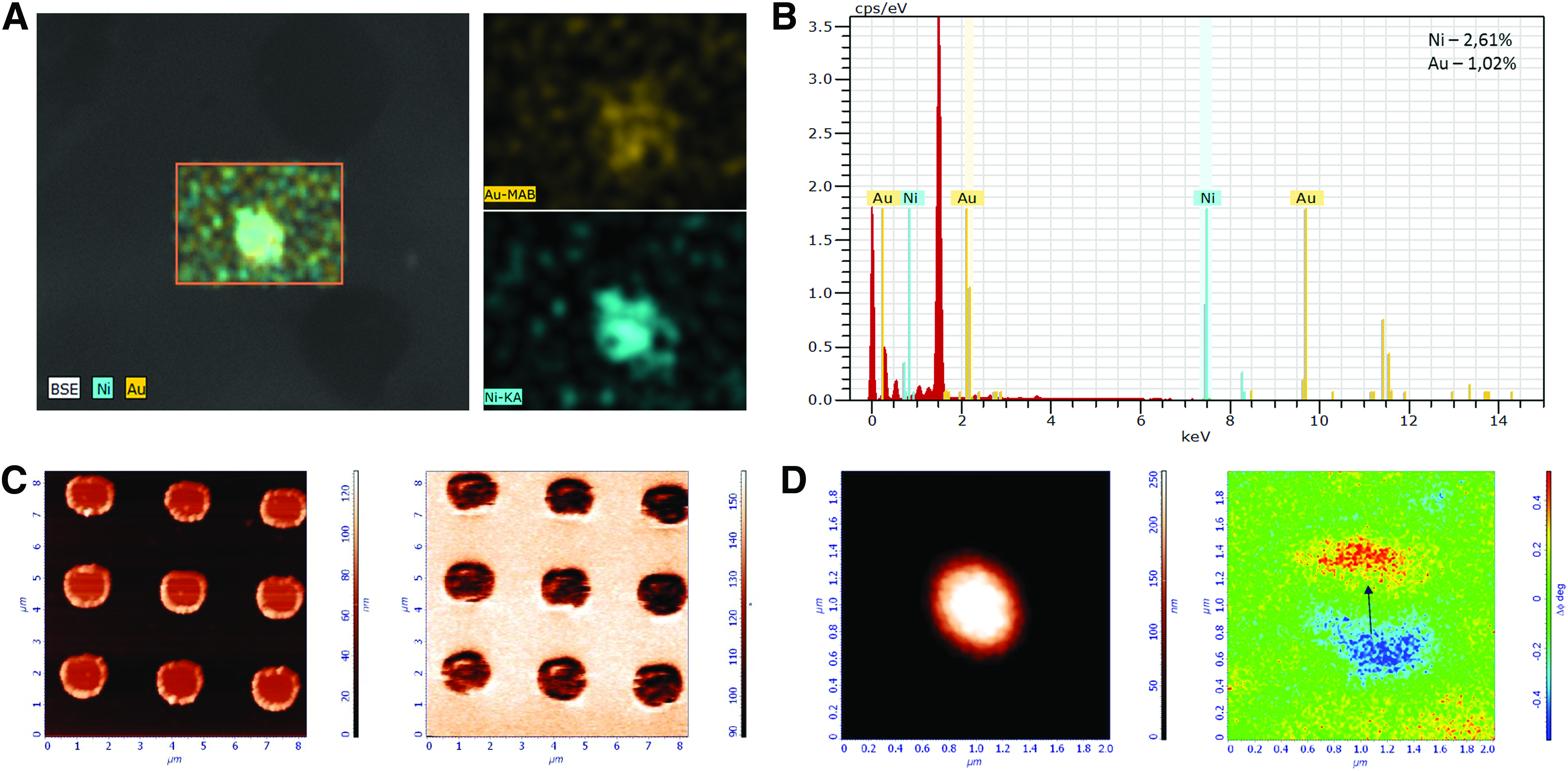
The magnetic structure of μMDs was observed using AFM with magnetic cantilevers by dynamic magnetic force microscopy (Fig. 2C, D). This method uses two passes across each raster scan line. On the first pass, topography was recorded using a semicontact mode of operation. On the second pass, the cantilever was lifted above the surface to a height of 100 nm and followed the same topographic contour. Simultaneously, the phase shifts in the cantilever oscillation caused by gradients in the magnetic force on the tip were recorded. In this study, the magnetic properties of the probes were checked with solid disks, and probe suitability for magnetic measurements was estimated by the image quality of magnetic tracks. It was revealed that the magnetic moment of μMD in constant magnetic field of 0.5 T is parallel to its plane (Fig. 2D).
In vitro antitumor activity of aptamer-modified μMDs after AMF treatment
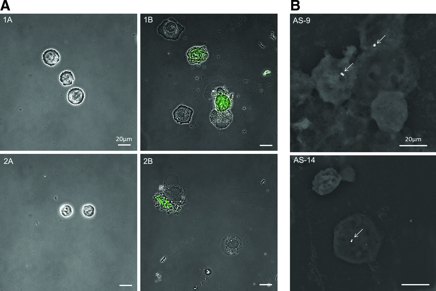
To observe the effect of aptamer-modified μMDs by confocal microscopy, we incubated ascite cells with Au-Ni-Au μMDs modified with FAM-labeled AS-14 and AS-9 and exposed them to AMF for 10 min. Control experiments were performed with intact ascite cells, ascite cells in AMF, and ascite cells incubated with aptamer-modified Au-Ni-Au μMDs without influence of AMF (Fig. 3A). AS-14- and AS-9-modified μMDs bound to ascite cells were observed by scanning EM as seen in Fig. 3B. The destruction of ascite cells incubated with AS-9- and AS-14-modified Au-Ni-Au μMDs began within the first hour after AMF exposure (Fig. 3, panel 2B).
Some of the ascite cells incubated with aptamer-modified Au-Ni-Au μMDs exposed to AMF lost their nucleus (Fig. 4, panel A2) and became smaller in size, indicating cell death. To estimate cell viability after the treatment, ascite cells were also stained with annexin V-Cy3 and 6-CFDA to estimate the number of apoptotic and living cells (Fig. 4B). The nonfluorescent compound 6-CFDA enters into the cells and hydrolyzes the esterases present in living cells to the fluorescent compound 6-carboxyfluorescein, indicating that the cells are viable. This combination of annexin V and 6-CFDA allows the differentiation among early apoptotic cells (annexin V positive, 6-CFDA positive), necrotic cells (annexin V positive, 6-CFDA negative), and viable cells (annexin V negative, 6-CFDA positive). We observed that the majority of ascite cells treated with aptamer-modified Au-Ni-Au μMDs became necrotic and apoptotic after 40 min of AMF exposure (Fig. 4, panel B2). A few viable cells were still observed in the sample. All nontreated ascite cells were viable (Fig. 4, panel B1). Importantly, aptamer-modified Au-Ni-Au μMDs were visibly attached to cell membranes after the influence of AMF (Fig. 4, panel C2).
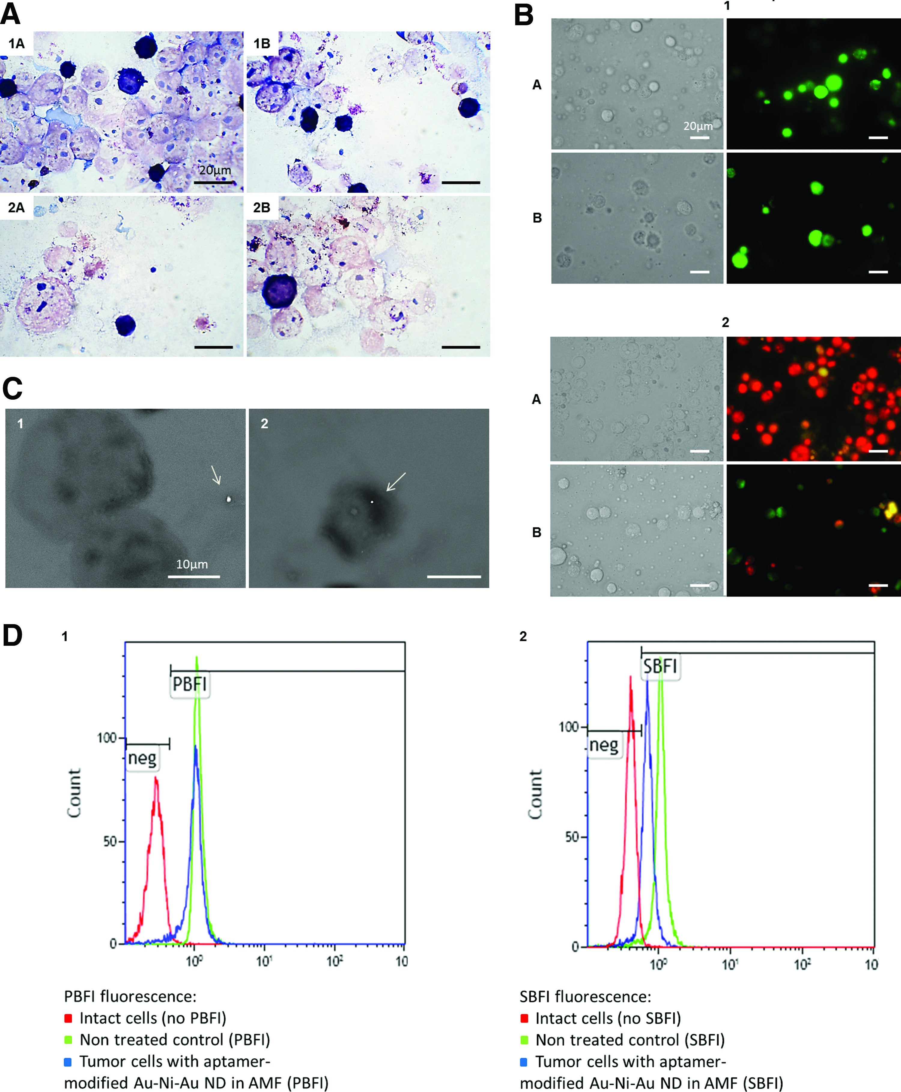
To estimate the effect of aptamer-modified μMDs in AMF, the functional state of treated ascite cells was evaluated. The levels of potassium and sodium were estimated using PBFI and SBFI probes by flow cytometry 40 min after exposure to AMF. Figure 4D presents the flow cytometry of nontreated ascite cells and ascite cells incubated with aptamer-modified μMDs stained with PBFI (Fig. 4, panel D1) and SBFI (Fig. 4, panel D2). The data showed that the treatment of ascite cells with AS-9- and AS-14-modified μMDs under the influence of AMF decreased the levels of sodium and potassium ions by 33.4% and 7.1%, respectively. This significant decrease in sodium concentration in ascite cells resulted in a reduction of the cell size, an increase in membrane potential, and, therefore, stimulated cell death.
It has been shown that AS-14 and AS-9 increased the effectiveness of influence of μMDs on ascite cells in AMF (Fig. 5A–C). However, there was no dependence on the number of μMDs and the percentage of viable and nonviable cells in the samples incubated with μMDs modified only with thiolated primer in DPBS and AS-14- and AS-9-modified μMDs. Even one μMD to a cell caused cell death in the same way as six μMDs to a cell.
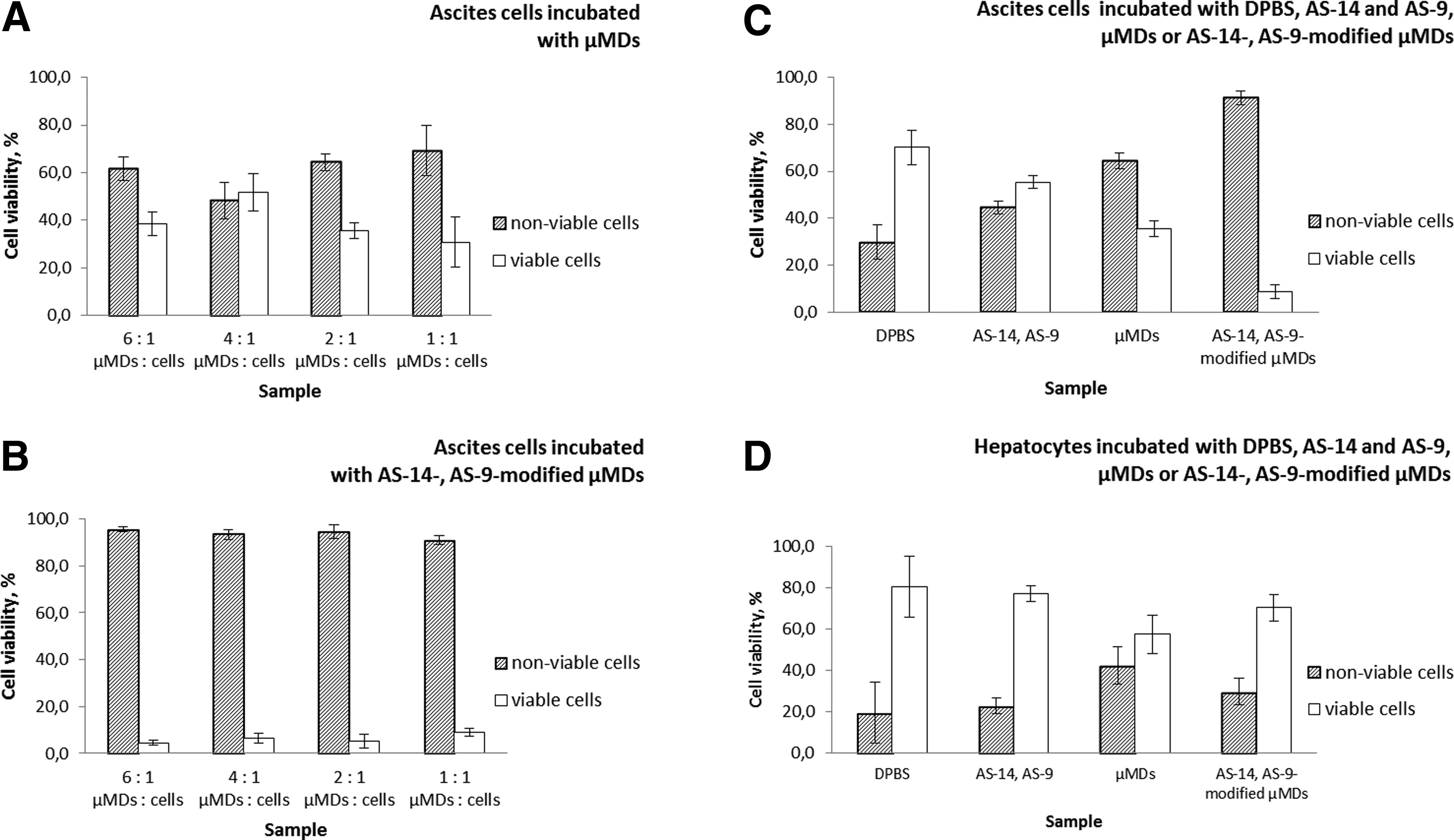
The percentage of viable cells and nonviable cells stained with trypan blue counted using fluorescent microscopy.
Interestingly, μMDs modified with thiolated primer had less impact on hepatocytes than on ascite cells. Nevertheless, AS-14 and AS-9 reduced off-target effect of μMDs on hepatocytes, resulting in increase of viable cells in the samples.
In vivo antitumor activity of aptamer-modified μMDs managed by AMF
It has been reported that Ehrlich ascite tumor cells lack H2 histocompatibility antigens [13], which apparently is the reason for their rapid proliferation in almost any mouse host. Since the description of Ehrlich ascites, we exploited it for the study of antitumor activity of aptamer-modified μMDs. There are various ways to evaluate the antitumor efficacy, such as (1) to examine the ascite cells histologically after AMF treatment, (2) to measure the increase in survival time, and (3) to measure the number of ascite cells formed after magnetomechanical therapy. For our studies, we used the method mentioned first to evaluate the efficacy of μMDs after AMF exposure.
To confirm in vivo antitumor activity of AS-14- and AS-9-modified μMDs after AMF exposure, tumor-bearing mice were treated with aptamer-modified μMDs, μMDs alone, or DPBS. One hour after the intratumoral injections of μMDs, mice were exposed to a 100 Hz AMF of 100 Oe for 10 min. It is important to note that no local heating of the tumor site during and after AMF treatment was observed by an infrared camera as shown in Fig. 1D. After 4 h, the tumors from all mice were isolated and histological sections with H&E staining were prepared. Another control group of mice was injected with aptamer-modified μMDs without exposure to AMF.
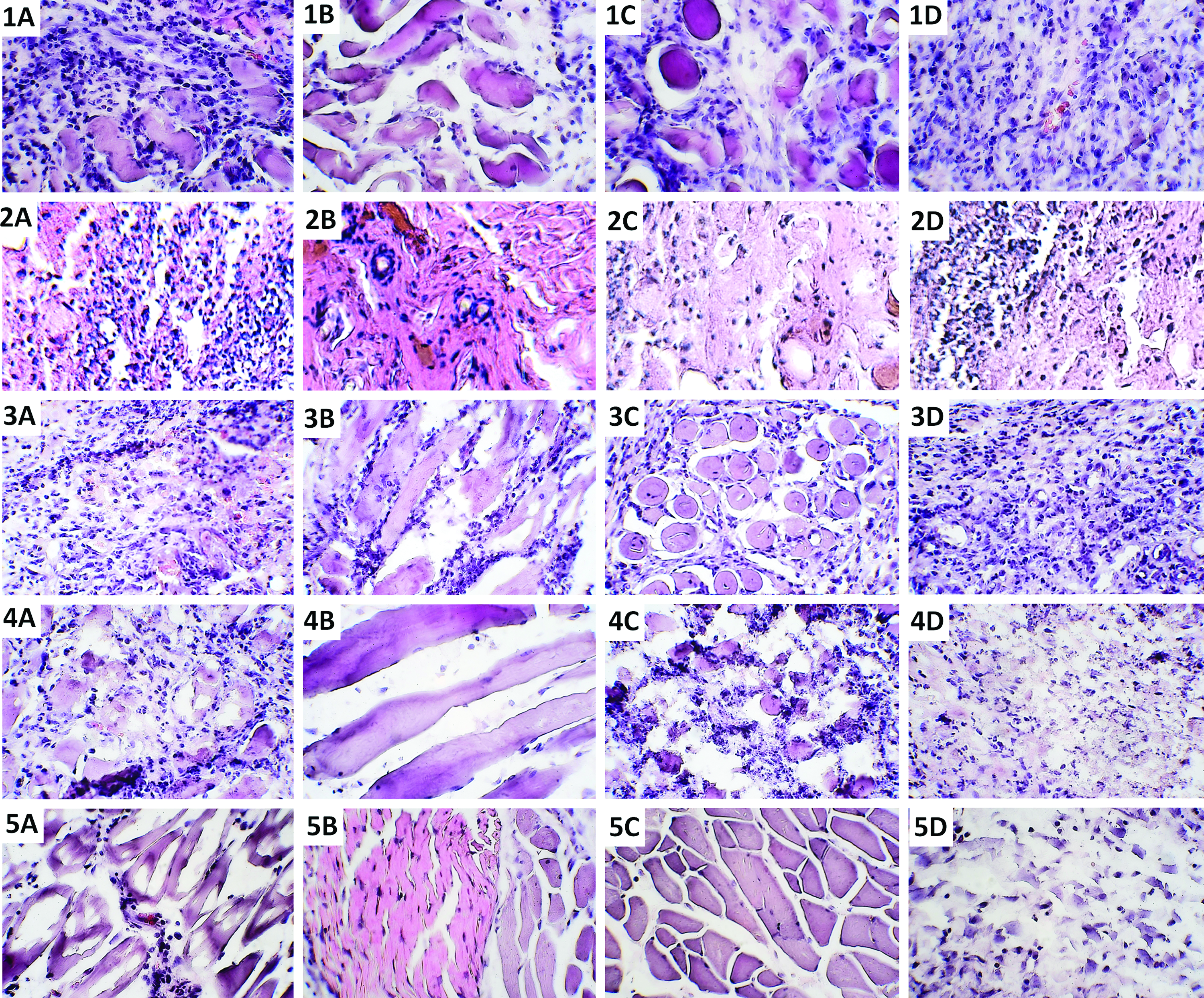
In DPBS control, swollen and deformed muscle tissue in a state of necrosis because of the malignant process was observed as shown in Fig. 6, panels 1A–C. Moderately deformed connective tissue is observed in Fig. 6, panel 1D. Tumor cells, seen in dark blue, are present in large numbers between deformed connective tissue and muscles. Injection of only aptamers caused some visible changes in tumor tissue (Fig. 6, panels 2A–D) such as disruption of tumor cells (Fig. 6, panels 2A, D). Injection of noncoated μMDs, stabilized with thiolated primers, did not change the pattern in the histological sections of tumor-bearing mice (Fig. 6, panels 3A–D). The photos also show strongly deformed muscle tissue (panels 3B and C) and moderately deformed connective tissue (panels 3A and D) with tumor cells around and inside of the tissue. AS-14- and AS-9-modified μMDs injection reduced a number of tumor cells in tumor-bearing mice even without AMF exposure (Fig. 6, panels 4A–D). Panels 4A and B in Fig. 6 show necrotic connective tissue with tumor infiltrates. Destroyed tumor cells are visible in the histological section in Fig. 6, panel 4C. Panel 4D in Fig. 6 shows connective tissue in a state of moderate necrosis. Injection of AS-14- and AS-9-modified μMDs in tumor-bearing mice treated with AMF reduced the number of tumor cells more significantly than injection of AS-14- and AS-9-modified μMDs without AMF treatment (Fig. 6, panels 5A–D), which may be attributed to cell apoptosis that started after the influence of AMF. In contrast, injection of only μMDs did not reduce the number of ascite cells in tumors of mice. Owing to the antitumor properties of AS-14 and AS-9, it was evident that the aptamer-modified μMDs caused destruction of tumor cells even without influence of AMF. Nevertheless, AMF action on mice injected with AS-14- and AS-9-modified μMDs led to a greater reduction in the number of tumor cells.
Visually monitoring of the treatment of mice has shown the effectiveness of the mice treatment with aptamer-modified μMDs in AMF (Fig. 7, panel 4). With regard to μMDs modified only with thiolated primer, it destroyed the tumor resulting in decrease of tumor size, but muscles and epithelium were also destroyed (Fig. 7, panel 3). Necrotic tissue lesions are seen in photos 3B–C from the first treatment procedure. Aptamers AS-14 and AS-9 injected into the tumor alone did not decrease the tumor size (Fig. 7, panel 2), nevertheless the tumor growth was not as fast as in control animals treated with DPBS (Fig. 7, panel 1).
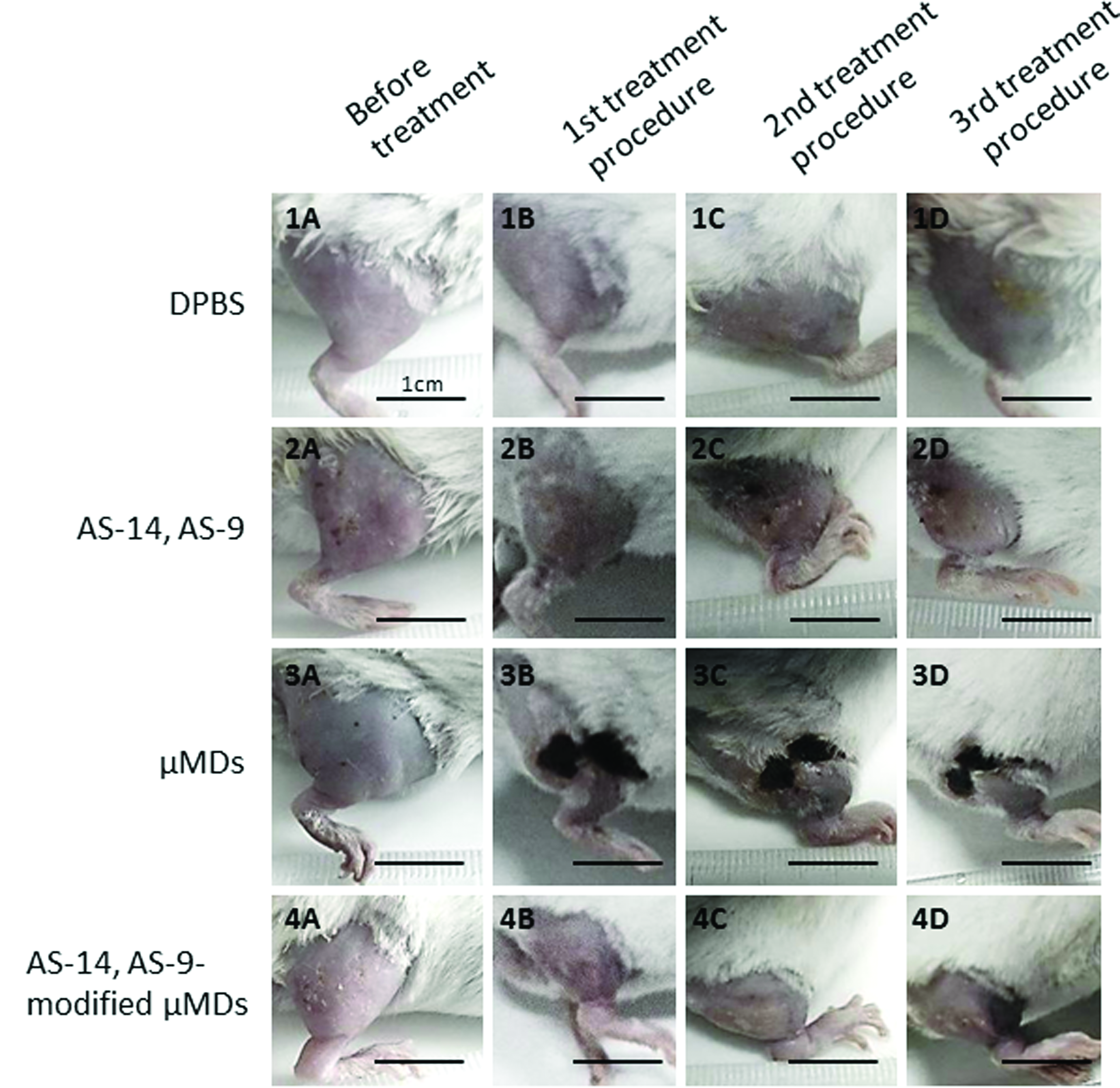
Treatment monitoring of mice injected with DPBS
Discussion
To date, despite the progress in medicine and biotechnology, cancer therapy is still an acute problem. The number of cancer patients is increasing every year, and this makes it necessary to create new effective methods of treatment. Microsurgery managed by magnetic field is one of the new perspective directions in treatment of malignancies. In our study, for the first time, the possibility of aptamer-modified Au-Ni-Au dipole magnetic μMDs managed by AMF for target destruction of tumor cells has been shown. Aptamers AS-9 and AS-14 with high selectivity to Erlich ascite adenocarcinoma cells were used to make a functional surface of μMDs. Previously, we have demonstrated that these aptamers display antitumor properties, causing apoptosis in mouse adenocarcinoma cells in vitro [12]. One of the likely targets of AS-9 is filamin A, involved in tumor cell proliferation, metastasis formation, and tumor progression. Aptamer AS-14 binds with fibronectin, participates in cell adhesion, migration, embryogenesis, wound healing, blood coagulation, and metastasis. Here, we used these aptamers as linkers to allow magnetic μMDs to bind to ascite cells for enhanced targeted microsurgery purposes.
We use mathematical simulations to explain aptamer-modified μMD interactions with cancer cells (see Supplementary Data; Supplementary Data are available online at www.liebertpub.com/nat). It has been shown that aptamers bound with μMDs tear target proteins from the cell membrane with violent forces of AMF. As there are only a few such protein molecules per disk, separation of target proteins from the cell membrane will more likely occur than the mechanical damage of the membrane itself. Experiments with a small number of aptamer-modified μMDs have shown the effectiveness of the treatment. The aptamers firmly attach to the disks and to the target proteins, and then pull target proteins out of the membrane, leading to its damage.
Cell death can be implemented in various ways: apoptosis, pyroptosis, oncosis, autophagy, and necrosis [14]. All of these processes related to changes in cell volume caused by modification of ion fluxes and primarily, modification of sodium and potassium fluxes. Apoptosis, in particularly, characterized by cellular compression, occurs because of the significant loss of sodium, potassium, and chlorine by the cell that are necessary for activation of caspases and nucleases [15]. Apoptosis can be induced by different factors: ligands such as CD95L (FASL) and tumor necrosis factor (TNF), or by an increase of intracellular concentration of calcium and active oxygen. To determine the ways of cell death induced by the action of aptamer-modified Au-Ni-Au μMDs managed by AMF, we evaluated the change of intracellular sodium and potassium using SBFI and PBFI probes, respectively, by flow cytometry. The data showed that sodium concentration decreased significantly (Fig. 4, panel D2) in the first 40 min after AMF exposure, whereas potassium concentration decreased slightly. It should be noted that the induction of apoptosis of tumor and normal cells occurs in various ways because tumor cells were characterized by membrane depolarization [16] caused by increasing of intracellular sodium. In our studies, sodium concentration in ascite cells incubated with AS-14- and AS-9-modified μMDs decreased directly after the influence of AMF, whereas potassium concentration remained almost unchanged. This resulted in a reduction of the cell size, an increase in membrane potential, and, therefore, stimulated apoptosis. It is well known that decreased cell volume, as observed in our experiments (Fig. 4, panels A2, C2), promotes cell death by apoptosis.
Mathematical simulations showed that conformation of target proteins bound with aptamer-modified Au-Ni-Au μMDs should be transformed under the influence of AMF. In our study, we used AS-9 and AS-14 aptamers, specific to filamin A and fibronectin, respectively, both proteins related to the function of sodium channels [12]. Cytoskeletal protein, filamin A, binds to sodium channels and inhibits their activity [17] that may cause a decrease in intracellular sodium concentration. Fibronectin can also influence the intracellular concentration of sodium, as sodium channels and the extracellular domain of the β2 subunit bound specifically to fibronectin type III [18]. Thus, it can be assumed that AS-14- and AS-9-modified μMDs could change the conformation of target proteins fibronectin and filamin A, which, in turn, decreased cellular sodium concentration and caused reduction of cells size, hyper polarization of cell membrane, and finally apoptosis.
μMDs damaged membranes of ascite cells and hepatocytes after 10 min of AMF exposure (Fig. 5). AS-14 and AS-9 increased effectiveness of μMDs (Fig. 5A, B) and reduced destructive influence on hepatocytes (Fig. 5D). AS-14 and AS-9 increased the number of nonviable ascite cells approximately two times compared with the control samples incubated with DPBS (Fig. 5C), whereas the aptamers almost did not show any influence on hepatocytes (Fig. 5C).
Four hours after exposure of tumor-bearing mice injected with aptamer-modified Au-Ni-Au μMDs to AMF, a number of tumor cells decreased (Fig. 6, panels 5A–D), which may be attributed to cell apoptosis that started after the influence of AMF. In contrast, μMDs alone injected in mice did not decrease the number of tumor cells at all (Fig. 6, panels 3A–C). Owing to the antitumor properties of AS-14 and AS-9, it was evident that the aptamer-modified μMDs caused destruction of tumor cells even without influence of AMF (Fig. 6 panels 3A–C). Nevertheless, AMF action on mice injected with AS-14- and AS-9-modified μMDs led to a greater reduction in number of tumor cells (Fig. 6, panels 5A–D).
Our study showed that aptamer-modified Au-Ni-Au μMDs trigger apoptosis of ascite cells within 40 min after 10 min exposure to 100 Oe AMF. Moreover, 4 h after the influence of AMF, the number of tumor cells significantly decreased. It was remarkable to observe that the antitumor properties of AS-14 and AS-9 aptamers were most likely enabled by coupling them with Au-Ni-Au magnetic μMDs with AMF exposure. All of our findings demonstrate the perspectives of aptamer-based microsurgery for treatment of oncological diseases and also the necessity of identifying the correct choice of ligands for modification of μMDs because only the strong interaction of μMDs with proteins involved in processes of apoptosis, tumor growth, and development would determine the outcome of treatment.
Obviously, μMDs unmodified by aptamers in AMF caused necrosis of tumor cells, leading to inflammation and wound formation (Fig. 7, panel 3). Targeting by μMDs with AS-14 and AS-9 apparently contributes to apoptosis of tumor cells or at least to a significant reduction of necrotic processes along with triggering of apoptosis. The fact that AS-14 and AS-9 alone have inhibitory effect on ascite cells (Fig. 7, panel 2 and Fig. 5C) suggests that these aptamers are useful, not only as target agents but also as therapeutics agents.
In this way, research has shown that application of μMDs in low-frequency magnetic fields modified with aptamers specific to tumor cells and suppressing tumor growth is an effective tool of magnetodynamic therapy that may be applicable in clinics in the future.
Footnotes
Acknowledgments
This work was supported by the Russian Scientific Fund (grant no. 14-15-00805).
Author Disclosure Statement
No competing financial interests exist.
References
Supplementary Material
Please find the following supplemental material available below.
For Open Access articles published under a Creative Commons License, all supplemental material carries the same license as the article it is associated with.
For non-Open Access articles published, all supplemental material carries a non-exclusive license, and permission requests for re-use of supplemental material or any part of supplemental material shall be sent directly to the copyright owner as specified in the copyright notice associated with the article.