
Abstract
Traumatic brain injury (TBI) involves the elaboration of oxidative stress that causes cerebrovascular dysfunction, including impairment of autoregulation of cerebral blood flow. Currently, there is no clinically effective antioxidant treatment for these pathologies. Most currently available antioxidants act through mechanisms in which the antioxidant either transfers the radical or requires regeneration, both of which are impaired in the toxic post-TBI environment. We previously reported that single-walled carbon nanotubes (SWCNTs) and ultrashort SWCNTs possess antioxidant activity, and their characteristics suggest that radical annihilation is the major mechanism. We have now developed a biologically compatible class of carbon-based nanovectors, poly(ethylene glycol)-functionalized hydrophilic carbon clusters (PEG-HCCs) that can be further functionalized with antibodies, and hence show promise as targeted drug delivery platforms. Here we report that PEG-HCCs possess innate antioxidant activity and can be rapidly targeted via an antibody to the P-selectin antigen in a model of injured cultured brain endothelial cells. One immediate application of this therapy is to vascular dysfunction that accompanies TBI and worsens outcome in the face of systemic hypotension. These in vitro results support the need for further investigation in animal models.
Introduction
R
The generation of oxidative radicals is rapid following TBI. Secondary insults such as hypotension, a major contributor to poor outcome in TBI, are additional sources of oxidative radical generation with several bursts of superoxide documented throughout the course of TBI and hypotension and reperfusion. Moreover, our previous work has shown that PEG-SOD has only small and transient effects. 6 Therapies to address oxidative stress must, therefore, be rapidly effective and able to address the site of radical generation or its consequences at a clinically realistic time point. There is little evidence that conventional antioxidants can achieve these goals. Hence, there is a need to find new classes of biologically compatible antioxidants. Nanoparticles are an emerging group of such materials. 14 Nanoparticles have markedly different biodistribution and metabolic profiles than small molecules or enzymes, and they also may afford improved in vivo efficacy.
We previously showed that single-walled carbon nanotubes (SWCNTs) and ultrashort SWCNTs are antioxidants, as measured by the oxygen radical absorbance capacity (ORAC) assay. 14 We have recently developed another class of biologically compatible carbon-based nanovectors, poly(ethylene glycol)-functionalized hydrophilic carbon clusters (PEG-HCCs). 15,16 PEG-HCCs can be readily functionalized with antibodies for targeted drug delivery. These PEG-HCCs can be loaded with hydrophobic drugs, and when mixed with an antibody, a noncovalent formulation capable of targeted drug delivery in vitro is formed. 17,18 PEG-HCCs are ∼50 nm in size at neutral pH based on dynamic light scattering (DLS) and have a zeta potential of −23.04 mV at pH 7.0, which is evidence for the negatively charge surface of the nanoparticle. This potential also indicates that the PEG-HCCs will flocculate with time. However, the PEG-HCCs can be easily re-suspended after vortexing the mixture for 5 min. Also importantly, ∼94% of the PEG-HCC is constituted by PEG, 17 which is a non-ionic hydrophilic polymer approved for certain uses by FDA, which is well-tolerated clinically, 19 –22 with the additional benefit that PEG can protect and restore the integrity of cell membranes. 23 –25 Our preliminary toxicity and biodistribution studies indicate that these PEG-HCCs nanoparticles are not acutely toxic in normal mice, with no evidence of chemical or pathological damage to major organs, and they have a circulating half-life of 2–3 h. 17
Here we report the in vitro characteristics of PEG-HCCs designed specifically to address cerebrovascular dysfunction that accompanies TBI. We show that PEG-HCCs possess antioxidant activity that is potent enough to reduce oxidative stress in vitro following injury, that its antioxidant mechanism does not involve metal chelation, and that PEG-HCCs can be targeted to a model of injured brain endothelial cells expressing P-selectin antigen. P-selectin expression is increased in brain endothelial cells following TBI, 26 and possibly systemically depending upon the trauma mechanism. These results suggest that targeted PEG-HCCs are potential therapeutics for oxidative stress-induced cerebrovacular dysfunction from mTBI.
Methods
For all these experiments, PEG-HCCs were prepared as previously described, and sterile filtered using a 0.20 μm pore size membrane. 17
ORAC assay
The following solutions were prepared daily: phosphate buffer at pH 7.4 (phosphate-buggered saline [PBS], 75 mM), fluorescein sodium salt (0.1 μM), α,α’-azodiisobutyramidine dihydrochloride (AAPH, 0.15 M), racemic 6-hydroxy-2,5,7,8-tetramethylchroman-2-carboxylic acid (Trolox, 400 μM). The experiment was performed in a 96-well plate and each sample was analyzed by triplicate as shown in Table 1. After the addition of the fluorescent probe or PBS and sample (first step) to the assigned wells, the plate was incubated at 37°C for 15 min in a Tecan plate reader. Then, the ice-cold AAPH or PBS was added (second step) and the fluorescent intensity at 530 nm (485 nm excitation) was measured every minute for 2 h. Data were analyzed as follows: control 2 (background) was subtracted from the assay and control 1. The assay well results were divided by the control 1, and the area under the curve (AUC) was determined by Equation 1. Trolox mass equivalents (TME) were determined by Equation 2.
17,27
–33
Trolox and phosphate buffer (blank) were run as samples. At least three standards of Trolox were run for the calibration curve.
ORAC, oxygen radical absorbance capacity; AAPH, α,α’-azodiisobutyramidine dihydrochloride; PBS, phosphate-buffered saline.
Chelation assays
It has been found that iron forms deposits in patients with mTBI; this iron accumulation can be involved in the pathology of a TBI as one of the major secondary events. 34 –36 Furthermore, free iron causes oxidative stress when it reacts with hydrogen peroxide to form hydroxyl radicals (Fenton reaction), and with lipids to produce alkoxy and peroxyl radicals (lipid peroxidation). Iron chelation therapy has been proposed in order to diminish the oxidative process. 35 Nonetheless, it is important to understand the effect that the chelator might have on the catalytic cycle of Fe+2/Fe+3 process. In order to explore the behavior of the iron when combined with our nanoparticles, we ran the following iron chelator assays: benzoate hydroxylation and ascorbate oxidation. Ascorbic acid (cat # AC40147), benzoic acid (cat # AC22180), iron(II) sulfate heptahydrate (cat # AC42373), iron(III) chloride hexahydrate (cat # AC21709), and the disodium ethylenediamine tetraacetate (EDTA, cat # E4884) were obtained from Fisher Scientific. Deferoxamine mesylate salt (DFO, cat # D9533) was obtained from Sigma-Aldrich.
Benzoate hydroxylation
This assay is based on the hydroxylation of benzoate when exposed to iron and H2O2, producing the fluorescent compounds 2,3,4-trihydroxybenzoate (308 nm excitation and 410 nm emission) 37 –40 with the major products 2-hydroxybenzoic acid salt or salicylic acid salt at pH 7.4. In brief, the following solutions were freshly prepared before each run according to reported procedures: 41 benzoic acid (3 mM, recrystallized from hot water), FeCl3 (1.80 mM), FeSO4 (1.80 mM), methoxypolyethylene glycol amine polymer (PEG MW 5,000, 0.02 mg/mL) and PEG-HCCs (0.02 mg/mL). EDTA (0.60 mM) and DFO (0.60 mM) were used as the positive and negative control, respectively. The benzoic acid was incubated at room temperature for 1 h in the phosphate buffer (pH 7.4) with 5 mM of H2O2 and the ferric or ferrous iron salt, in the presence of one of the controls or the PEG-HCCs. The reaction was started by the addition of any the iron salts and kept in dark. Salicylate was used to determine the quenching effect of the materials. 41
Ascorbate oxidation
This assay was performed in a similar way to that described previously. 42 –46 Briefly, the following solutions were prepared prior to each experiment: ascorbic acid (0.1 mM), FeCl3 (0.60 mM), potassium phosphate buffer (50 mM, pH 7.4, cleaned with Chelex resin), EDTA (0.02 mg/mL previously recrystallized three times from hot water), DFO (0.02 mg/mL), and PEG-HCCs (0.02 mg/mL). The ascorbate absorbance at 265 nm was measured after 1, 5, and 30 min using a Tecan plate reader.
Cell culture
Murine brain endothelioma (bEnd.3, CRL 2299, ATCC, Manassas, VA) were cultured in Dulbecco's Modified Eagle Medium (DMEM)-F12 (50:50) with 4 mM L-glutamine media, supplemented with 10% fetal bovine serum (both from HyClone) and 1% Pen Strep (+10,000 u/mL penicillin/+10,000 μg/mL streptomycin, from Gibco®). During the second week, between the fourth and sixth passage, cell experiments were performed.
P-selectin targeting
bEnd.3 cells were grown on glass covers to 60–70% confluence in six-well plates. P-selectin-targeted PEG-HCCs (ps/PEG-HCCs) were synthesized by adding 0.01 μg anti-P-selectin antibody (Santa Cruz Biotechnology Inc., Santa Cruz, CA) to 0.49 μg PEG-HCCs and incubating at room temperature (RT) for 1 h. The antibody possesses hydrophilic and hydrophobic domains that allow the nanovector to sequester it. The bEnd.3 cells were treated with histamine (20 μM) to induce P-selectin expression or with 1× PBS (histamine diluent) as a control. 47 After a 15 min incubation at 37°C, the cells were treated with vehicle (1× PBS), PEG-HCCs (0.49 μg), or ps/PEG-HCCs. Cells were incubated for 15 min and then washed twice and fixed with methanol. Cells were washed and stained with anti-PEG (Epitomics, Burlingame, CA) and anti-P-selectin antibodies (Santa Cruz Biotechnology Inc, Santa Cruz, CA). After washing, cells were probed with fluorescent secondary antibodies to the anti-PEG and anti-P-selectin antibodies (Invitrogen, Carlsbad, CA). Glass covers were mounted on slides and fluorescent images were captured on an epifluorescent microscope.
Intracellular superoxide cell culture model
bEnd.3 cells were cultured to 50–80% confluence in six-well plates in 2 mL of culture media. The cells were treated with vehicle (1× PBS), PEG-HCCs, ps/PEG-HCCs, or P-selectin antibody alone (same concentration as found in ps/PEG-HCCs). After 15 min, the cells were treated with 10 μL of 2 mM antimycin A or 10 μL ethanol (control). Antimycin A has been shown to induce intracellular superoxide radical production.
48
The cells were incubated at 37°C for 40 min, at which point 2 μL of 10 mM dihydroethidine (DHE, in 50%/50% dimethyl sulfoxide [DMSO]/1×PBS)
49
was added to wells or 50%/50% DMSO/1×PBS (control). DHE reacts with ROS to form 2-hydroxyethidium, which possesses red fluorescence when excited near 480 nm; therefore, the increase in red fluorescence is proportional to the ROS in the sample.
49
The cells were then placed on ice, trypsinized, and washed twice. Cells were subsequently counted and stained with SYTOX Red (viability stain); 10,000 cells were analyzed per treatment group using the TXRED channel (DHE) and SYTOX Red to assess cell viability. A higher dose of antimycin A was also studied to confirm that PEG-HCCs were able to protect against oxidative cell death (Fig. S1)(See supplementary Figure 1 at
Results
ORAC assay
The antioxidant capacity of the PEG-HCCs was compared by ORAC to known antioxidants vitamin C, caffeic acid, and catechin. ORAC measures antioxidant capacity by the ability of a substance to inhibit the loss of fluorescence caused by the oxidation of a fluorescent dye by peroxyl radicals formed during the thermal decomposition of the AAPH,
27
–33
without the assistance of any metal.
31
The linear relationship between the net area and antioxidant concentration was evaluated by using a set of Trolox and PEG-HCCs standards (Figs. S2-S4) (See supplementary Figures 2–4 at
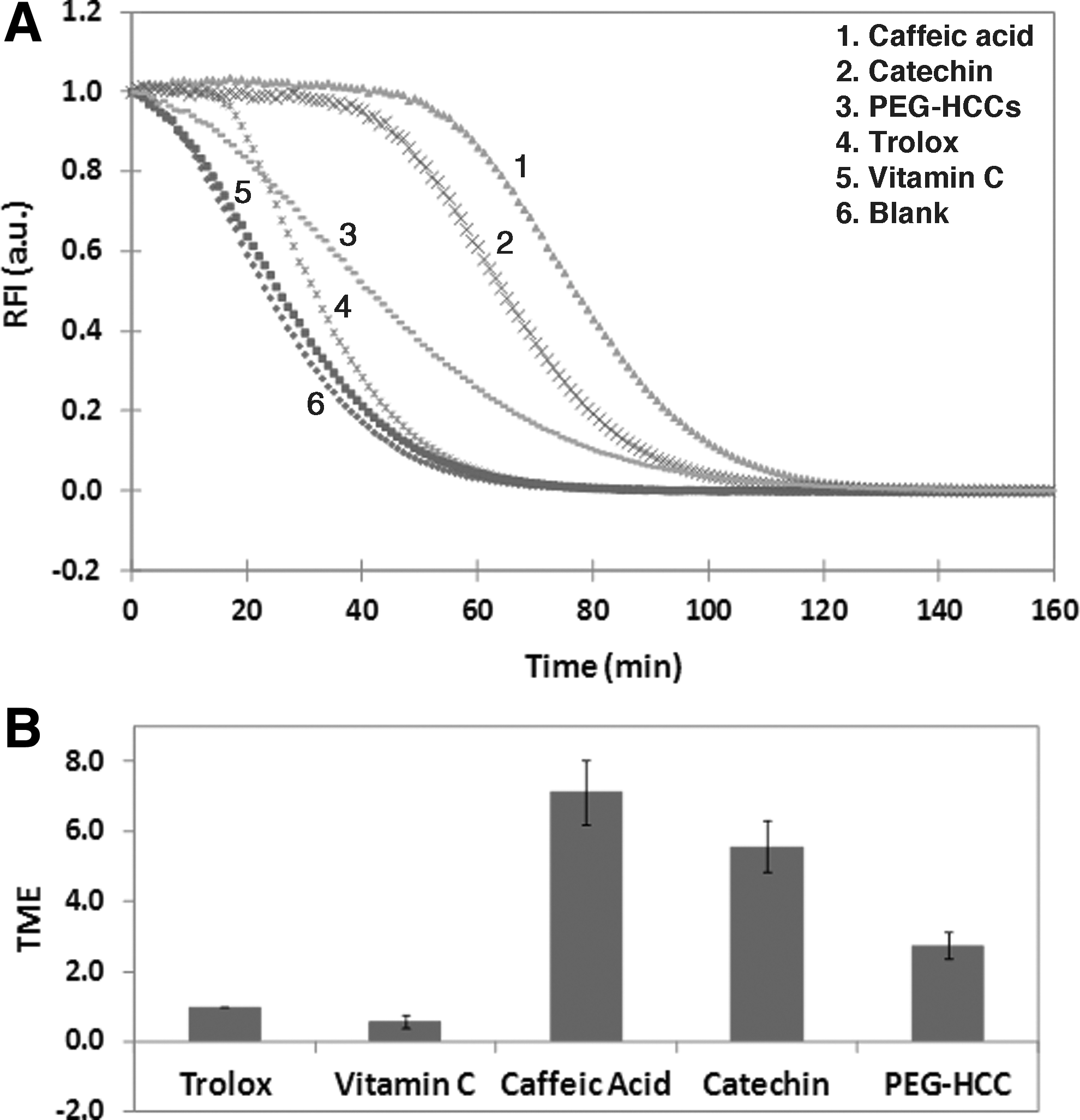
Typical fluorescence decay and calibration curves obtained.
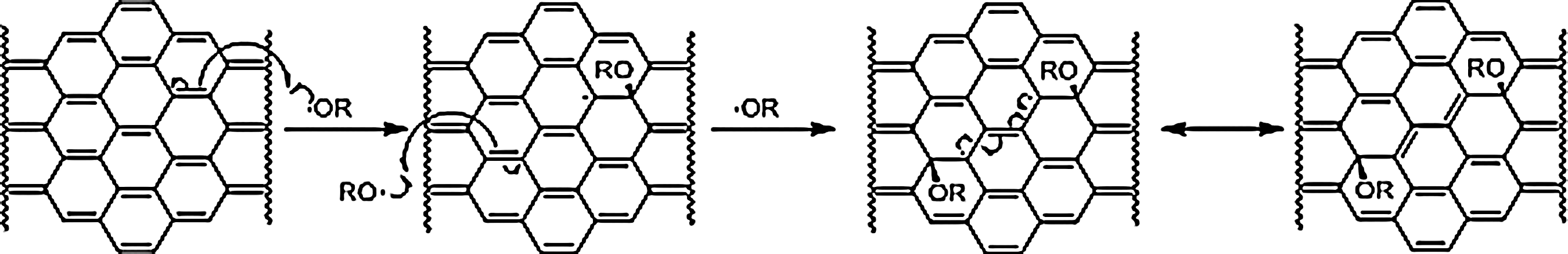
Suggested radical scavenging mechanism. Two additions of ROS result in the loss of two C–C pi-bonds and the formation of two new C–O sigma bonds and one new C–C pi bond without any radical species remaining. In RO·, R=H or alkyl.
Chelation as a potential mechanism
Iron accumulation in the pathology of TBI has been reported for mice 50 and humans 34 based on magnetic resonance imaging. It is known that Fe+2 and Fe+3 form lipid alkoxy radicals and peroxyl radicals, respectively, which induce the production of the neurotoxic aldehydes. 14 Therefore, it is important to determine if the nanoparticles stimulate the iron redox cycle (similar to EDTA) or inhibit it (similar to DFO). In order to assess this property, chelation assays were performed.
Benzoate hydroxylation assay
The iron chelating assay was used to evaluate the antioxidant capacity of PEG-HCCs when exposed to hydroxyl radicals produced by the reaction between iron and H2O2. In this assay, we monitored the oxidation of benzoate to salicylate by hydroxyl radicals produced by the Fenton reaction. To ensure that this measurement would be robust, sodium salicylate (SA) was mixed with PEG or PEG-HCCs every time that the experiment was run, in order to estimate how the fluorescence intensity was affected by the nanomaterial or the polymer, and then we proceeded to correct it (Fig. 3A). In general, it was observed that PEG-HCCs quench ∼20% of the fluorescence, and that PEG can enhance the fluorescence up to ∼10%. In the actual assay, EDTA and DFO were included as positive and negative controls, respectively. PEG-HCCs decreased the hydroxylation process as well as DFO in comparison to the case in which no chelating agent was added (Fig. 3B) for both iron systems. Interestingly, both PEG-HCCs and PEG reduced the hydroxylation of benzoic acid when the highest concentration of Fe+2 was used. We speculate that this could be because of the radical scavenger capacity of the carbon core (Fig. 2), the degradation of the polymer chains as previously reported, 51 –53 or both. Iron/PEG-HCCs systems do not induce the Fenton reaction.

Ascorbic acid assay
Ascorbic acid is able to serve as a donor antioxidant in free radical-mediated oxidative processes. But it is also able to reduce redox-active metals such as copper and iron, thereby increasing the oxidant properties of those metals. In general, at low concentrations, ascorbic acid is prone to be a pro-oxidant, and at high concentrations serves as an antioxidant. 44 In this assay, the oxidation of ascorbic acid by Fe3+ is monitored in the presence or absence of substances. EDTA again enhanced the oxidative activity of the Fe+3, whereas DFO inhibited it. We tested the PEG-HCCs in this system to determine if their antioxidant capacity could be because of interactions with trace iron content, and if there is such an interaction, the oxidation of the ascorbic acid should be inhibited. We followed the oxidation of the ascorbic acid after 5 min and 30 min of metal addition (Fig. 4A and B) and we found that in this system the PEG-HCCs function as potent oxidants, regardless of the presence or absence of Fe3+. PEG-HCCs and ascorbic acid show similar pro-oxidant and antioxidant behaviors; the difference is that for PEG-HCCs, no metal participation is necessary.
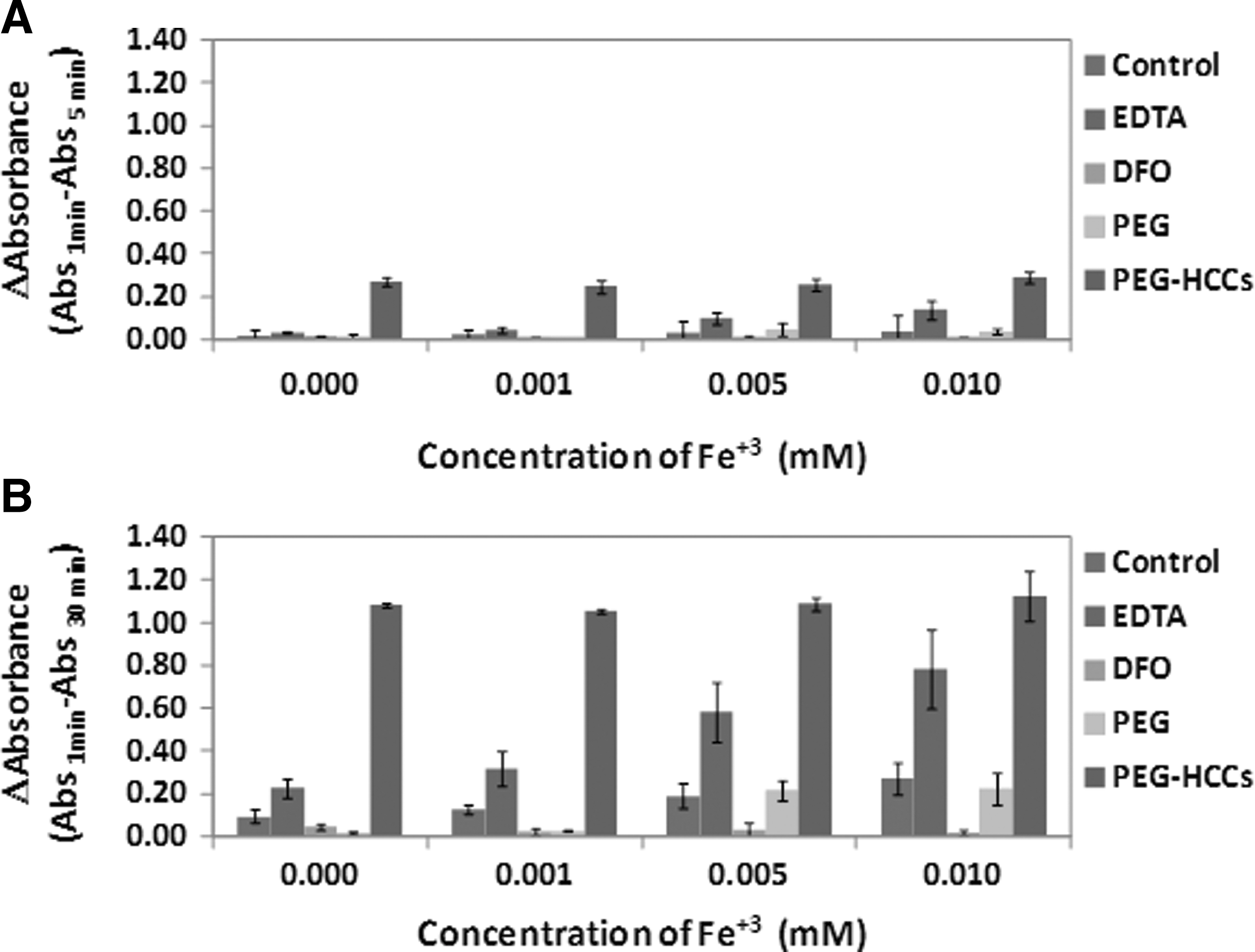
Oxidation of the ascorbic acid after
Targeted binding of PEG-HCCs to cultured brain endothelial cell line
Having demonstrated that the PEG-HCCs possessed antioxidant capacity in cell free systems, but that it could be dependent on the presence of reductants such as ascorbate, we evaluated if the PEG-HCCs could be targeted to cultured brain endothelial cells and if they could also alleviate oxidative stress in a biological system. A brain endothelial cell line was chosen because endothelial dysfunction is apparent even after mTBI, 54 and in the case of mTBI, there are minimal detectable brain injury and functional deficits unless hypotension is superimposed. 7 bEnd.3 cells were treated with histamine, which is known to rapidly induce the expression of P-selectin. PEG-HCCs were targeted to P-selectin. In endothelial cells, P-selectin, a cell adhesion molecule, is involved in recruiting leukocytes to sites of inflammation and injury. P-selectin is rapidly expressed on endothelial cells following activation by histamine. 48 In this experiment, we treated cells with histamine or 1X PBS to induce P-selectin expression (Fig. 5). After 15 min, the cells were treated with 1X PBS, PEG-HCCs, or ps/PEG-HCCs. The cells were incubated for another 15 min and after washing and fixing, they were stained with fluorescent antibodies for the presence of PEG to detect PEG-HCCs using an anti-PEG antibody (green), anti-P-selectin antibody(red) to detect P-selectin, and DAPI (nuclear stain; blue). In the cells that are not stimulated by histamine, there is little binding apparent with the P-selectin targeted PEG-HCCs. However, there was a dramatic increase in binding with the targeted P-selectin antibody- bound PEG-HCCs after stimulation by histamine (Fig. 6, bottom panel). This experiment demonstrates greatly enhanced binding of the targeted PEG-HCCs to endothelial cells in a model that induces expression of a protein, P-selectin.
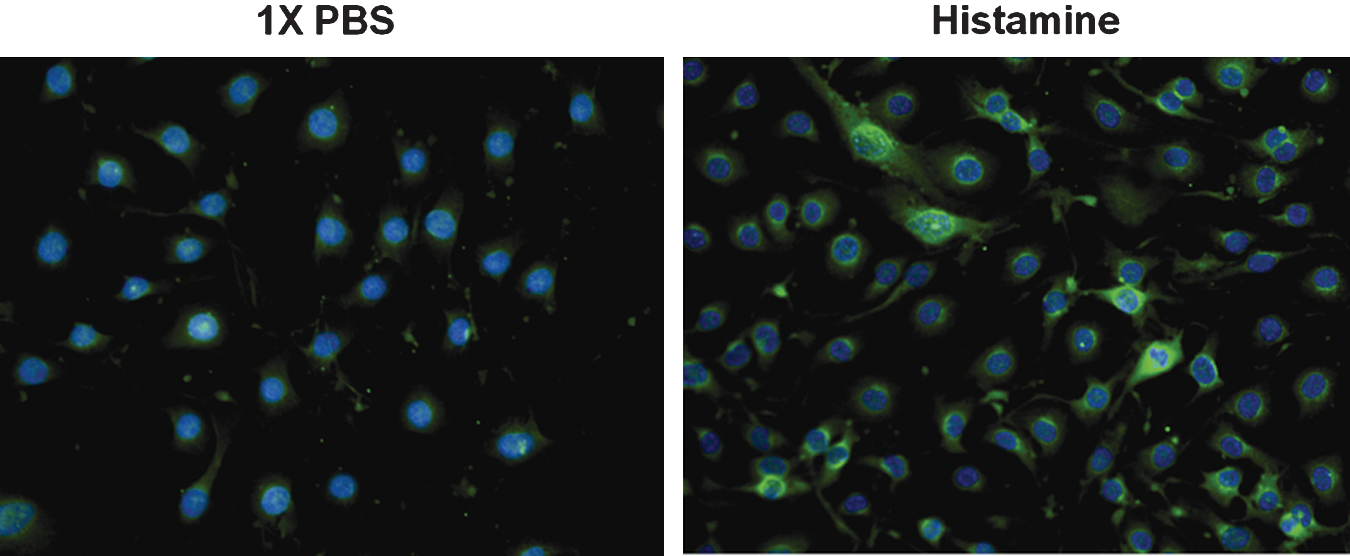
b.End3 cells express p-selectin when stressed by histamine. Cells were treated with histamine or the diluent 1× phosphate-buffered saline (PBS), fixed, and stained for the presence of p-selectin (green) and DAPI (blue). There was considerably enhanced binding of p-selectin antibody following histamine treatment, confirming increased expression of p-selectin. Color image is available online at
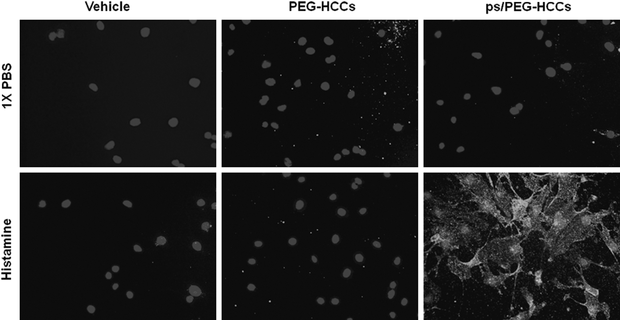
b.End3 cells oxidatively stressed by treatment with histamine or 1× phosphate-buffered saline (PBS) were exposed to vehicle, poly(ethylene glycol)-functionalized hydrophilic carbon clusters (PEG-HCCs) or the targeted variant ps/PEG-HCC and stained for the presence of PEG and DAPI. There is considerably enhanced binding of anti-PEG antibody following histamine treatment, indicating enhanced binding of the ps/PEG-HCCs to stimulated b.End3 cells.
We then evaluated whether applying the targeted P-selectin antibody to the PEG-HCCs would reduce its antioxidant ability. We tested the protective capacity of the nanoparticles by stressing the cells with antimycin A and determining the effectiveness of the native or antibody- bound PEG-HCCs at reducing levels of oxidative radicals. The level of oxidative stress was measured by DHE fluorescence. An increase in fluorescence is proportional to oxidative stress and is roughly proportional to superoxide levels, although not specific under all conditions. 46 In this study, we replicated the ability of the PEG-HCCs to reduce DHE fluorescence, 55 even when applied after the antimycin A, and in this study found that ps/PEG-HCCs reduced the level of DHE fluorescence to comparable levels as non-antibody-bound PEG-HCCs, and to background (DHE) (Fig. 7). By comparison, P-selectin antibody by itself had no significant effect on DHE fluorescence.
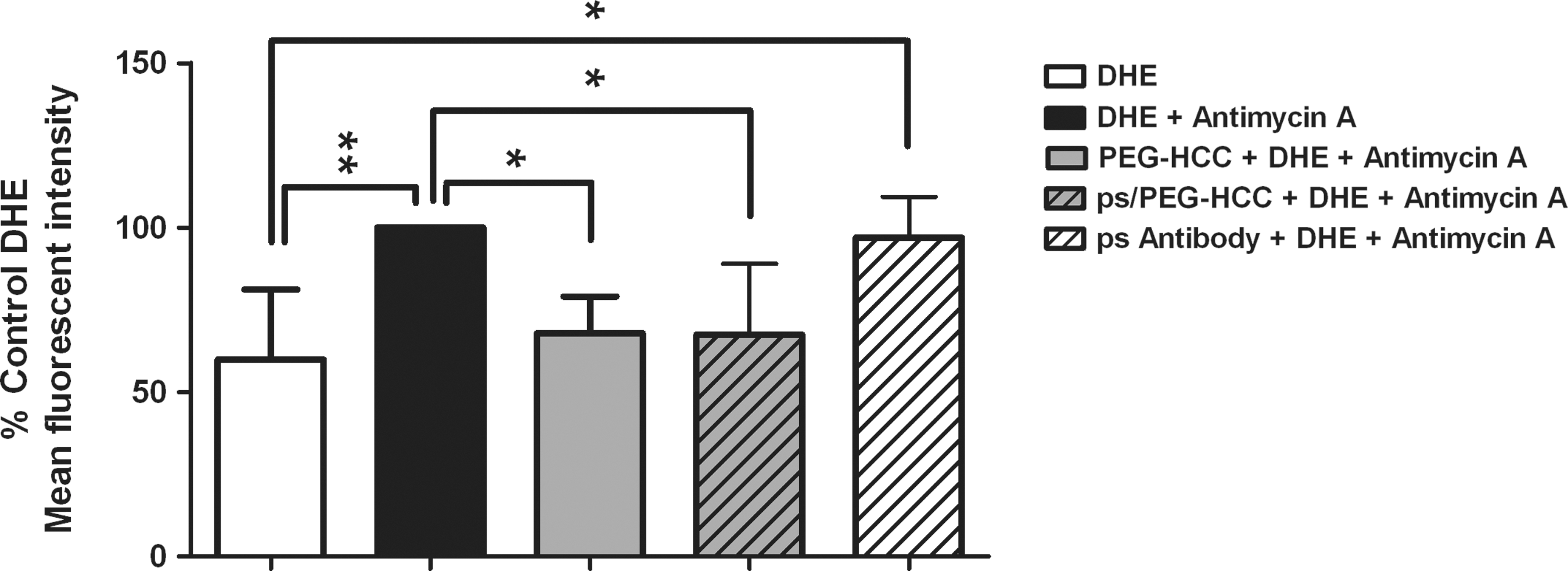
Poly(ethylene glycol)-functionalized hydrophilic carbon clusters (PEG-HCCs) and targeted ps/PEG-HCCs effectively reduce intracellular oxidative stress. Antimycin A was used to induce intracellular superoxide production. Dihydroethidine (DHE) mean fluorescence intensity (indicative of oxidative stress) was measured in cells treated with antimycin A to induce superoxide production (mean of five separate experiments). Results are given as % control (DHE+antimycin A, black bar) to account for minor differences in DHE concentrations and laser fluctuations. Untargeted (solid gray) PEG-HCCs and targeted ps/PEG-HCCs (striped gray) were effective at reducing DHE fluorescence after antimycin A treatment, whereas treatment with a similar amount of p-selectin antibody in the absence of PEG-HCCs (striped white) was not effective.
Discussion
These results indicate that PEG-HCCs possess antioxidant capacity sufficient to be biologically relevant and, further, that the mechanism does not involve chelation. They were effective in cultured brain endothelial cells in vitro when administered following administration of a mitochondrial toxin that induces oxidative stress. They could also be readily targeted to oxidatively stressed brain endothelial cells, and when functionalized for targeting, they retained the ability to alleviate oxidative stress in cultured brain endothelial cells. Overall, these results suggest that targeted PEG-HCCs are potential therapeutics for the cerebrovascular dysfunction from mTBI, and the results open additional possibilities for targeting a variety of pathological processes.
Because of the deleterious effects of loss of cerebrovascular responsiveness, even in mild injuries, a therapy that mitigates oxidative injury to endothelial cells should hold promise for the improvement of outcome following mTBI. Furthermore, because there is a major burst of superoxide radical at the time of resuscitation following TBI and hemorrhagic hypotension, 6 treatment at the time of resuscitation may provide a clinically realistic time point that can address at least one pathological event in the care of the TBI patient. Our previous work also showed that the enzymatic antioxidant, PEG-SOD, had only transient and minimal effect in an in vivo model of experimental TBI complicated by hypotension and resuscitation, 6 consistent with lack of clinical benefit, and we have preliminary results that PEG-SOD was effective in this antimycin A assay only when administered at high doses prior to the toxin, whereas post-treatment with PEG-HCCs was quite effective. The effectiveness remained in the targeted ps/PEG-HCCs. This characteristic may be a major advantage in considering the use of this agent for treating TBI. Our preliminary results indicate that PEG-HCCs are able to restore cerebral blood flow following resuscitation in an experimental TBI model 55 when treated during resuscitation. A more fully defined time course, both in vitro and in vivo, will be necessary to confirm the treatment time window and effect on outcome.
We are encouraged that targeting PEG-HCC using a P-selectin antibody demonstrated a more rapid binding to stressed bEnd.3 cells. This characteristic may have advantages as a therapeutic agent with enhanced binding to molecules expressed at the site of injury, and possibly to reduce the total dose administered to the patient. There are many potential such targets, but P-selectin appears particularly promising, as there is some added therapeutic benefit in experimental TBI of the antibody itself 56,57 perhaps providing added benefit in addition to the antioxidant ability of the PEG-HCCs. Further comparisons of these different therapeutic approaches will be pursued.
Conclusion
In summary, we have described a new class of antoxidant materials that were developed to specifically address the cerebrovascular dysfunction that follows TBI. We have shown that PEG-HCCs bound to a P-selectin antibody are rapidly targeted to stressed endothelial cells and are antioxidants capable of reducing oxidative stress-related fluorescence even when administered after a mitochondrial toxin. It is possible that their effectiveness following injury may be because of the unique mechanism of annihilating radicals, rather than relying on downstream antioxidants that may be depleted in the toxic post-TBI environment. These cumulative characteristics support further testing in TBI models, particularly those that involve cerebrovascular dysfunction, such as mTBI and hypotension/resuscitation, in which loss of cerebral autoregulation and poor reperfusion cause brain injury far beyond that of the TBI alone. 7
Footnotes
Acknowledgments
Funding came from the Alliance for NanoHealth through a subcontract from the University of Texas Health Science Center, Houston (Department of Defense, W8XWH-09-2-0139); the Traumatic Brain Injury Consortium, funded by the United States Army (W81XWH-08-2-0141 and W81XWH-08-2-0143); and the Nanoscale Science and Engineering Initiative of the National Science Foundation (NSF) under NSF Award EEC-0647452 for funding through the NSF Center for Biological and Environmental Nanotechnology.
Author Disclosure Statement
No competing financial interests exist.
References
Supplementary Material
Please find the following supplemental material available below.
For Open Access articles published under a Creative Commons License, all supplemental material carries the same license as the article it is associated with.
For non-Open Access articles published, all supplemental material carries a non-exclusive license, and permission requests for re-use of supplemental material or any part of supplemental material shall be sent directly to the copyright owner as specified in the copyright notice associated with the article.