




Abstract
Childhood is one the highest risk periods for experiencing a mild traumatic brain injury (mTBI) from sports-related concussions, motor vehicle accidents, and falls. In addition, many children experience lingering symptomology (post-concussion syndrome) from these closed head injuries. Although the negative sequel of mTBI has been described, a clinically reliable animal model of mild pediatric brain injury has not. The purpose of this study was to examine the validity of a modified weight-drop technique as a model for the induction of mTBI/concussion in juvenile rats following a single impact. Male and female rats (P30) were exposed to a single mTBI or a sham injury followed by a behavioral test battery. Juvenile rats who experienced a single mTBI displayed significant motor/balance impairments when tested on the beam walking task and in the open field, as well as deficits of executive functioning as measured with the novel context mismatch task and the probe trial of the Morris water task. In addition, both male and female rats showed depression-like behavior in the forced swim task, with male rats also exhibiting decreased anxiety-related behaviors in the elevated plus maze. The results from this study suggest that the modified weight-drop technique induces a clinically relevant behavioral phenotype in juvenile rats, and may provide researchers with a reliable animal model of mTBI/concussion from which clinical therapeutic strategies could be developed.
Introduction
T
In designing modeling systems for translational studies, the meaningfulness of the findings are only as good as the model implemented. Although several animal models of TBI have been developed, many of them produce injuries that are much more severe and often do not represent the etiological mechanisms (i.e., biomechanical forces) or the pathophysiology of mild brain injury or concussions. 5 The availability of an applicable animal model of mTBI would foster a greater knowledge of the underlying mechanisms, as well as the behavioral and neuropsychological outcomes, associated with this common injury. 1,6,7 In-depth analysis of the characteristics that lead to mild concussive-like outcomes indicate that a high velocity impact and head acceleration are key factors for injury generation. 8 –10 The research indicates that in order to model this effectively, two constraints need to be met: 1) the simulation needs to impact a head that is permitted to move freely, and 2) a high velocity impact is needed to produce translational acceleration and deceleration of the head. 9,11 The clinical relevance and the experimental potential of an experimental setup that could model this type of closed head injury from a single impact are invaluable.
Although it is important for the model of mTBI/concussion to represent the etiology of the injury, if it is going to be useful for therapeutics and pathophysiology, the resulting symptomology must also be consistent with the symptoms reported by the clinical population. Concussions and PCS are characterized by headaches, dizziness, balance or motor disturbances, decreased concentration, memory problems, depression, anxiety, deficits of executive function, and sleep disturbances (for review see 12,13 ). In addition, an important hallmark characteristic of human TBI is symptom heterogeneity; individual responses to similar injuries vary substantially as a result of genetic, epigenetic, and environmental influences. 7 Although it would be extremely difficult to assess some of these outcomes in experimental animals, a majority of the indicators can be examined with carefully designed behavioral testing. The purpose of this study was to investigate the relevance of a new animal model of mTBI 6 for the study of pediatric concussion induced by a single injury incident. Juvenile (P30) Sprague Dawley rats were subjected to a single mTBI/concussion or sham injury followed by an extensive behavioral test battery that included measures of balance and motor coordination (beam walking and the open field), cognitive functioning (novel context mismatch [NCM]), spatial learning (Morris water task [MWT]), and emotionality (elevated plus maze [EPM] and forced swim [FS] task). Results from this study indicate that the modified weight-drop technique may be a reliable tool for the induction and study of mTBI/concussion in rodent populations.
Methods
Subjects and mTBI procedure
All experiments were conducted in accordance with the Canadian Council of Animal Care and were approved by the University of Calgary Conjoint Faculties Research Ethics Approval Board. Sprague Dawley rats were ordered from Charles Rivers Laboratories (Charles River Laboratories, Wilmington, MA) to arrive at our facility on postnatal day 21 (P21). Upon arrival, animals were housed in groups of 3 or 4 and maintained on a 12:12 h light:dark cycle in a quiet temperature-controlled (21°C) husbandry room. Animals had access to food and water ad libitum, and were handled daily by an experimenter in order to reduce the stress associated with transportation, and to familiarize the animals to the experimenter.
On P30, animals received a single mTBI, similar to that described by Kane et al., 6 or a sham injury (male mTBI n=7, male sham n=8, female mTBI n=8, female sham n=8). Briefly, the mTBI was induced using a modified weight-drop technique. Animals were anesthetized lightly until there was an absence of peripheral nociceptive response, that is, lack of toe pinch response, and quickly placed chest down upon a stage consisting of a slit piece of aluminum foil 10 cm above a sponge cushion. A weight (150 g, tethered to the instrument with commercially available knotless Nylon 3.6 kg angler fishing line) was dropped from 0.5 m through a PVC guide tube (20 mm diameter ×1.5 m length) and permitted to impact the head of the anesthetized rat. Upon impact, the rat underwent a complete 180 degree horizontal rotation and fell freely onto the sponge cushion. The weight was tethered to the instrument with fishing line to ensure that the weight did not travel further than 1cm beyond the dorsal surface of the head and could not produce “re-hits” to the head or body of the rat. (See Fig. 1 for photographic representation of the injury device.) Immediately after impact, topical lidocaine was applied to the heads of the rats and they were placed in a clean cage set upon a warm heating pad to recover. Animals experiencing a sham injury were lightly anesthetized, placed chest down on the aluminum foil stage, but were removed without the impacting weight or the rotational free-fall. They also received application of topical lidocaine to the head and were placed in clean cages on warm heating pads to recover. The time each rat took to wake from anesthetic and elicit an appropriate righting reflex was recorded. Time to right was measured as the time from weight impact/sham impact until the rat had flipped from a supine position to a prone/standing position. Once the rats were behaving in a typical manner (walking, exploring) they were returned to their home cages.
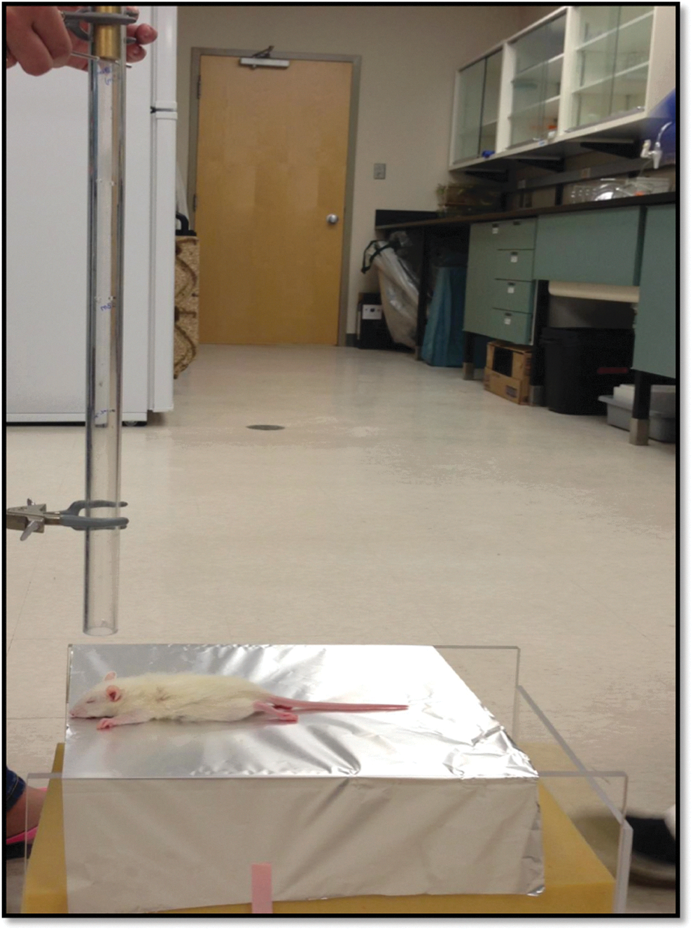
Apparatus used for the induction of the mild traumatic brain injury. The juvenile rat is placed chest down on the scored tin foil; the head is positioned directly below the falling weight. Color image is available at
Behavioral testing
For 16 days following the mTBI, all animals were subjected to a comprehensive behavioral test battery to examine balance and motor coordination, cognitive functioning, spatial learning, and emotionality. The behavioral test battery included beam walking, open field (locomotor activity), EPM, NCM, MWT, and the FS task.
Beam walking
Twenty-four hours post-mTBI (P31) animals were tested in a beam walking task similar to that described by Schallert et al. 14 The rat's home cage was placed at one end of the 165 cm long tapered beam, and the start point was the other wider end of the beam. The tapered beam had ledges (2 cm wide) that provided “safety” when the rat's foot slipped off of the beam. The rat was given a single trial to learn to walk from the start of the beam to its home cage. Once the rat reached its home cage, it was permitted to remain in the cage for 60 sec to reinforce the target location. The rat was given four additional videotaped trials, each separated by the 60 sec reinforcement. The video camera was set to record from the starting point of the beam, and positioned to view down the length of the beam. A researcher blinded to the experimental groups scored the videos for hind leg foot slips off the tapered beam onto the safety ledges.
EPM
One day post-mTBI (P31) animals were also tested in the EPM. The EPM was constructed from black Plexiglas® and consisted of two open arms and two closed arms that were elevated 55 cm above the ground. The maze was located in an empty room, and video recording occurred with the lights on. The video camera was placed at the end of one of the open arms in a slightly elevated position. Rats were placed in the center of the platform with their front paws facing a closed arm. Rats were videotaped for 10 min and scored by a research analyst blinded to the experimental conditions for the amount of time rats spent in the closed and open arms of the platform, as well as time spent in the center of the maze. The EPM was cleaned with Virkon after each rat completed the task.
Open field
Two days post-mTBI (P32) animals were tested in the open field task. Rats were individually placed into a 100×100×50 cm square and allowed to explore their environment for 10 min. An overhead tracking camera was used to track the rat's movement and measure distance travelled as well as time spent in the outer portion of the square and the inner portion of the square. The open field box was cleaned with Virkon after each rat completed the task.
NCM
From P35 to P38, animals were tested in the NCM task similar to that described by Spanswick and Sutherland. 15 Rats were exposed to two different contexts for 5 min each day, one immediately after the other, for 3 consecutive days. Context A was a clear plastic rectangular box (70×40×33 cm) and context B was a dark circular bin (diameter 47 cm×36 cm high). For the 3 days of context learning, each context housed a unique pair of identical objects (context A – two Rubik's cubes, context B – two 50 mL conical tubes), securely attached to the floor of the context, which the rats explored for 5 min. On the probe day, rats were permitted to explore each of the normal contexts, one after another for 5 min. After a 5 min delay, the rats were videotaped while they were exposed to a modified context (context A with one object from context A and one from context B; or context B with one object from context B and one object from context A) for 5 min. A research analyst blinded to the experimental conditions scored the amount of time the rat spent with the old object and the amount of time spent with the new/novel object.
MWT
Animals were trained in the MWT from P40 to P43, and tested on P44 in a protocol similar to that described elsewhere. 16 The water task was conducted in a circular pool (165 cm diameter and 0.5 m deep) that was filled with water (∼20°C). A clear Plexiglas® platform was placed in the middle of a specified quadrant. The water level was adjusted so that the platform was ∼2 cm below the surface of the water and not visible to the rat. The rat was given eight training trials per day over 4 days of learning. A trial consisted of placing the rat in the water at the perimeter of the pool from one of four locations (north, south, east or west) and allowing it 60 sec to find the platform. If the rat found the platform, it was permitted to remain on the platform for 10 sec to visualize the existing spatial cues. If the rat failed to find the platform in the 60 sec, it was placed on the platform by the experimenter and given 10 sec to visualize the spatial cues. The order of the trials (north, south, east, west) varied from day to day to prevent the rats from developing a swimming pattern. On the 5th day (the probe trial), the platform was removed from the pool and rats were placed in the pool from the south location and permitted to swim for 60 sec. The amount of time the rat spent in each quadrant during this trial was recorded.
FS
On P45 animals were tested in a modified FS task similar to that described by Yadid et al. 17 A cylindrical tank (30 cm diameter ×60 cm deep) was filled with warm water (∼25°C) to a level such that the rat's tail could not touch the bottom of the tank. The rat was placed in the tank and videotaped for 7 min. Upon completion of the task, the rat was dried with a warm towel and returned to its home cage. The tank was emptied and refilled with clean warm (∼25°C) water between each test session. A research analyst blinded to the experimental conditions scored the videos for the amount of time each rat spent immobile (not swimming, just floating).
Statistical Analysis.
As described, all scoring of behavioral measures was completed by a research analyst blinded to the experimental conditions. Two way ANOVAs with sex (male and female) and injury (mTBI and sham) as factors were run for each of the behavioral outcomes measured and p values<0.05 were considered statistically significant. SPSS 19.0 for Mac was used to complete all analyses.
Results
Animal characteristics
Although both groups (mTBI and sham) were lightly anesthetized, there was a significant difference in the time it took the animals to right themselves following the injury; both male and female rats that experienced the single mTBI took significantly longer to right themselves when compared to the sham injury group (Fig. 2). The two way ANOVA revealed a main effect of injury – F(1, 30)=36.65, p<0.01, and of sex, F(1, 30)=4.45, p=0.04 – but the interaction was not significant; F(1, 30)=3.58, p=0.07 (see Fig. 3).

Illustrative representation of the average time rats took to right themselves from a supine position following the mild traumatic brain injury (mTBI) or sham injury; animals with an mTBI took significantly longer to right themselves (*p<0.01).
There were no significant within-sex differences in body weight for mTBI and sham animals at the time of injury or the time of euthanasia. There was, however, a significant difference in body weights between male and female rats at the time of euthanasia. A two way ANOVA demonstrated a main effect of sex – F(1,30)=397.73, p<0.01, but not of injury, F(1, 30)=2.00, p=0.17 – and the interaction was not significant, F(1, 30)=3.55, p=0.07.
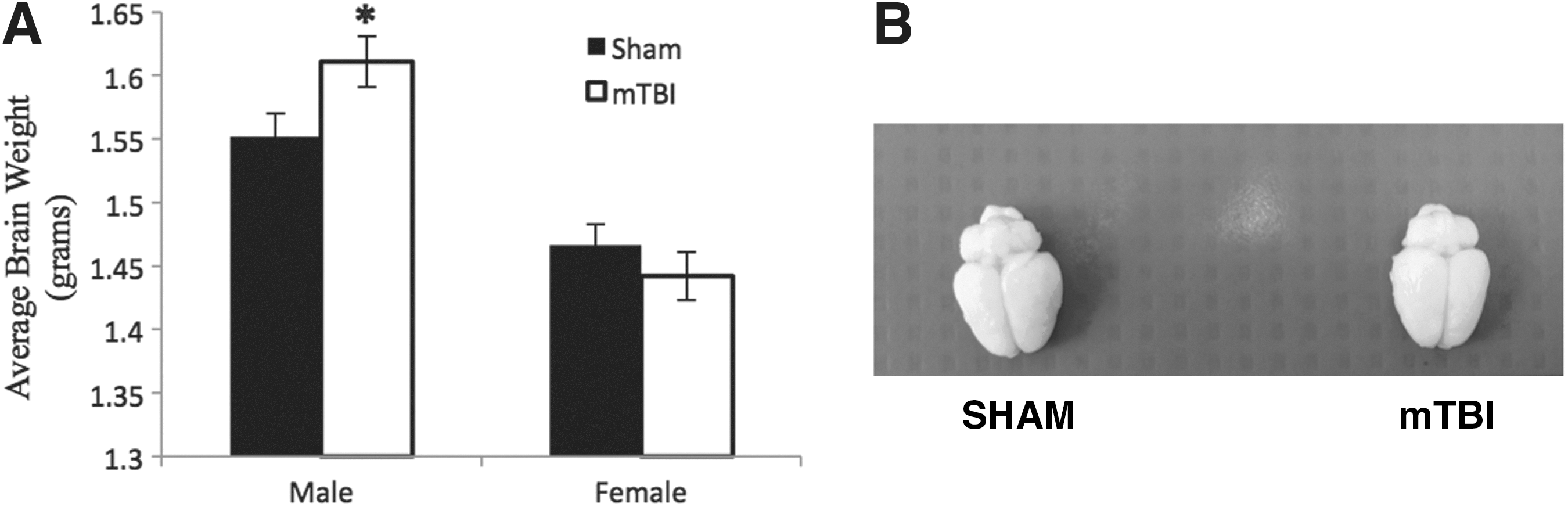
Male rats that experienced an mTBI also had significantly heavier brains than male rats in the sham group at the time of euthanasia, but there was no group difference in brain weights between female rats. The two way ANOVA demonstrated a main effect of sex – F(1, 30)=46.99, p<0.01, but not of injury, F(1, 30)=0.88, p=0.36; however, the interaction was significant, F(1, 30)=4.96, p=0.04 (see Fig. 3). Examination of the brain tissue at the time of extraction revealed no hemorrhaging or structural damage, and there was no overt damage that was distinguishable between the mTBI and sham animals (see Fig. 2).
Behavioral testing
Beam walking
Both male and female rats that experienced an mTBI displayed significant deficits in the beam walking test as measured by increased hind leg foot slips onto the safety ledge. The two way ANOVA revealed a main effect of injury – F(1, 30)=34.20, p<0.01, but not of sex, F(1, 30)=0.13, p=0.72. The interaction was also not significant (p=0.72; see Fig. 4).
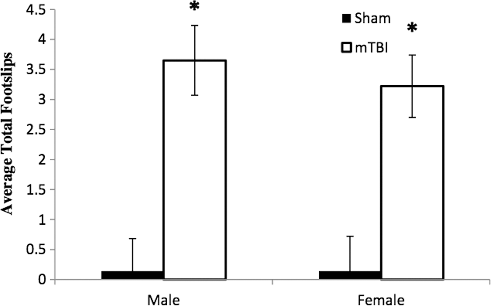
Graphic representation of the increase in average total number of hind leg foot slips made by both male and female rats with mild traumatic brain injury (mTBI) when compared with the sham control group 24 h post-injury (*p<0.01).
EPM
The behavior of male rats that experienced an mTBI differed from that of the sham group and the female rats. Although male mTBI rats spent less time in the closed arms than did their sham comparison group, they did not spend more time in the EPM open arms. Male mTBI rats spent significantly more time in the center of the platform (see Fig. 5). The two way ANOVA for time spent in the center of the platform demonstrated a main effect of injury – F(1, 30)=5.44, p=0.03 – and a significant interaction – F(1, 30)=5.63, p=0.02 – but no effect of sex – F(1, 30)=2.35, p=0.14.

Graphic representation of the average time rats spent in the closed arms and center of the elevated plus maze (EPM). Male rats with mild traumatic brain injury (mTBI) spent less time in the closed arms of the EPM than did the male sham injury group or the either of the female groups, which was representative of the significant increase in the time spent in the center, not the open arms of the EPM (*p<0.05).
Open field
When tested in the open field box, both male and female rats that experienced an mTBI showed significant reductions in their overall activity levels – F(1, 30)=9.87, p<0.01, with no sex effect or significant interaction ( p's>0.05). In addition, male mTBI rats also spent significantly more time in the center of the open field box than did the sham comparison group (see Fig. 6). The two way ANOVA for percentage of time in the outer portion of the box demonstrated a main effect of injury – F(1, 30)=7.61, p=0.01; a main effect of sex – F(1, 30)=7.73, p=0.01; and a significant interaction – F(1, 30)=6.56, p=0.02.
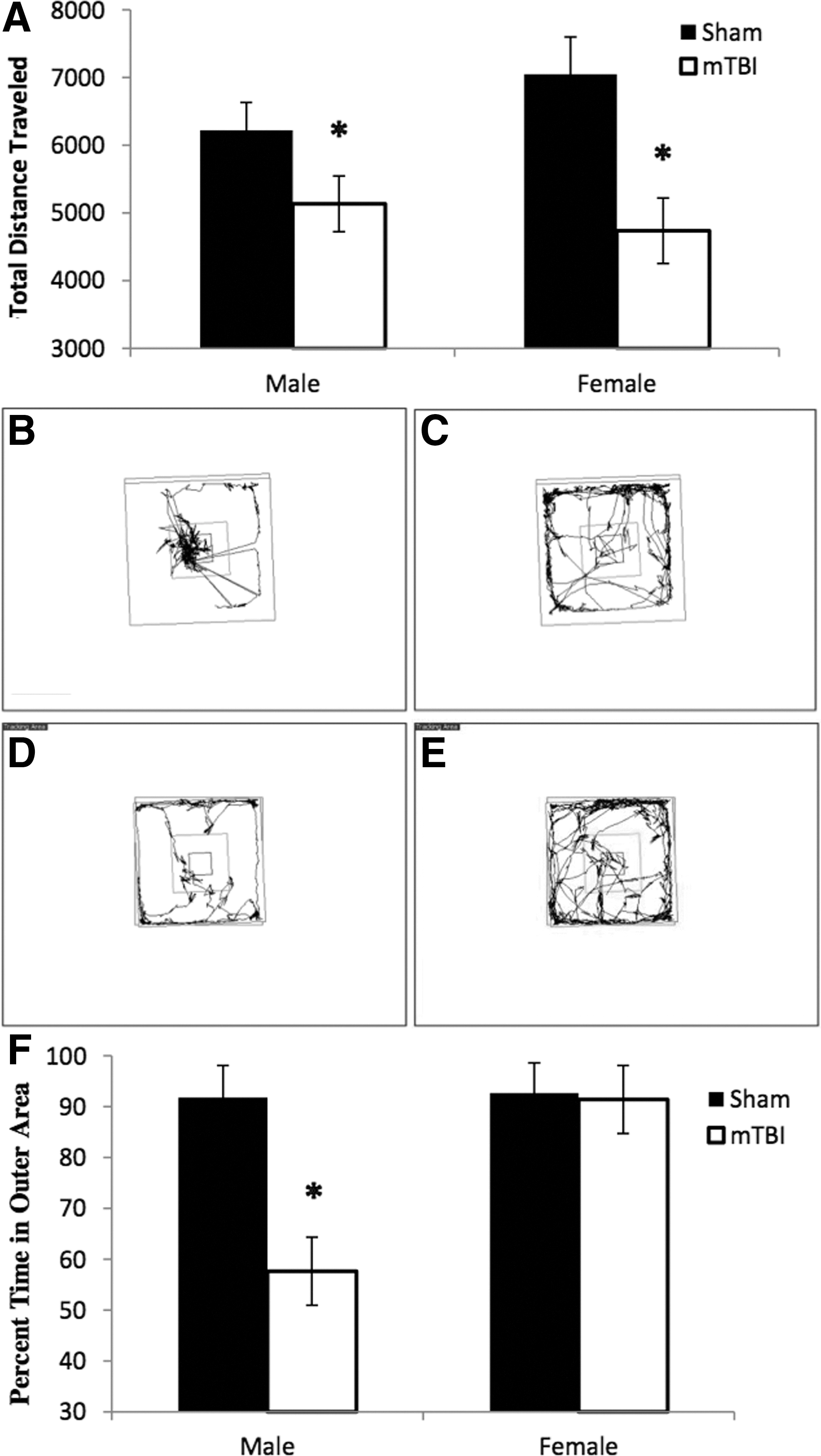
Illustrative representation of average behavior in the open field test for juvenile rats (P31) with or without a mild traumatic brain injury (mTBI).
NCM
Animals that experienced an mTBI spent significantly less time with the novel object on the probe trial in the NCM paradigm than did animals with a sham injury (as illustrated in Fig. 7). The two way ANOVA demonstrated a main effect of injury – F(1, 30)=5.96, p=0.02; but not of sex – F(1, 30)=0.71, p=0.41; and there was no significant interaction – F(1, 30)=0.09, p=0.76.
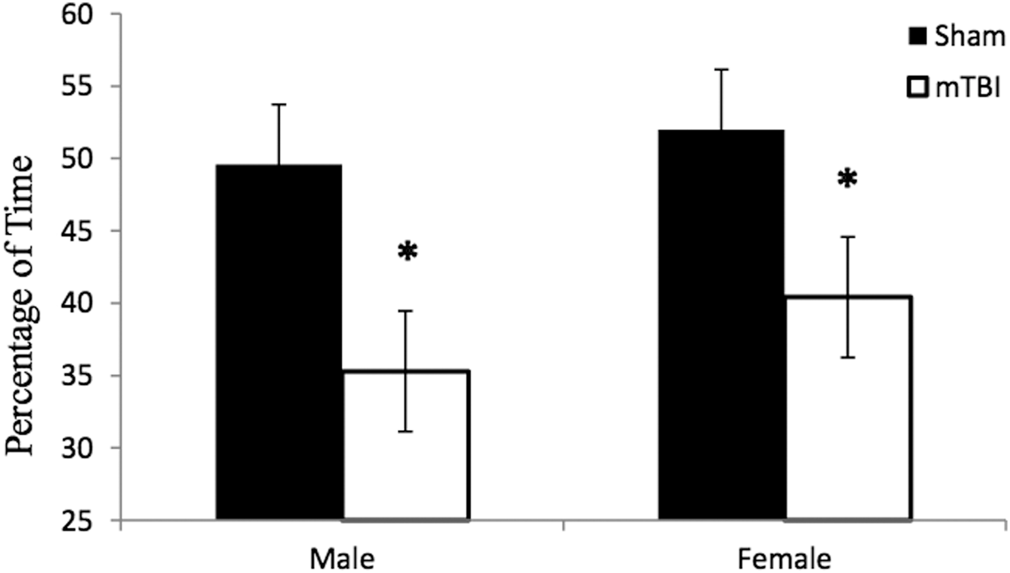
Illustrative representation of the decreased time rats with a mild traumatic brain injury ( mTBI) spent with the novel object during the 5 min probe trial of the novel context mismatch task (*p<0.05).
MWT
The mTBI did not interfere with spatial learning, as animals with the injury learned the location of the platform at the same rate as the sham animals. The repeated measures ANOVA with sex and injury as factors and trial days as the latency factor revealed no main effect of injury – F(3, 27)=1.61, p=0.21; or of sex – F(1, 27)=0.86, p=0.47. The interaction was also not significant (p>0.05). However, in the probe trial, both male and female rats that experienced a brain injury were unable to switch their problem-solving strategy, and spent significantly more time in the quadrant that had previously held the platform. The two way ANOVA demonstrated a main effect of injury – F(1, 30)=8.06, p=0.01; but not of sex – F(1, 30)=0.05, p=0.82; nor was there a significant interaction – F(1, 30)=2.04, p=0.17 (see Fig. 8).
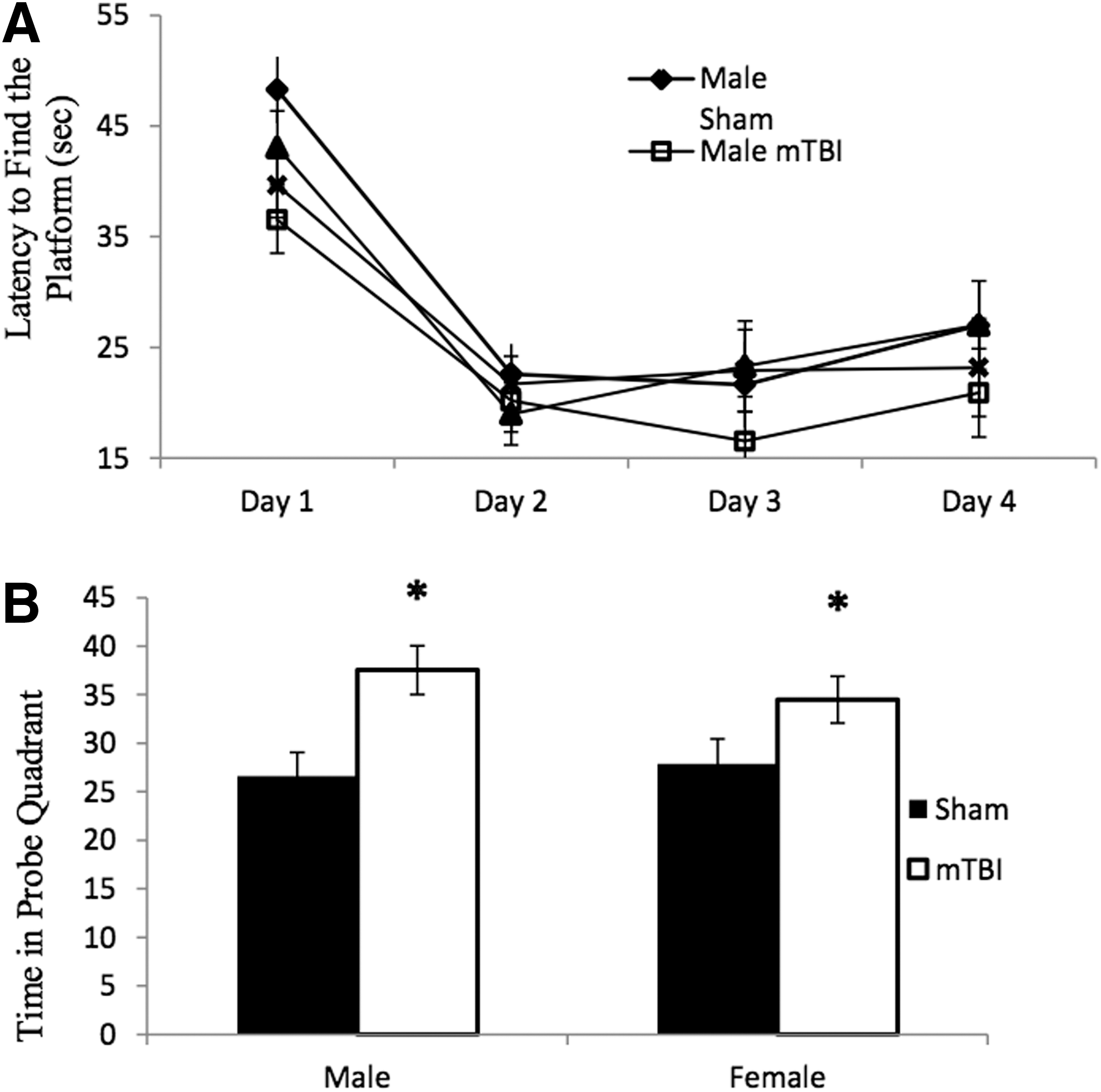
Graphic representation of the rat's performance on the Morris water task (MWT).
FS
Both male and female animals that experienced an mTBI demonstrated greater immobility than did the rats in the sham comparison group (Fig. 9). The two way ANOVA for percentage of time immobile exhibited a main effect of injury – F(1, 30)=5.70, p=0.03 – but not of sex – F (1, 30)=0.42, p=0.53. The interaction was not significant (p=0.90).
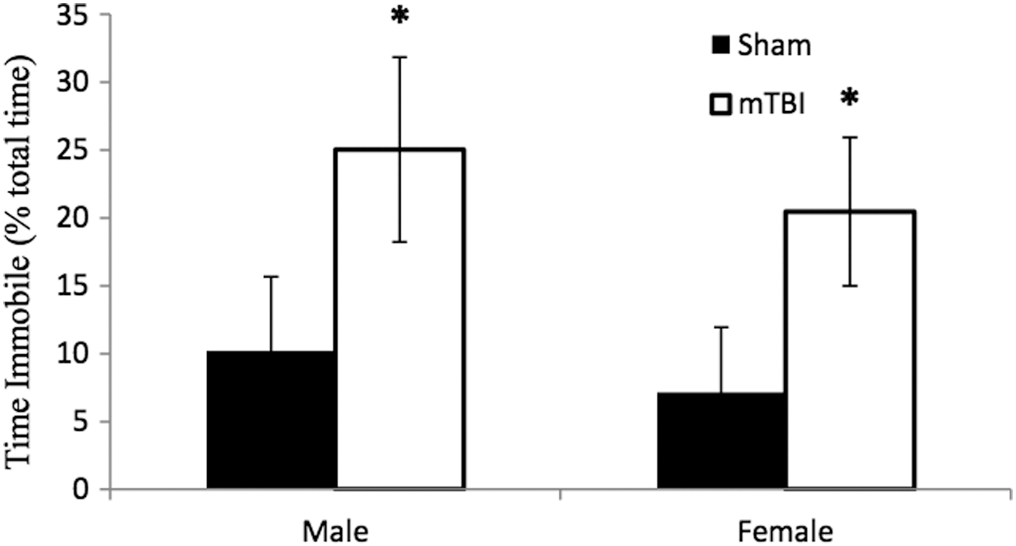
Representative results demonstrating that rats with a mild traumatic brain injury (mTBI) spent a greater percentage of time in the force swim task immobile than did the sham injury group (*p<0.05).
Discussion
The validity of basic science research focused on translational outcomes is highly dependent on the modelling system implemented. For example, despite decades of research and an overwhelming need, there has been little advancement in therapeutic strategies that reduce or reverse the negative outcomes associated with mTBI or concussion. 1 This lack of translational innovation likely arises from a discrepancy between the mechanisms and pathology between actual and model-induced trauma. To prevent a complete cessation of discovery in this particular field, novel models of mTBI need to be developed and validated. The definition of mTBI in humans specifies that the brain injury results from blunt trauma as well as acceleration and deceleration forces, with one or more of the following symptoms: 1) self-reported transient confusion or disorientation, 2) memory impairment near the time of the injury, 3) observed signs of neurological or neuropsychological dysfunction, or 4) any period of observed or self-reported loss of consciousness (LOC). 18 In this study, we used a modified weight-drop technique to produce a closed head injury that involved acceleration/deceleration forces induced by a single transient impact to the head, but one that did not cause any overt cranial or cerebral injury. The injury itself caused what appeared to be a brief LOC, but not any significant morbidity or mortality. Furthermore, we demonstrated behavioral symptoms that were representative of the neurological and neuropsychological dysfunction reported by persons with an mTBI.
The modified weight-drop model employed in this study induced a glancing impact to an intact skull, and because the platform offered minimal resistance, the brain was subsequently exposed to rapid acceleration/deceleration forces that resulted from movement of the head. 6 Critical to the study of mTBI, and in congruence with clinical populations, 13 the modified weight-drop technique induced heterogeneous outcomes from a single injury experience. Although there were significant behavioral differences between juvenile rats that experienced an mTBI and those that received a sham injury, there was also substantial variation within the mTBI group for the measured outcomes. Based on the focus of this study, it was not possible to decipher the reason underlying these within-group differences, but it is a clinically relevant and noteworthy finding, as significant heterogeneity exists between patient outcomes, despite similar injuries. 19 Instead, the results from this study will be discussed within the context of induced symptomology (e.g., affect, cognition, executive function) and model validation for pediatric mild to moderate TBI.
Of the four traits that define the presence of a concussion, LOC is one of the most difficult symptoms to measure in a rodent. Although time to wake and right following the injury is not a perfect measure of LOC, the significantly increased time period for righting between the sham and the mTBI groups suggests that there are injury-related differences in responsiveness. It was difficult to ascertain if the increased time to wake resulted from an interaction between the anesthetic and the injury, or from the injury alone. Although the juvenile rats that experienced the mTBI appeared “stunned” for a short period of time following the injury, the injury did not interfere with basic skills, and the rats resumed normal grooming and eating patterns within 1 h. Furthermore, because the incidence of LOC is not necessary for classification of a concussion, 12,18 and the occurrence of LOC has not been associated with neurological outcome, 20 the inability to prove LOC in this model does not negate its value.
In addition to LOC, individuals experiencing an mTBI who have PCS often report balance and motor disturbances. 13 Although balance and motor disturbances often exhibit timely recovery, 21 their initial presence has been identified as a reliable indicator of mTBI or concussion. 12 In this study, when tested 24 h post-mTBI, significant deficits were found in balance and motor control as measured by the number of hind leg foot slips on a tapered beam. Rats that had experienced an mTBI were significantly more likely to slip off of the tapered beam and catch themselves on the safety ledge than were their sham injury counterparts. In human populations, whether analyzed with sophisticated technology or rudimentary tests, deficits in postural stability have been identified up to 72 h post-concussion. 12 However, this study is limited because beam walking was only measured 24 h post-mTBI. As such, ongoing studies will measure the temporal persistence of this motor/balance deficit to determine if it exhibits the same recovery time that has been identified in human cohorts, and conversely, whether its persistence occurs in combination with other PCS symptoms.
Along with the assessment of balance and motor deficits, impairments in neuropsychological function and affect were measured using three distinct behavioral tests: EPM, open field, and FS. PCS in particular is associated with increased prevalence of depression- and anxiety-related disorders. 13 Results from this study indicate that following an mTBI, both male and female juvenile rats displayed depressive-like behaviors, as suggested by increased immobility in the FS task. Interestingly, although male and female rats exhibited abnormal behaviors in the open field and EPM, there was a sex-dependent effect in the phenotypic manifestations. Whereas both male and female juvenile rats displayed reduced locomotor activity in the open field 48 h after an mTBI, the male rats spent a substantial amount of time in the center of the enclosure, suggesting a decrease in general anxiety usually expressed by animals placed in open areas. Consistent with this result, when tested in the EPM 24 h after induction of the injury, male rats spent less time in the closed arms of the EPM than did the sham controls and female mTBI groups. Although this behavior is often attributed to decreased anxiety, and is associated with increased time in the open arms of the maze, this did not appear to be the case in the present study. Rather, male rats with an mTBI spent more time in the center of the EPM, not venturing into either arm. Although in conjunction with the open field data, it is tempting to conclude that this also demonstrates a reduction in anxiety, reviews of the EPM literature do not provide conclusive explanations for when the animal spends the majority of their time in the center of the platform, 22,23 making it difficult to interpret this result. It did not, however, appear that the mTBI animals were overtly stressed as compared with other animals, or that they “froze” in the center, as they would often venture into the openings of the closed arms.
In comparing the above-described findings from this study to results published in previous research using rodent models of mTBI, it is difficult to reach a consensus from the current literature on either of the neuropsychological traits. Although two prior studies found that mTBI (induced with fluid percussion or conventional weight drop) increased anxiety in the EPM, 24,25 a third study of mTBI using a conventional weight drop found no change in measures of anxiety between groups. 26 Similarly, two studies of mTBI in mice demonstrated increased immobility in the FS task, an indicator of depression, 26,27 but a third study of mTBI in rats failed to replicate the findings. 24 Because of the complexity of brain injury, the dynamic nature of neuropsychological function, and the multitude of variables that differ among studies (i.e., sex, age, strain, and species of the rodent; mode of injury induction; time lapse between injury and testing) it is perhaps not surprising that results were so variable. Even within our experimental groups, we found significant variation in the magnitude of the behavioral measures we investigated, suggesting innate differences in susceptibility or resilience among individual animals. Overall, however, findings from this study of increased depressive-like behaviors, decreased locomotor activity, and altered neuropsychological functioning, are important for the validation of this particular model of mTBI/concussion in juvenile rats.
As memory impairments are one indicator of a concussion, and PCS is associated with memory problems and deficits of executive function, the rats were subjected to the MWT, and the NCM paradigm. The MWT is a very effective spatial memory task that requires that the rat learn the location of a hidden platform with respect to the location of visual cues. 28,29 In this experiment, animals that received a mTBI did not show an impaired ability to learn the location of the hidden platform, therefore demonstrating normal spatial learning and cognitive ability. Rats with an mTBI were also indistinguishable from sham injury rats on measurements of swim speed and path length, indicating that they did not have motor deficits that may have impacted their ability to complete the task. As overt damage to the hippocampus is generally associated with impaired functioning on this particular spatial memory task, 30 the absence of impairment suggests that there was no extensive hippocampal damage in this model.
These results are consistent with the human literature demonstrating minimal: 1) hippocampal structural differences in individuals with and without concussions as measured by MRI, 31 2) changes in H-MR spectroscopy of neurometabolic function, 31 or 3) changes in functional MRI (fMRI) blood oxygen level dependent (BOLD) signalling during working memory tasks. 32 Although the human literature is consistent with the findings from this study, prior research with rodent models of mTBI has identified deficits in the acquisition of spatial memories. 33 –35 The discrepancy between other animal studies in the literature and our results likely arises from the differences in injury model and, therefore, injury pathophysiology. The model used in this study employed acceleration/deceleration forces and a glancing impact to an intact skull. In contrast, the majority of previous studies involve models in which the skull is fixed, the brain is exposed through a craniotomy, and a device is use to induce a controlled focal injury. Therefore, the modes of injury between ours and the other studies are quite different, and it seems reasonable to presume that the outcomes would also differ.
Another intriguing finding from our assessment with the MWT was that when the rats learned the location of the platform and the platform was removed on the probe trial, rats with an mTBI remained in the learned quadrant, failing to engage new strategies to search for the platform. In contrast, control rats explored the quadrant that had previously housed the platform, but when it could not be located, typically moved into other areas of the pool. This finding suggests a form of perseveration that can often be seen following brain injury and frontal lobe/executive function impairment. 36 Similarly, in the NCM task, rats with mTBIs displayed deficits of executive function exhibited by poor working memory and increased time spent with the old rather than the new objects; possibly perseveration. Many human studies of concussion symptomology have identified persistent impairments in executive function, with specific deficits in task switching and short-term memory. 37 –40 Consistent with the behavior exhibited by the rats with an mTBI, individuals with a history of concussion are reported to have deficits of short-term memory and difficulty ignoring irrelevant stimuli, and are less effective at altering their responses in a context-dependent manner. 37,39,41,42 Importantly, we found persistence of these symptoms in animals following mTBI; present at 8 (NCM) and 15 (MWT) days post-mTBI. Although human and rat time scales differ significantly, and there are several different estimation techniques based on different physiological variables. One technique for estimating age based on neurodevelopment 43,44 would suggest that 2 weeks of a prepubescent rodent's life is roughly equivalent to 3 human years. Based on this species conversion technique, our finding of prolonged symptomatology could be in keeping with the human phenomena of PCS, and our findings of prolonged memory impairment and neurological dysfunction following mTBI provide further support for the translational validity of this model.
Conclusion
In summary, this study found that when applied to young rats, the modified weight-drop technique impaired executive functioning to a greater extent than cognition, altered balance and motor behaviors, and produced sex differences in affect whereby males were more impaired than females. It is particularly interesting that male mTBI rats had impaired performance in the open field and EPM and also had increased brain weight. Although largely conjecture at this point, it is possible that rather than being less anxious, the male rats had a form of malignant cerebral edema and were encephalopathic, thereby impairing their judgment of the environmental conditions. To further explore this question, however, ongoing studies in our laboratory are examining the anatomical and molecular pathophysiology of the brain following induction of our mTBI paradigm. This includes a quantitative analysis of cerebral edema using the brain weight technique ([wet-dry]/dry brain weight), along with detailed animal MRI studies of water diffusivity in the brain and analysis of the differential expression of molecular targets such as aquaporin-4 (AQP4). The AQP4 hypothesis is particularly intriguing, as previous studies have demonstrated an association between altered expression of AQP4 and cerebral edema in a different TBI model, 45 and AQP4 knockout animals have decreased cerebral edema in several different models of central nervous system (CNS) injury. 46 In addition, it has recently been shown that small interfering RNA targeted toward AQP4 improved behavioral outcomes and reduced cerebral edema in a juvenile TBI model. 47 However, the physiological role of AQP4 is complex, and there are several conflicting studies on its relationship to CNS pathology. 48 Therefore, additional studies are warranted to confirm the presence of cerebral edema in this model of mTBI, and ascertain the potential role of AQP4 in injury etiology.
Finally, based on our current findings and in conjunction with the study conducted by Kane and colleagues, 6 we feel that the modified weight drop technique is a reasonable model of concussion-like mTBI that overcomes many of the limitations of other rodent models of TBI, including the need for multiple hits. Unlike previous studies, 6,49 –51 the model employed here was able to induce concussive-like symptomology following a single impact in juvenile rats. Moreover, a majority of existing literature has focused on adult outcomes associated with TBI; this model allows us to examine the effects of mTBI/concussion on the pediatric brain. Further characterization of this rodent model may aid in the advancement of effective treatment strategies for mTBI and concussion.
Footnotes
Acknowledgments
The authors thank Irene Ma, Rose Tobias, and Dr. Jong Rho for their technical and organizational assistance. Funding for this study was provided to Dr. Esser by the Department of Pediatrics at the University of Calgary, the Alberta Children's Hospital Foundation (ACHF), and the Alberta Children's Hospital Research Institute (ACHRI). Dr. Mychasiuk's postdoctoral fellowship was funded by ACHF.
Author Disclosure Statement
No competing financial interests exist.