




Abstract
Sports-related concussion is a major public health issue; however, little is known about the underlying changes in functional brain networks in adolescents following injury. Our aim was to use the tools from graph theory to evaluate the changes in brain network properties following concussion in adolescent athletes. We recorded resting state electroencephalography (EEG) in 33 healthy adolescent athletes and 9 adolescent athletes with a clinical diagnosis of subacute concussion. Graph theory analysis was applied to these data to evaluate changes in brain networks. Global and local metrics of the structural properties of the graph were calculated for each group and correlated with Immediate Post-Concussion Assessment and Cognitive Testing (ImPACT) scores. Brain networks of both groups showed small-world topology with no statistically significant differences in the global metrics; however, significant differences were found in the local metrics. Specifically, in the concussed group, we noted: 1) increased values of betweenness and degree in frontal electrode sites corresponding to the (R) dorsolateral prefrontal cortex and the (R) inferior frontal gyrus and 2) decreased values of degree in the region corresponding to the (R) frontopolar prefrontal cortex. In addition, there was significant negative correlation between degree and hub value, with total symptom score at the electrode site corresponding to the (R) prefrontal cortex. This preliminary report in adolescent athletes shows for the first time that resting-state EEG combined with graph theoretical analysis may provide an objective method of evaluating changes in brain networks following concussion. This approach may be useful in identifying individuals at risk for future injury.
Introduction
S
Approaches such as diffusion tensor imaging (DTI) are showing promise in being able to detect changes in the microstructural properties of the white matter of the brain 9 –12 following concussion. Several recent studies show that critical white matter regions in the frontal association and commissural pathways, such as the anterior corona radiata and the corpus callosum, are highly susceptible to microstructural changes. 11 –15 These tracts are involved in key cognitive functions, such as executive function, attention, memory, and learning.
Although increasing evidence shows diffuse axonal injury after concussion, little is known about the consequences of such injury to functional brain networks, particularly in the adolescent population. In the context of adult concussion, the resting state of the brain (i.e., in the absence of task performance or stimulation) is associated with decreased functional connectivity. Slobounov and colleagues 16 reported that interhemispheric connectivity was reduced in the primary visual cortex, hippocampus, and dorsolateral prefrontal cortex (DLPFC) networks in concussed adults who were clinically asymptomatic at the time of scanning. Tang and colleagues 17 reported that, in comparison to control subjects, adults with mild traumatic brain injury (mTBI) showed widely distributed functional connectivity between thalamic and cortical regions during the resting state. This pattern of connectivity was correlated with decreased neurocognitive function. Reduced functional connectivity has also been reported in the posterior cingulate and parietal cortices during resting state in concussed athletes who were clinically asymptomatic. 18 In addition, reduced connections between the left DLPFC and the left parietal cortex were found in athletes who had multiple concussions, but were also clinically asymptomatic.
In contrast, task-related functional networks are associated with increased functional activations within bilateral DLPFC and in parietal areas in adults 19 and decreased activation in the same regions in adolescents. 20,21 Though both resting state and task based activations have revealed important functional changes in the underlying brain networks, little is known about the relationships and network dynamics within these networks.
Recently, mathematical models derived from graph theory have been used to analyze brain network organization. 22 Graph theory characterizes the brain as a set of networks. Each network is made up of distinct brain regions called nodes and edges that delineate pathways connecting these regions and nodes. The relationship between nodes and edges provides quantitative information about the organization and efficiency of the network and characterizes the network according to both global (whole brain) and local (specific brain regions) attributes. The healthy human brain generally functions as an efficient “small-world” network, characterized by connections between nodes that allow for both local specialization and global integration. 23 In contrast, a “random” network is characterized by connections that promote global integration, but not local specialization, and an “organized” network promotes local specialization, but not global integration. Using this approach, the topological organization and structure of brain networks have been successfully investigated in brain development, aging, and brain disease. 24 –26 Graph analysis of functional brain networks in adult concussion/mTBI suggests a shift toward suboptimal network organization 27 –29 ; however, such analysis has not yet been done with an adolescent population.
In this preliminary study, we asked: 1) Are there differences in the global and/or local connectivity measures of resting-state electroencephalography (EEG) networks following concussion in adolescent athletes?, and 2) Are connectivity measures correlated with scores on a widely used concussion assessment tool (Immediate Post-Concussion Assessment and Cognitive Testing (ImPACT).
Methods
Participants
Nine adolescent athletes (all male; mean age, 16.5 years) with a clinical diagnosis of subacute (≤3 months previously) sports-related concussion and 33 healthy, adolescent soccer players (all male; mean age, 16 years) participated in this study. Control athletes were recruited from the Whitecaps FC Residency soccer program in Burnaby, British Columbia, Canada. Concussed athletes were recruited from the same soccer program and through minor league ice hockey teams in Vancouver. Exclusion criteria for all subjects included focal neurologic deficits, pathology, and/or those on prescription medications for neurological or psychiatric conditions. All participants were right-handed. Parents signed an informed consent form that was approved by the University of British Columbia, and all participants provided assent.
Immediate Post-Concussion Assessment Testing
ImPACT is a commercially available computerized assessment tool designed to measure different aspects of neurocognitive function in individuals suspected of having a concussion. It includes a 22-item PCS scale and the following composite scales: verbal memory, visual memory, visual motor speed, reaction time, and impulse control (a measure of errors on testing). Higher scores on the verbal memory and visual motor speed composite scales reflect better performance, whereas higher scores on the reaction time scale reflect slower or worse performance. A total symptom score is also reported. ImPACT includes a PCS questionnaire and collects information regarding various historical and demographic characteristics.
Electroencephalography recording protocol
EEG was recorded using a 64-channel Hydrogel Geodesic SensorNets (EGI, Eugene, OR). EEG was recorded with a Net Amps 300 amplifier at a sampling rate of 250 Hz. Scalp electrode impedances were usually less than 50 kΩ. Five minutes of resting data were collected while participants had their eyes closed. The signal was subsequently filtered from 4 to 40 Hz. 24 EEG signals were interpolated at 27 locations on the scalp using BESA's Virtual Standard 10-10 Average montage.
Brain connectivity network modeling
The Brain Connectivity Toolbox 30 running MATLAB (The MathWorks, Inc., Natick, MA) was used to perform the graph theoretical analysis and a network learning model described in Li and Wang. 31 The network structure learning algorithm is based on a Bayesian network learning algorithm. A Bayesian network is a probabilistic graphical model, representing regions of interests as nodes and the stochastic interactions between regions of interests as edges. Brain connectivity networks were computed using the false discovery rate controlled PC (PCFDR) algorithm. 31 PCFDR is a computational efficient and reliable network learning method that is based on the conditional dependence/independence testing. It estimates the existence of connections by testing the conditional independence of each pair of regions conditional on all other regions. This method is proven to asymptotically control the false discovery rate (FDR) under predefined levels.
In network learning, FDR is defined as the expected ratio of falsely detected connections to all those detected. Compared with the traditional type 1 and 2 error rates, FDR has more reasonable error rate criteria in brain connectivity modeling given that it is directly related with the uncertainty of the learned networks. The details and pseudocode of the PCFDR algorithm can be found in Li and Wang. 31 In this study, the FDR threshold was set at 5%. The binary undirected connectivity networks were computed for each individual subject and each condition independently.
Graph theoretical analysis
Based on the learned connectivity networks, graph theoretical analysis has been used to extract the structural features from learned networks. 22 Here, traditional graph theoretical measures were used to characterize the network features in terms of density, global efficiency, clustering coefficient, and modularity. Density is defined as the fraction of present connections to all possible connections. Global efficiency describes the communication ability of the entire graph, 32 defined as the average of the inverse shortest path. Clustering coefficient describes the degree to which nodes in a graph tend to cluster together. Modularity of the network is used to measure how well the network can be divided into the submodules. 33 A higher value of modularity demonstrates that the graph is better divided with tighter connections within modules.
In addition to looking at overall network features, traditional graph theoretical measures were used to characterize the local nodal features, with particular interest in degree and betweenness centrality. 34 Local metrics are designed to measure the structural properties of each node, such as the degree, efficiency, and betweenness centrality. 35 The degree is the number of all the links for a specified node. The efficiency is the mean of the inverse of the minimum path length between a particular node and all other nodes in the network. The betweenness centrality of a node is the number of the shortest paths between any two nodes. 36 The hub/authority measure was first proposed by Kleinberg to identify important pages in the World Wide Web. 37 The hub/authority value of one node is related to the structure of its neighbors, where a high value means that more important nodes are connected with it.
Results
Immediate Post-Concussion Assessment Testing scores
Table 1 lists the demographics and scores on individual sections of the ImPACT. There were no statistically significant differences in visual memory, visual-motor, reaction time, or impulse control scores between the two groups. Both total symptom score and verbal memory score were significantly higher for the concussed group.
SD, standard deviation.
Network topology
Figure 1 shows the group-averaged plots of network topology of each group. The overall network functional connectivity for the two groups is similar. However, note that there are increased connections between regions, such as the frontal and temporal areas (e.g., F10–T8) in the control group that are not significant in the concussed group. In addition, there are connections in the concussed group that are not present in the control group (e.g., Fp1–F7).

Group-averaged plots of network topology in the control adolescents (
Global network properties
Graph theoretical analysis showed no significant differences for the global network properties of density, global efficiency, modularity, or clustering coefficient (Table 2). There was no change in the overall network organization following concussion.
Adjusted p values for multiple comparisons (Bonferroni's correction) are reported.
SD, standard deviation.
Local network properties
A number of local measures of network function showed significant differences between groups. The measure of betweenness was significantly higher for the F4 (p=0.05) and F10 nodes (p=0.02) in the concussed group (Fig. 2A,B). Betweenness measures the centrality importance of the node in the network. It is the ratio of the number of shortest paths from all vertices to all others that pass through that node. The higher value of betweenness means that the node plays a more important role in the network with regard to the flow of information. There was a significant increase in degree centrality for the F10 node (p=0.01) and a significant decrease in degree centrality for the Fpz node in the concussed group (p=0.01; Fig. 2C). In addition, node O2 showed a significant decrease in the hub value in the concussed group (p=0.033; Fig. 2D).
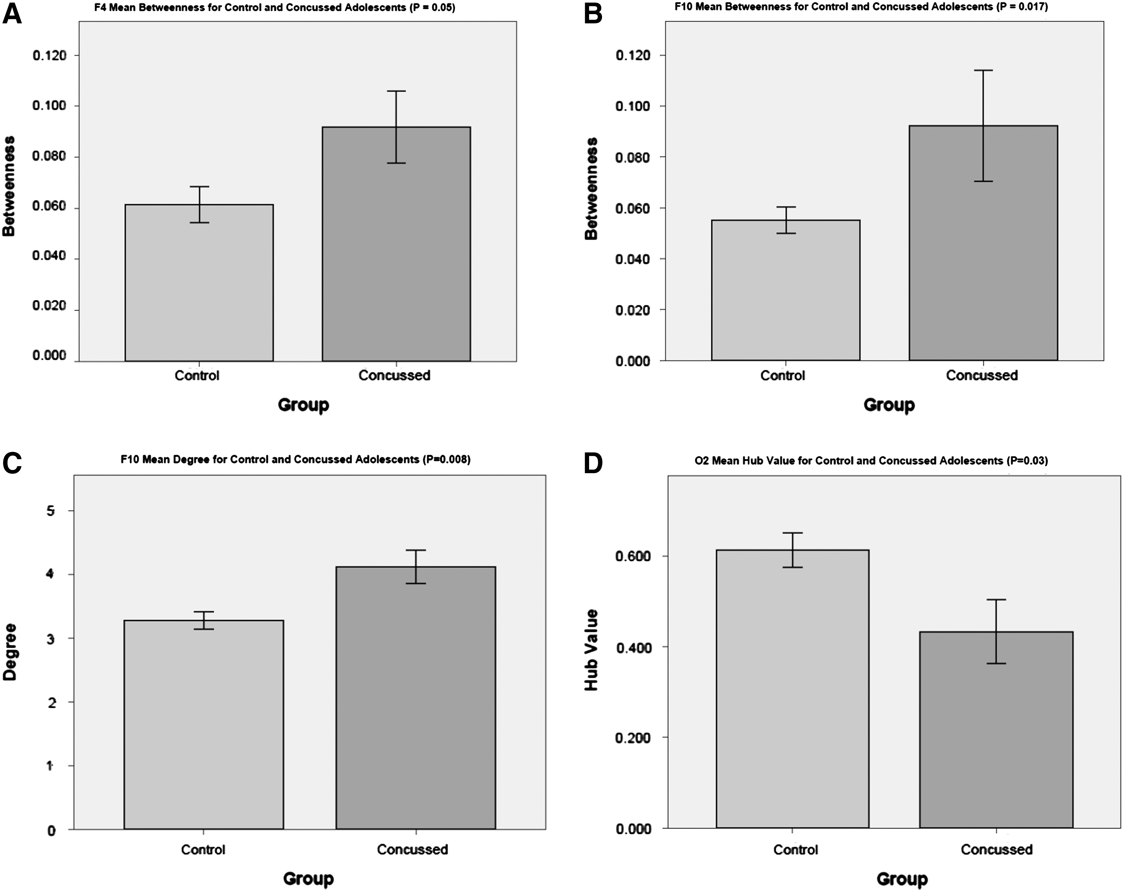
Bar graphs showing significant differences in local metrics between control and concussed adolescents. Error bars represent 1 standard error.
Correlation analyses
Multivariate linear regression was used to explore the relationships between the four global metrics and the scores on ImPACT. Additionally, the correlations between the node-wise centrality metrics were calculated for each subject/group. There was significant negative correlation between both degree (–0.525; p=0.009) and hub value/authority (–0.504; p=0.012) with total symptom score at the Fpz node (Fig. 3).

Correlations between degree and hub value of Fpz and total symptom score.
Discussion
In the present study, we set out to investigate differences in brain networks in a group of adolescent athletes with a sports-related concussion and healthy, active control adolescent athletes, using a graph theoretical approach. Our preliminary results show that concussion does not alter resting-state global network efficiency. However, we found significant changes in the local networks in the concussed group.
Following concussion, we observed local increases in both betweenness and degree in F4 and F10. These electrodes correspond to Brodmann area 838 or the (R) DLPFC and (R) inferior frontal gyrus. The (R) DLPFC is known to play a significant role in executive functions, such as working memory, cognitive flexibility, abstract reasoning, and attention. 39,40 The significant increase of betweenness and degree in this area suggests that the DLPFC becomes a key hub region following concussion, with increased connectivity with neighboring regions. Dettwiler and colleagues 19 recently reported increased activity in bilateral DLPFC during performance of an n-back task in varsity athletes up to 2 months postinjury, suggesting that altered activation in functional networks related to memory and attention may be a distinguishing feature of concussion that is present in both resting and task-based conditions. In addition, Palacios and colleagues 41 reported increased functional connectivity within the frontal lobe in patients with chronic TBI, suggesting that increased connectivity may be reflective of mechanisms compensating for the loss of structural connectivity. Caeyenberghs and colleagues 27 also recently reported increased connectivity in adult patients with TBI using graph theory analysis. They suggest that increased connectivity may in fact be “nonfunctional” and may reflect increased effort in recruiting the appropriate neural networks. It is possible therefore that the disruption we have found in the overall organization in the right frontal regions may reflect an ongoing dynamic process, reflecting both the response to initial injury as well as recovery mechanisms. Abnormally increased connectivity within top-down attention networks may reflect increased distractibility. It has been proposed that changes in attention network connectivity may be related to PCS, such as increased cognitive fatigue, headache, and increased distractibility.
We also observed a local decrease in degree for Fpz, which corresponds to Brodmann area 10 or the (R) frontopolar prefrontal cortex. It has been hypothesized that the frontal poles play a key role in executive function and act as a gateway to coordinate and evaluate information from stimulus-oriented and stimulus-independent thought. 42 Altered frontal polar activation may provide a plausible mechanistic underpinning for disrupted capacity to switch between the constructs of mindfulness and mind wandering following mTBI.
Node O2 also showed a significant decrease in the hub value in the concussed group. The node corresponds to the middle occipital gyrus 38 and is considered to be a critical area involved in visual-spatial attention. 43 These results are consistent with reports of deficits in spatial attention following concussion/mTBI. 44,45
Of particular interest is that the (R) frontopolar prefrontal cortex shows a significant negative correlation with the ImPACT symptom score, such that a decrease in connectivity is associated with a higher number of symptoms. Further investigation into the functional consequences of these disruptions is warranted to characterize the behavioral consequences of altered resting network connectivity after concussion.
This is a preliminary report with a small number of concussed adolescents. A larger study to confirm these results is clearly needed. In addition, given that the majority of changes were observed in brain regions related to attention and cognition, neurocognitive tests that focus on these higher-level functions may be more useful in identifying the nature of deficits following concussion.
In summary, resting-state EEG networks analyzed using graph theoretical approaches shows that concussion most likely affects the functioning of local networks subserving cognitive function and symptoms. Graph theoretical analysis provides a novel method of describing the impact of concussion on brain network organization. Our results provide evidence for local changes in the frontal and occipital regions of the brain, with the DLPFC taking on a more prominent role accompanied by a less prominent role of the frontopolar prefrontal cortex. These changes are likely to affect the efficient processing of cognitive functions following concussion.
Footnotes
Acknowledgments
This research was supported by the University of British Columbia and the Brain Research Center, University of British Columbia. The authors thank the Whitecaps Residency program for their support and all the participants, trainers, and coaches who took part in this study. Jenna Smith-Forrester is supported by a Canadian Institutes of Health Research (CIHR) Frederick Banting and Charles Best Canada Graduate Scholarship. Dr. Naznin Virji-Babul had full access to all the data in the study and takes responsibility for the integrity of the data and the accuracy of the data analysis.
Author Disclosure Statement
No competing financial interests exist.