
Abstract
Large-scale network analysis characterizes the brain as a complex network of nodes and edges to evaluate functional connectivity patterns. The utility of graph-based techniques has been demonstrated in an increasing number of resting-state functional MRI (rs-fMRI) studies in the normal and diseased brain. However, to our knowledge, graph theory has not been used to study the reorganization pattern of resting-state brain networks in patients with traumatic complete spinal cord injury (SCI). In the present analysis, we applied a graph-theoretical approach to explore changes to global brain network architecture as a result of SCI. Fifteen subjects with chronic (> 2 years) complete (American Spinal Injury Association [ASIA] A) cervical SCI and 15 neurologically intact controls were scanned using rs-fMRI. The data were preprocessed followed by parcellation of the brain into 116 regions of interest (ROI) or nodes. The average time series was extracted at each node, and correlation analysis was performed between every pair of nodes. A functional connectivity matrix for each subject was then generated. Subsequently, the matrices were averaged across groups, and network changes were evaluated between groups using the network-based statistic (NBS) method. Our results showed decreased connectivity in a subnetwork of the whole brain in SCI compared with control subjects. Upon further examination, increased connectivity was observed in a subnetwork of the sensorimotor cortex and cerebellum network in SCI. In conclusion, our findings emphasize the applicability of NBS to study functional connectivity architecture in diseased brain states. Further, we show reorganization of large-scale resting-state brain networks in traumatic SCI, with potential prognostic and therapeutic implications.
Introduction
T
More recently, the utility of graph theory, which models the brain as a network comprising nodes and edges, has been demonstrated in the assessment of normal and diseased populations. 8 The analysis involves mass-univariate testing to check for temporal correlation between each pair of nodes. The resulting large number of multiple comparisons inherent in this approach requires correcting for familywise error (FWE) rate. The present article applies a statistical approach called network-based statistic (NBS) to control for FWE to evaluate changes to large-scale brain networks in SCI patients based on the premise of providing a gain in statistical power. 9
Based on prior animal and human rs-fMRI studies, we proposed changes in the resting-state functional connectivity architecture in patients with traumatic SCI compared with intact controls, using NBS. 8,10 –16
Methods
Fifteen subjects with complete cervical SCI (all males; age, 45.1 ± 15.1 years) and 15 neurologically intact controls (12 males, 3 females; age, 41.9 ± 19.0 years) were scanned at the Center for Imaging Research (CIR), Medical College of Wisconsin, Milwaukee, Wisconsin (Table 1). The procedures followed for enrolling and scanning subjects were subject to approval by the Institutional Review Boards of the Medical College of Wisconsin and the Veterans' Administration health system, including signing written informed consent forms. 17
MVA, motor vehicle accident; MCC, motorcycle crash.
The enrollment of SCI subjects involved a chart review and included: 1) those with American Spinal Injury Association (ASIA) Impairment Scale A (AIS A); 2) those 18–75 years old; 3) those with a cervical SCI level; and 4) those whose injury duration was >24 months. Exclusion criteria for the study were: 1) associated traumatic brain injury or seizure disorders; 2) reduced cognition or inability to give consent; 3) active bladder or other infections, or severe contractures; 4) cardiac arrhythmias with pacemakers; 5) history of gunshot wounds or eye injuries; and 6) history of non-magnetic resonance (MR) approved implanted materials. 17
The rs-fMRI scans were acquired with a whole-body 3.0 T Signa GE scanner (Waukesha, Wisconsin) using a multi-channel head and neck coil. No cognitive tasks were performed during scanning, and the participants were told to relax, close their eyes, and stay awake. The resting-state data were obtained in 8 min using gradient-echo echo-planar imaging (EPI) pulse sequence with repetition time (TR) = 2000 ms, echo time (TE) = 25 ms, field of view (FOV) = 24 cm2, image matrix = 64 × 64, bandwidth = 250 kHz, slice thickness of 3.5 mm with no gaps, sagittal image orientation = sagittal, and images with voxel resolution of 3.75 × 3.75 × 3.5 mm3.
Following image acquisition, preprocessing of raw imaging data was conducted using Analysis of Functional Neuroimaging (AFNI) (
The whole brain was parcellated into 116 anatomically defined regions of interest (ROI) based on the Automated Anatomical Labeling (AAL) atlas in MNI space. 20 The time course extracted from each constituent voxel was calculated for the averaged time series for each particular ROI. Each possible ROI pair was evaluated for strength of temporal association using the Pearson correlation coefficient (r). The r values generated an association matrix for each subject, which was averaged across both the groups to generate group association matrices. Differences in network connectivity across the groups were assessed using NBS with 5000 iterations performed to identify any variations. 9
Results
Temporal association between every possible pair of ROIs was calculated to generate association matrices for individual subjects. Individual matrices, comprising of correlation values, were averaged to generate group association matrices (Fig. 1). The connectivity analysis involved actual correlation values without the application of a threshold. Further, undirected matrices were compared, because the strength of association between pairs of ROIs was used for computation.
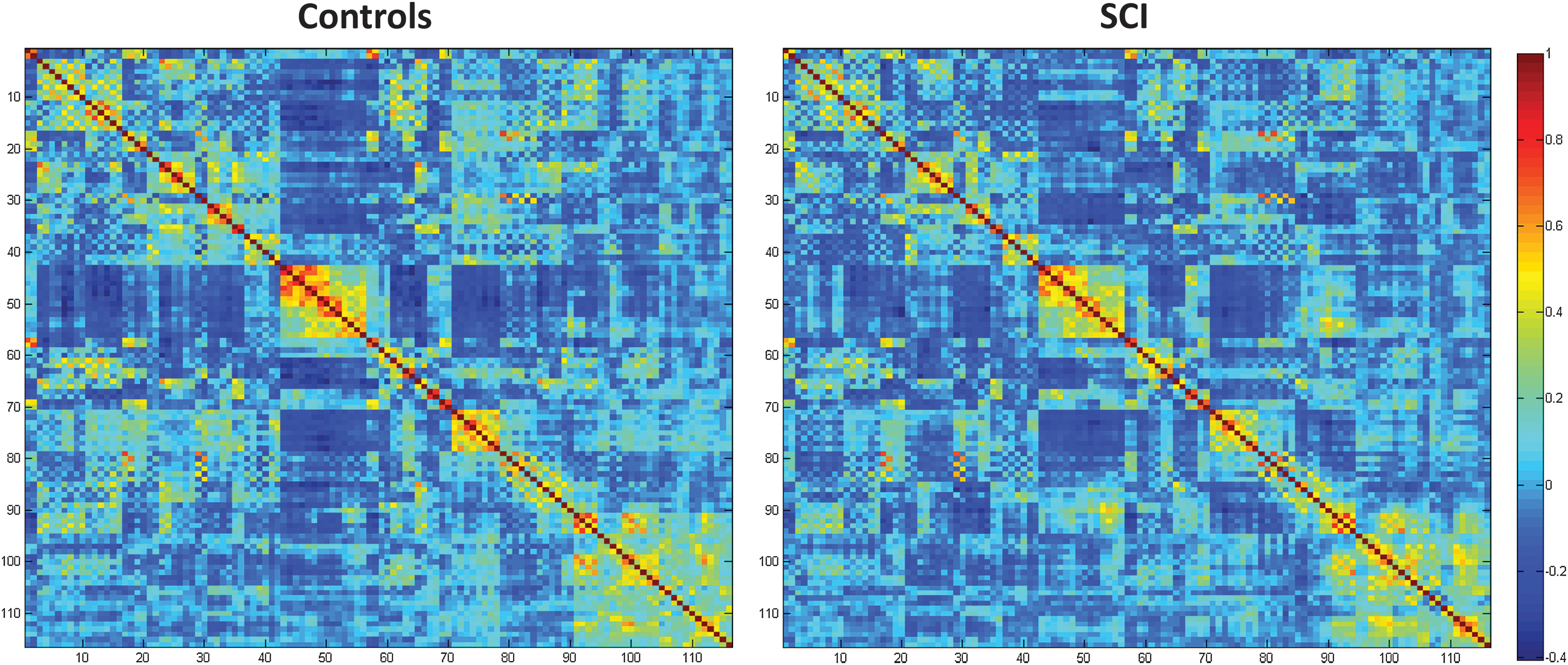
Correlation coefficient matrices, following parcellation of the whole brain network into 116 regions of interest (ROIs), in control and spinal cord injury (SCI) groups. Color image is available online at
Following the generation of group matrices, NBS was applied to assess connectivity differences. The application of this statistical methodology resulted in significant findings in whole-brain network as well as a network comprising the sensorimotor cortex and cerebellum at p < 0.05 (Fig. 2). The resting-state connectivity architecture of the whole brain showed a subnetwork with decreased connectivity in SCI patients (p = 0.02). Further, the comparison of the network containing the sensorimotor cortex and cerebellum showed a subnetwork with increased connectivity in the SCI subjects compared with controls (p = 0.02). The ROIs of the sensorimotor cortex that showed increased connectivity to the cerebellum included left and right paracentral lobule (numbers 69 and 70 in the AAL template classification scheme).
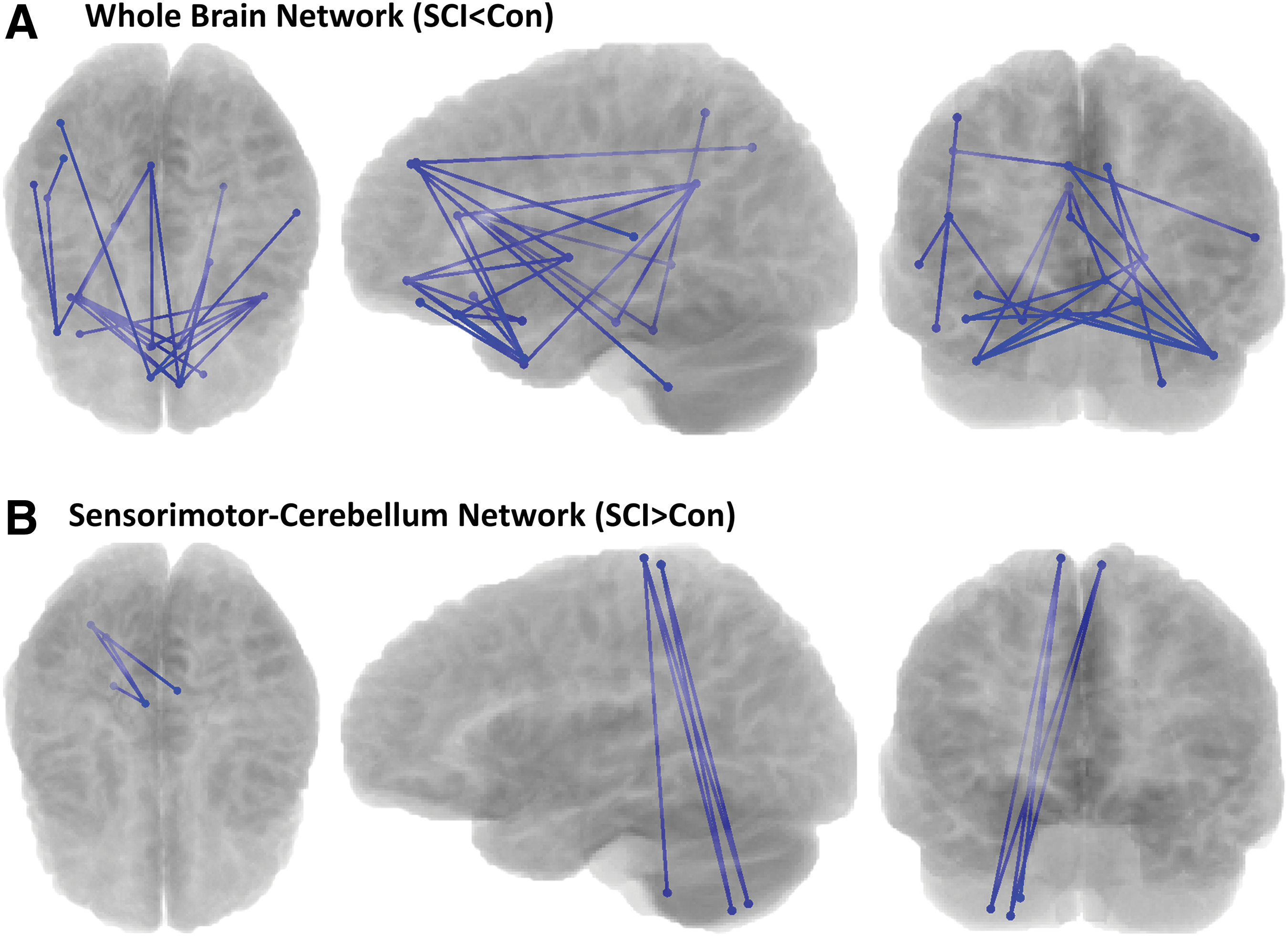
Depiction of differences in resting-state functional connectivity subnetworks between spinal cord injury (SCI) subjects and controls as identified with network-based statistics (NBS) for networks comprising
Discussion
To our knowledge, this is the first study to analyze the alterations to the whole-brain network in patients with traumatic SCI using the NBS approach. The demonstration of significant differences in the resting-state connectivity networks between the SCI and the control groups underscores the utility of NBS in multivariate comparisons to highlight changes to brain network topology in distant neural pathologies such as SCI.
To correct for FWE on account of the enormous number of multiple comparisons, the false discovery rate (FDR) was applied to the data, which resulted in no significant findings. Following this, we applied NBS to the group association matrices to compare network connections exhibiting a structure. The rationale behind using this approach was to generate greater statistical power compared with independent correction of p values for each link, using a generic procedure such as FDR to control for FWE. 9 The application to our data set uncovered significant differences in the whole-brain functional connectivity network as well as the sensorimotor-cerebellum network not highlighted by FDR previously.
The results of the present data analysis demonstrated a subnetwork with reduced functional connectivity in the whole brain in SCI subjects. The decrease in the resting-state connectivity pattern in patients with complete SCI could be caused by the imbalance in transmission of afferent and efferent neural impulses following cord trauma. Further, atrophic changes throughout the neuraxis caused by retrograde degeneration after distant cord injury might influence functional modifications because of the dependence of function on structure. 21 –23
The functional connectivity of a subnetwork of the sensorimotor cortex and cerebellum network was increased in patients with SCI. This increase in connectivity seems to suggest strong neural synchrony between these brain regions, which might serve to facilitate recruitment of neural substrates to compensate for neural deficits in SCI. Upon closer inspection, the ROIs comprising the left (69) and right (70) paracentral lobule as part of the sensorimotor cortex formed this subnetwork, which showed differences across groups. The paracentral lobule communicates with both the sensory and the motor cortex, and the increase in connectivity with the cerebellum might serve to highlight passage of more neuronal traffic between these two areas in traumatic SCI.
The present study has a number of limitations that warrant further consideration to better understand alterations to brain connectivity following a distant central nervous system (CNS) insult such as SCI. Widening the scope to include SCI patients with varying grades and levels of injury might improve on the characterization of connectivity alterations at supraspinal levels post-SCI. This would allow for correlation analysis to check for the relationship between the extent of clinical impairment and resting-state functional connectivity findings The pain experienced in SCI is neuropathic, with the thalamus serving as an important conduit, which warrants further exploration. 24 The exclusive focus on network reorganization in the brain does not account for changes to the spinal cord network configuration, and their contribution to the cortical findings and needs to be studied separately. 23 The present study contains female controls, whereas the patient group is solely comprised of males. This was done to ensure age matching, but needs to be accounted for in future analysis to check for the effect of gender on resting state connectivity. Comparative studies using alternate parcellation schemes might add to the present AAL template-based classification for defining functional regions.
Conclusion
In conclusion, our results emphasize the applicability of NBS to study functional connectivity architecture in diseased brain states. Further, we highlight differences in resting-state functional connectivity using NBS in patients with traumatic SCI. The presence of altered connectivity in various subnetworks is indicative of reorganization of large-scale resting-state brain networks in traumatic SCI, with potential prognostic and therapeutic implications.
Footnotes
Acknowledgments
The authors thank Moriah Iverson, MS, and Judeen Richlen, RN, (research coordination); Dana Seslija, MD, MS, William Waring, MD, and Merle Orr, MD, (patient recruitment support); and Yu Liu, MS (MRI technical support). This study was funded by the Research Administration Committee of the Department of Physical Medicine and Rehabilitation at the Medical College of Wisconsin, Marquette University; Bryon Riesch Paralysis Foundation; and The Falk Foundation.
Author Disclosure Statement
No competing financial interests exist.