




Abstract
There are no drugs to manage traumatic brain injury (TBI) presently. A major problem in developing therapeutics is that drugs to manage TBI lack sufficient potency when dosed within a clinically relevant time window. Previous studies have shown that minocycline (MINO, 45 mg/kg) plus N-acetylcysteine (NAC, 150 mg/kg) synergistically improved cognition and memory, modulated inflammation, and prevented loss of oligodendrocytes that remyelinated damaged white matter when first dosed 1 h after controlled cortical impact (CCI) in rats. We show that MINO (45 mg/kg) plus NAC (150 mg/kg) also prevent brain injury in a mouse closed head injury (CHI) TBI model. Using the CHI model, the concentrations of MINO and NAC were titrated to determine that MINO (22.5 mg/kg) plus NAC (75 mg/kg) was more potent than the original formulation. MINO (22.5 mg/kg) plus NAC (75 mg/kg) also limited injury in the rat CCI model. The therapeutic time window of MINO plus NAC was then tested in the CHI and CCI models. Mice and rats could acquire an active place avoidance task when MINO plus NAC was first dosed at 12 h post-injury. A first dose at 12 h also limited gray matter injury in the hippocampus and preserved myelin in multiple white matter tracts. Mice and rats acquired Barnes maze when MINO plus NAC was first dosed at 24 h post-injury. These data suggest that MINO (22.5 mg/kg) plus NAC (75 mg/kg) remain potent when dosed at clinically useful time windows. Both MINO and NAC are drugs approved by the Food and Drug Administration and have been administered safely to patients in clinical trials at the doses in the new formulation. This suggests that the drug combination of MINO plus NAC may be effective in treating patients with TBI.
Introduction
T
The combination of minocycline (MINO) plus N-acetylcysteine (NAC) limited brain injury when rats were first dosed 1 h after controlled cortical impact (CCI) in rats. 4 MINO plus NAC improved cognition and memory in an active place avoidance (APA) task, limited white matter injury by inducing remyelination, and modulated neuroinflammation. 5,6 MINO plus NAC synergized to produce many of these therapeutic effects. 4,6
MINO or NAC as individual drugs has shown efficacy in animal models of TBI. MINO is a tetracycline antibiotic agent that crosses the blood–brain barrier. 7 MINO inhibits microglial activation, blocks apoptosis, reduces excitotoxicity, inhibits metalloproteinase activity, scavenges reactive oxygen species, and prevents myelin loss. 6,7 These neuroprotective effects occurred with a first dose of MINO no later than 1 h post-injury (PI). 5,6, 8 –13 MINO improved performance in Barnes maze when first dosed 4 h after mild blast injury. 14 The efficacy of MINO is unknown when first dosed later than 4 h PI.
NAC is a potent antioxidant that undergoes first-pass metabolism to cysteine and cystine. Cysteine and cystine, potent antioxidants, can also increase glutathione levels in the brain that protect mitochondria. 15,16 Reduction of cellular redox by NAC also modulates neuroinflammation. 3,17 In addition to its antioxidant action, NAC increases activity of the xc-cysteine-glutamate antiporter that elevates extracellular levels of glutamate. 18 NAC has antiapoptotic and anti-inflammatory action when dosed within 1 h PI. 19,20 One-hour dosing of NAC (100 mg/kg) to rats improved performance and retention of Morris water maze, Y-maze, and novel object recognition. 21
In a small cohort of patients, treatment with NAC within 24 h after mild blast TBI improved neuropsychological outcomes. NAC was effective when first dosed within 72 h PI, but its potency was less than with 24 h dosing. 22 NAC has been shown, in small clinical trials, to limit anxiety, attention deficit hyperactivity disorder, and amyotrophic lateral sclerosis. These findings have not yet been replicated with larger patient cohorts. 23
This study uses the mouse closed head injury (CHI) and the rat CCI models to examine the efficacy and therapeutic time windows of MINO, NAC, and MINO plus NAC. The CHI and CCI models used in this study differ in the species used and in the method to injure the brain. CHI involves striking the head using an electromagnetically controlled piston that both deforms the skull and induces a rapid acceleration and deceleration of the head. 24 Injury produced by CHI can be heterogeneous, yet injury severity can be predicted by the duration of post-injury apnea and the time needed to restore the righting reflex. 24 The CCI model uses an electromagnetically controlled piston to directly strike the dura through a unilateral craniotomy. 25 At the impact site, CCI produces a more uniform and localized damage to gray and white matter than CHI. 25 The craniotomy in CCI is a potential confound for TBI studies because it produces vascular changes, inflammation, cell death, and prevents changes in intracranial pressure. 26
Both CHI and CCI produce transient motor deficits and long-lasting behavioral deficits. 24,25 This study uses Barnes maze and APA to examine the behavior of injured mice or rats. Barnes maze is a purely spatial task in which rodents escape a brightly lit, open maze surface by learning the location of the escape hole. 27 APA, in contrast requires learning to avoid a stationary shock zone on a rotating arena. 28 The location of the shock zone is learned by sensory segregation of relevant distal visual spatial cues and irrelevant proximal olfactory cues deposited on the arena. 28
Many drugs prevent brain injury when dosed within hours after an experimental TBI. 4 Most either lose efficacy as the time to first dose after injury is increased or the therapeutic time window is unknown. 4 MINO plus NAC is highly effective when dosed 1 h PI, but its efficacy or the efficacy of the individual drugs is not known if first dosed more than 1 h after injury. This study titrated the concentrations of MINO and NAC in the drug combination to find a more potent combination. The therapeutic time window of MINO plus NAC alone and in combination was then examined in the mouse CHI model and the rat CCI models.
Methods
Experimental TBI
Experiments using the CHI model were performed on male C57/BL6 mice (15 to 17 weeks old, 26–28 g). Baseline weights were obtained before sham-CHI or CHI. Anesthesia was induced for 2 min with isoflurane (3.5% in O2 (1.0 L/min) administered via a nose cone that was maintained (3% in O2 (1.0 L/min)) until after the impact. CHI was produced as described by Grin'kina and associates. 24 The top of the head of the mouse was shaved and placed in a Kopf stereotaxic apparatus modified by placing a single 12.7 mm sheet of polyurethane foam on the bed of the adaptor and two 12.7 mm sheets of polyurethane foam wrapped around the ear bar holders.
CHI was produced using a 5.0 mm diameter impactor tip controlled by an electromagnetic impactor (Leica Microsystems, Buffalo Grove, IL). The impactor tip was placed 3 mm lateral from the midline and 5 mm caudal from the eyes. The impactor produced single 6.3 m/sec impact to a depth of 3 mm with a 1 sec dwell time. Sham-injured mice received identical treatment without the impact. The entire procedure was completed in less than 3 min.
The head of the mouse was able to move freely after impact as described previously in Grin'kina and colleagues. 24 The ability of the mouse to spontaneously breathe was assessed after injury. If spontaneous breathing did not begin within 30 sec, cardiopulmonary resuscitation was initiated with chest compressions at a rate of 150–160 per min, while the mouse was ventilated with 100% O2. Mice were then assessed for restoration of righting reflex before return to their home cage. The average righting reflex of injured mice was 8.64 ± 0.47 min. Of these injured mice, 83.6% had righting reflexes greater than 5 min.
CCI was performed as described by Haber and coworkers 5 on male Long Evans rats (250–300 g, Charles River, Wilmington, MA). Anesthesia was induced with isoflurane 5% in O2 and maintained as 3% in O2. A craniotomy (5.0 mm) was made midway between lambda and bregma over the right parietal lobe without disrupting the dura. Sham-CCI rats received identical treatment without the impact. After restoration of righting reflex, rats were returned to their home cage.
Only male rats or mice were included because these studies were initiated before the National Institutes of Health (NIH) guidelines on scientific rigor and reproducibility. On arrival at SUNY-Downstate, mice or rats were assigned a code number that was used during all determinations. Therefore, experimenters were unaware of the injury or treatment status of the mouse or rat. All animal experiments complied with the Animal Research: Reporting of In Vivo Experiments guidelines and were performed in accordance with the NIH guide for the care and use of laboratory animals (NIH Publications No. 8023, revised 1978). All studies were approved by the Institutional Animal Care and Use Committee of the State University of New York-Downstate Medical Center (protocol #14-10406).
Drug treatments
In the titration studies, mice received different concentrations of MINO (in mg/kg; 10, 22.5, 45, and 90) and NAC (in mg/kg; 10, 37.5, 75, 150, and 300) in NaPO4 (10 mM), NaCl (138 mM), and KCl (2.7 mM). All drugs were from Sigma (St. Louis, MO). Mice received three intraperitoneal injections of MINO plus NAC or saline at 1 h, one and two days after sham-injury or injury. The dose titration studies examined the total shock zone entrances in eight trials of APA (see below for detail). MINO plus NAC was only titrated using mice. In the studies examining therapeutic time window, mice or rats received three intraperitoneal injections of MINO (22.5 mg/kg), NAC (75 mg/kg), saline, or MINO (22.5 mg/kg) plus NAC (75 mg/kg). The first dose was given 6, 12, or 24 h after injury. The second and third doses were administered two and three days after sham-injury or injury.
Barnes maze
Mice were tested on Barnes maze with slight modifications of the method of Sunyer and coworkers. 27 Barnes maze was performed in a rectangular room (3 m × 5 m) containing a circular platform (92 cm diameter) elevated 105 cm above the floor with 20 evenly spaced holes (5 cm diameter; 7.5 cm between holes) along its perimeter. The center of the maze was 1.5 m from the walls of the room that contained prominent visual landmarks. A Tecknet C016 720p HD webcam located 1.5 m above the maze tracked the position of the mouse. Innate aversion to the open space and gently moving air from a nearby fan motivated the mouse to find the escape hole.
Seven days after sham-CHI or CHI, mice received a single 5-min habituation session. Immediately after habituation, mice received four 3-min trials with a 15-min intertrial interval for four days. Any Maze software (Stoelting) assessed the video records of each trial for path length, time elapsed to reach the target hole (latency), and entrances into holes other than the escape hole (errors). Latency and errors measured task acquisition and path length assessed for motor function. Sham-CHI–treated mice receiving a first saline dose at 12 h or 24 h had similar latency and errors; thus, the 12 h and 24 h groups were combined. CHI-saline mice first dosed at 12 h or 24 h also had similar latency, and errors and were combined into one group.
Seven days after sham-CCI or CCI, rats were tested on Barnes maze using the same protocol as mice but with an apparatus consisting of a circular platform 194 cm in diameter and elevated 90 cm above the floor, with 17 equally spaced holes (12 cm diameter; 15 cm between holes) along the perimeter.
APA
Eleven days after sham-CHI or CHI, mice were tested on APA with modifications of the method of Burghardt and associates. 29 Behavioral assessments were performed in a rectangular room (4 m × 3 m) with prominent visual landmarks on the walls. APA was conducted on a 40-cm diameter circular behavioral apparatus that rotated at one revolution per minute. The position of the mouse was tracked with an infrared Firewire camera located 1.2 m above the arena computer. Track analysis software (Bio-Signal Group Corp., Brooklyn, NY) analyzed the movement of the mouse and the entries in the shock zone.
Mice were habituated for 10 min to the rotating arena with an inactivated shock zone. Total distance traveled was assessed. Mice then received 4 × 10-min sessions of APA with an active shock zone with a 50-min intertrial interval. Total distance traveled, speed, linearity, number of shock zone entrances, shocks/entrance, and time to first entrance were assessed. Time to first entrance was assessed initially on the final trial of APA in a similar manner as with rats. Mice moved more than rats during APA that led to more apparently inadvertent entries into the shock zone. This increased the variability in time to first entrance compared with rats. 30 Therefore, the time to first entrance in the last two trials of APA were averaged and then analyzed. Mice were returned to their home cages after testing.
Beginning 11 days after sham-CCI or CCI, rats were tested on APA using the protocol of Abdel Baki and colleagues. 31 Testing consisted of an open-field test, passive place avoidance, and APA. Distance traveled on open field measured innate exploratory behavior. On the same day, rats received four 10-min passive avoidance trials with a 10-min intertrial interval on a nonrotating arena with an active shock zone. The total number of shock zone entrances measured the ability to sense and avoid shock, and the distance traveled assayed the conditioned ability to inhibit movement. The following day, rats received six 10-min trials for one day on a rotating arena with the shock zone on with a 10-min intertrial interval. 32 Shock zone entrances, speed, linearity, shocks/entrance, and time to first entrance were assessed.
Histological analysis
Mice or rats were transcardially perfused with 4% (w/v) paraformaldehyde (PFA) 14 days after sham-injury or injury, post-fixed for 48 h at 4°C., and paraffin-embedded sagittal sections were prepared (Histowiz, Brooklyn, NY). Difficulties with fixation and sectioning prevented analysis of the injured rats that first received MINO at 12 h. Contusion volume was not measured because MINO plus NAC first dosed 1 h after CCI had no effect on contusion volume. 4 Parasagittal sections (9 μm) located between 10 μm and 100 μm from the midline were stained using mouse monoclonal antibodies against macrotubule-associated protein 2 (MAP2) (Abcam, 1:1000) and the appropriate fluorescent secondary antibody (Alexa Fluor 568). The amount of MAP2 immunofluorescence subtracted from background fluorescence was assessed in specific regions of interest (ROIs) using ImageJ v.1.48 software. Myelin content was determined by staining with Luxol fast blue (LFB) according to the manufacturer's instructions (American Mastertech, Lodi, CA).
Digital images were prepared and the amount of LFB staining was assayed using Image J software in ROIs within the body of the corpus callosum, splenium, cingulum, and fimbria in sagittal sections located 1.5–1.9 mm from midline for mice and 2.0–3.0 for rats. To determine the amount of LFB staining that was from myelin staining, stain intensity in the ROIs was subtracted from stain intensity values from the stratum radiatum of the hippocampus that contained the unmyelinated Schaffer collateral. 4 Each determination consisted of one section per animal.
Statistical analysis
Group differences in total shock zone entrances during titration of MINO and NAC were analyzed by one-way analysis of variance (ANOVA) with Student-Neumann-Keul post hoc test. In the studies of therapeutic time window, latency and primary errors on Barnes maze, and shock zone entrances on APA were analyzed by repeated two-way ANOVA on factors of group and trial for each day assessed. Pairwise comparisons were then made using the Sidak post hoc test. One-way ANOVA with Student-Neumann-Keul post hoc test analyzed all other APA parameters and Barnes path length. Histological parameters of gray and white matter injury were analyzed by two-way ANOVA followed by Scheffe post hoc test. Statistical significance was set at 0.05 for all tests. All values are presented as mean ± standard error of the mean.
Results
Titration of MINO and NAC in the mouse CHI model
The original formulation of MINO (45 mg/kg) and NAC (150 mg/kg) was developed using drug concentrations that limited brain injury in animal models of TBI. 10,17 Few studies have examined different doses of the drug, so we therefore tested different formulations of MINO and NAC for improved efficacy over the original formulation. The MINO concentration was systematically varied from 10 mg/kg to 90 mg/kg, and the NAC concentration was varied from 10 mg/kg to 300 mg/kg. Mice received a first dose of saline or drugs 1 h after CHI (Fig. 1A). They also received a second and third dose two and three days after CHI or CCI.
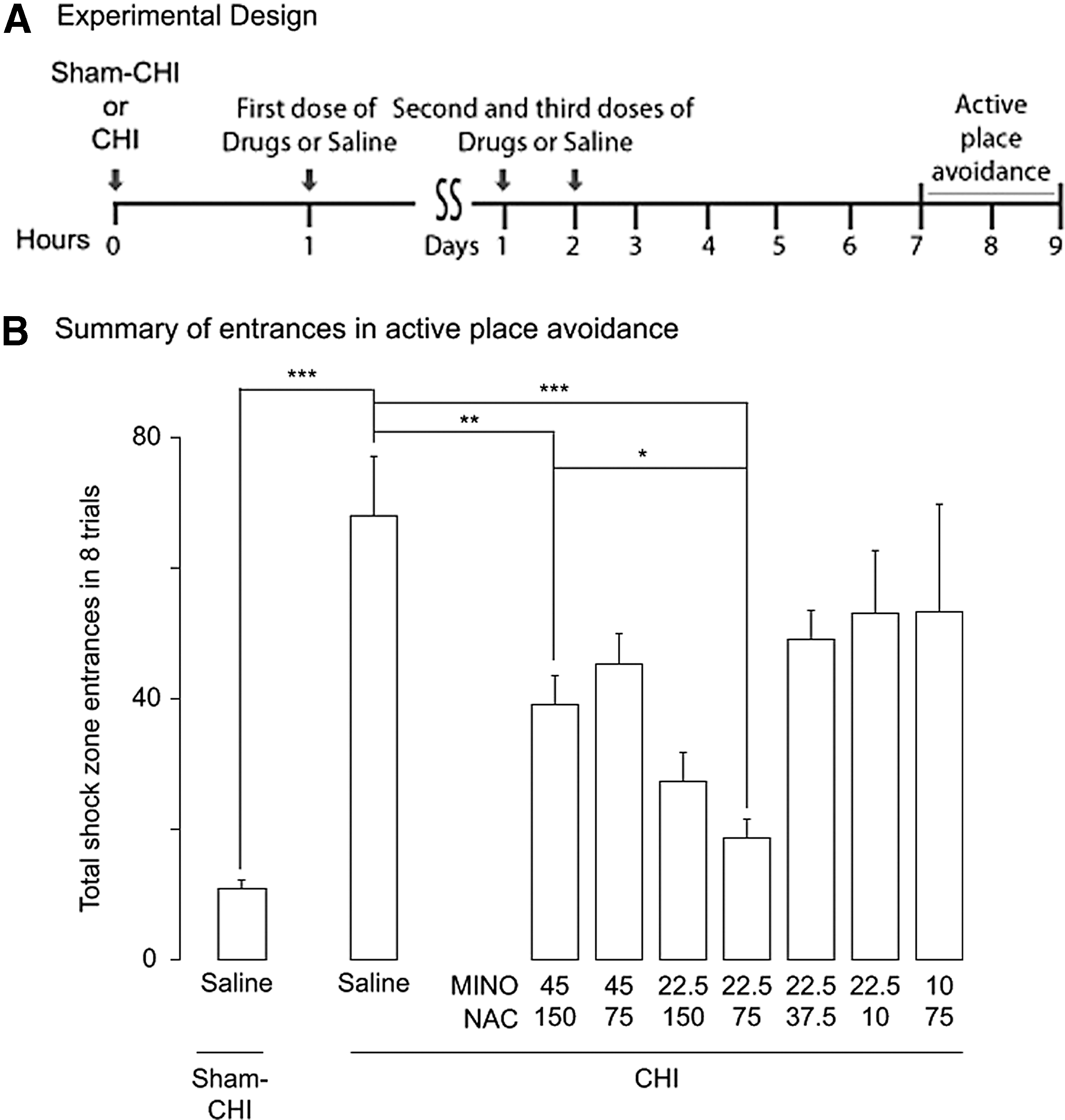
Titration of minocycline (MINO) or N-acetylcysteine (NAC) in the MINO plus NAC combination in the mouse closed head injury (CHI) model. (
The highest MINO or NAC concentrations in the formulations tested were toxic to injured mice. Dosing with MINO (90 mg/kg) plus NAC (150 mg/kg) resulted in the death of six of 10 mice within 2.7 ± 0.3 days PI. Dosing with NAC (300 mg/kg) with MINO (45 mg/kg) resulted in the death of six of six mice 2.4 ± 0.6 days PI. CHI was shown previously to have a mortality rate of 9.1%.
24
These data suggest that combinations containing MINO (90 mg/kg) or NAC (300 mg/kg) were toxic. The toxicities of MINO (90 mg/kg) or NAC (300 mg/kg) agree with previous reports that the LD50 in mice of a single IP dose is 299 mg/kg for MINO (
APA testing began one week after sham-CHI or CHI (Fig. 1B). The total number of shock zone entrances in APA assessed the drug efficacy (Fig. 1B). Shock zone entrances showed a significant effect of treatment (F8,61 = 8.10, p < 0.0001). CHI significantly increased shock zone entrances (post hoc, p < 0.0001); this increase was significantly decreased by MINO (45 mg/kg) plus NAC (150 mg/kg) (post hoc, p < 0.005). Lowering of the concentrations of MINO to 22.5 mg/kg and NAC to 75 mg/kg further decreased shock zone entrances suggesting this formulation is more potent than MINO (45 mg/kg) plus NAC (150 mg/kg) (post hoc, p < 0.05). A further lowering of MINO to 10 mg/kg or NAC to 37.5 mg/kg increased shock zone entrances compared with MINO (22.5 mg/kg) plus NAC (75 mg/kg) combination (post hoc, p < 0.05). All groups traveled a similar distance on open field/habituation, suggesting similar motor skills and ability to habituate (Supplementary Table 1; see online supplementary material at
Time to first entrance, a parameter of APA task retention, also showed a significant treatment effect (F8,67 = 7.61 p < 0.0001). All groups increased time to first entrance except the injured groups receiving saline, MINO (10 mg/kg) plus NAC (150 mg/kg), MINO (22.5 mg/kg) plus NAC (37.7 mg/kg), MINO (22.5 mg/kg) plus NAC (10 mg/kg), or MINO (10 mg/kg) plus NAC (75 mg/kg) (Supplementary Table 1). These data suggest that the better performance produced by MINO plus NAC in APA was because of improved cognition and memory rather than because of changes in motor or sensory function. On the basis of these experiments, the remaining studies further tested the therapeutic time window of MINO (22.5 mg/kg) plus NAC (75 mg/kg).
Analysis of the therapeutic time window in a mouse CHI model
The mouse CHI model of TBI was used to test the therapeutic time window of MINO, NAC, or MINO plus NAC. Control groups included sham-CHI and CHI-injured mice treated with saline (Fig. 2A). Drugs or saline was first dosed at 12 h or 24 h PI. Mice were tested on Barnes maze beginning at 7 PI. Primary errors and latency were measured to assess drug efficacy; path length was measured to ensure similar motor ability of all groups (Fig. 3, Supplementary Table 2; see online supplementary material at

Experimental designs to test the therapeutic time window of minocycline (MINO) plus N-acetylcysteine (NAC). (
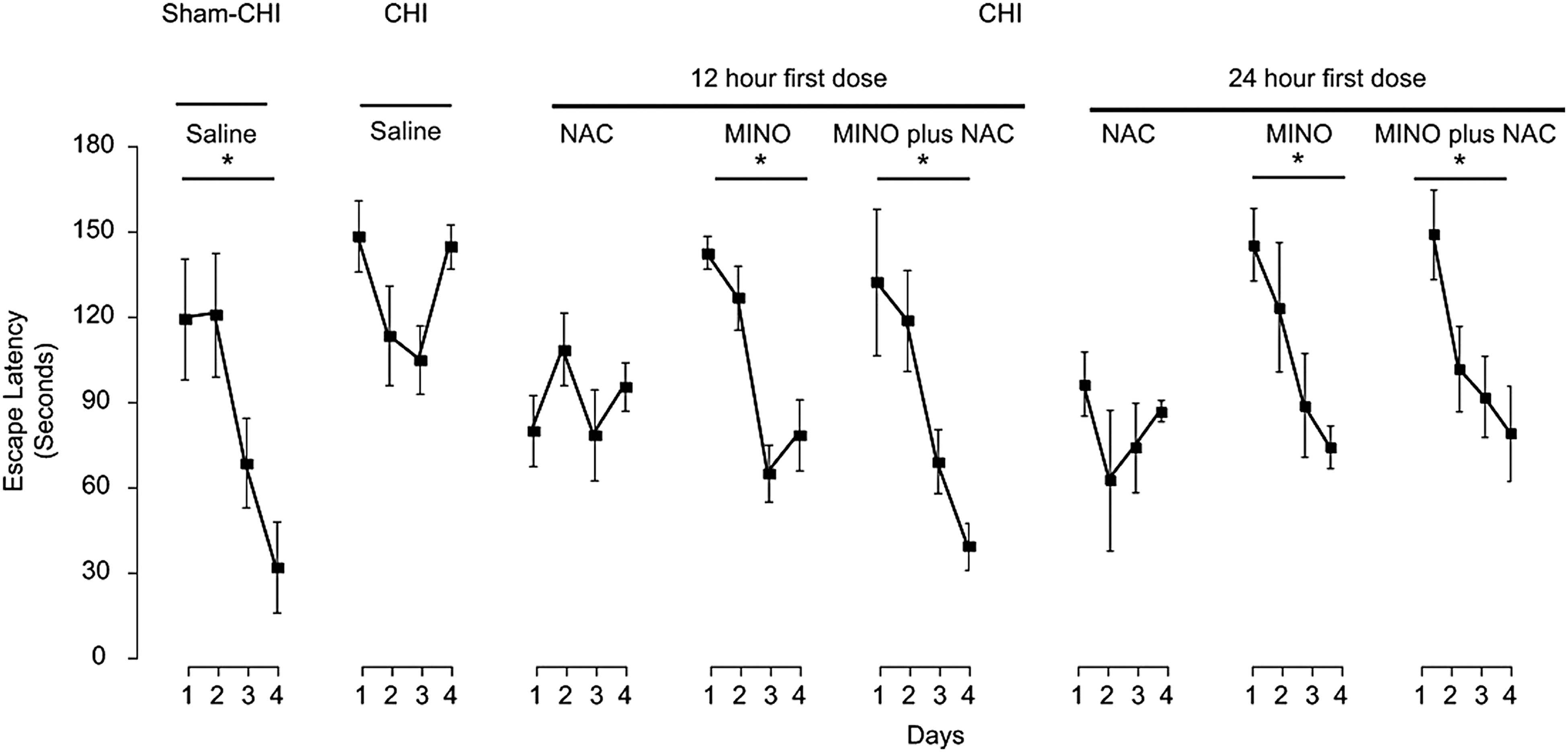
Assessment of drug efficacy on Barnes maze in mice after closed head injury (CHI). Mice received either sham-CHI or CHI. Mice were first dosed with saline or drugs 12 h or 24 h post-injury (PI) and were tested on Barnes maze beginning seven days PI. Latency decreased in the sham-CHI group treated with saline or the injured groups treated with minocycline (MINO) or with MINO plus N-acetylcysteine (NAC) (*p < 0.05). Injured mice treated with saline or NAC did not decrease latency.
Latency had a significant effect of treatment (F7,32 = 4.52, p < 0.01) and day (F3,32 = 16.18, p < 0.001) with a significant interaction of treatment and day (F10,128 = 4.62, p < 0.0005) (Fig. 3). The latency of the sham-CHI group was similar to that of injured mice first dosed with MINO or MINO plus NAC at 12 h or 24 h PI. The latency of the injured mice was similar whether they were treated with saline or first dosed with NAC at 12 h or 24 h PI. Only the sham-CHI group lowered the number of errors (Supplementary Table 2; see online supplementary material at
After completion of Barnes maze testing, the same groups were tested on APA (Fig. 2A, 4). Drug efficacy was assessed using shock zone entrances, a measure of task acquisition; and time to first entrance, a measure of task retention. Shock zone entrances showed a significant effect of treatment (F7,32 = 14.28, p < 0.0001) and trial (F3,32 = 3.84, p < 0.05) with no interaction between treatment and trial (F10,128 = 4.66 p < 0.001) (Fig. 4). The number of shock zone entrances of the sham-CHI group was similar to that of injured mice first treated with drugs at 12 h, but not at 24 h PI (post hoc, p < 0.01). These data suggest that a first dose of drugs at 12 h allowed CHI-injured mice to acquire APA. Sensory and motor parameters were similar among all the groups, suggesting the ability of the drug combination to lower shock zone entrances was because of improved cognition (Supplementary Table 3; see online supplementary material at
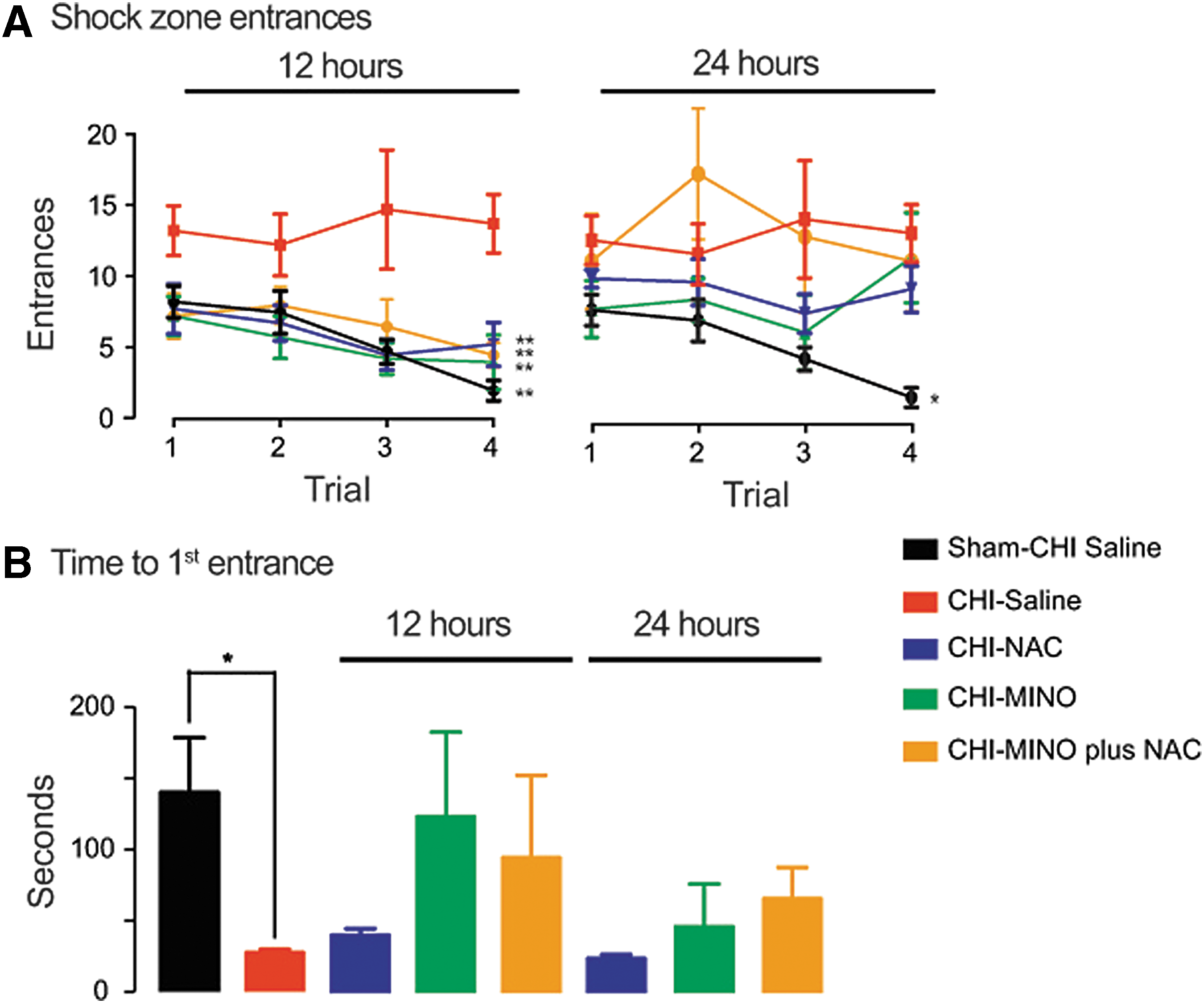
Assessment of drug efficacy on active place avoidance (APA) in mice after closed head injury (CHI). Mice received either sham-CHI or CHI. Saline or drugs were first dosed at 12 h or 24 h post-injury (PI), and mice were tested on APA 11 days PI. (
MAP2 immunoreactivity was examined in parasagittal brain sections ipsilateral to the impact site from sham-CHI treated with saline or CHI-injured mice first dosed either at 12 h or 24 h PI with saline, NAC, MINO, or MINO plus NAC (Fig. 5). MAP2 immunoreactivity was examined in the hippocampal CA3, CA1, and DG regions (Fig. 5A). The CA3 and CA1 regions showed a significant effect of treatment and time to first dose (treatment: CA3, F4,32 = 16.42, p < 0.0001; CA1, F4,32 = 9.93, p < 0.01; DG, F4,32 = 3.37, p > 0.05; time to first dose: CA3, F1,32 = 5.38, p < 0.05; CA1, F1,32 = 3.12, p < 0.05; DG, F1,32 = 0.19, p > 0.5). None of the hippocampal regions had a significant interaction between treatment and time to first dose (CA3, F4,32 = 2.33, p > 0.05; CA1, F4,32 = 1.05, p > 0.1; hilus, F4,32 = 0.26, p > 0.5).
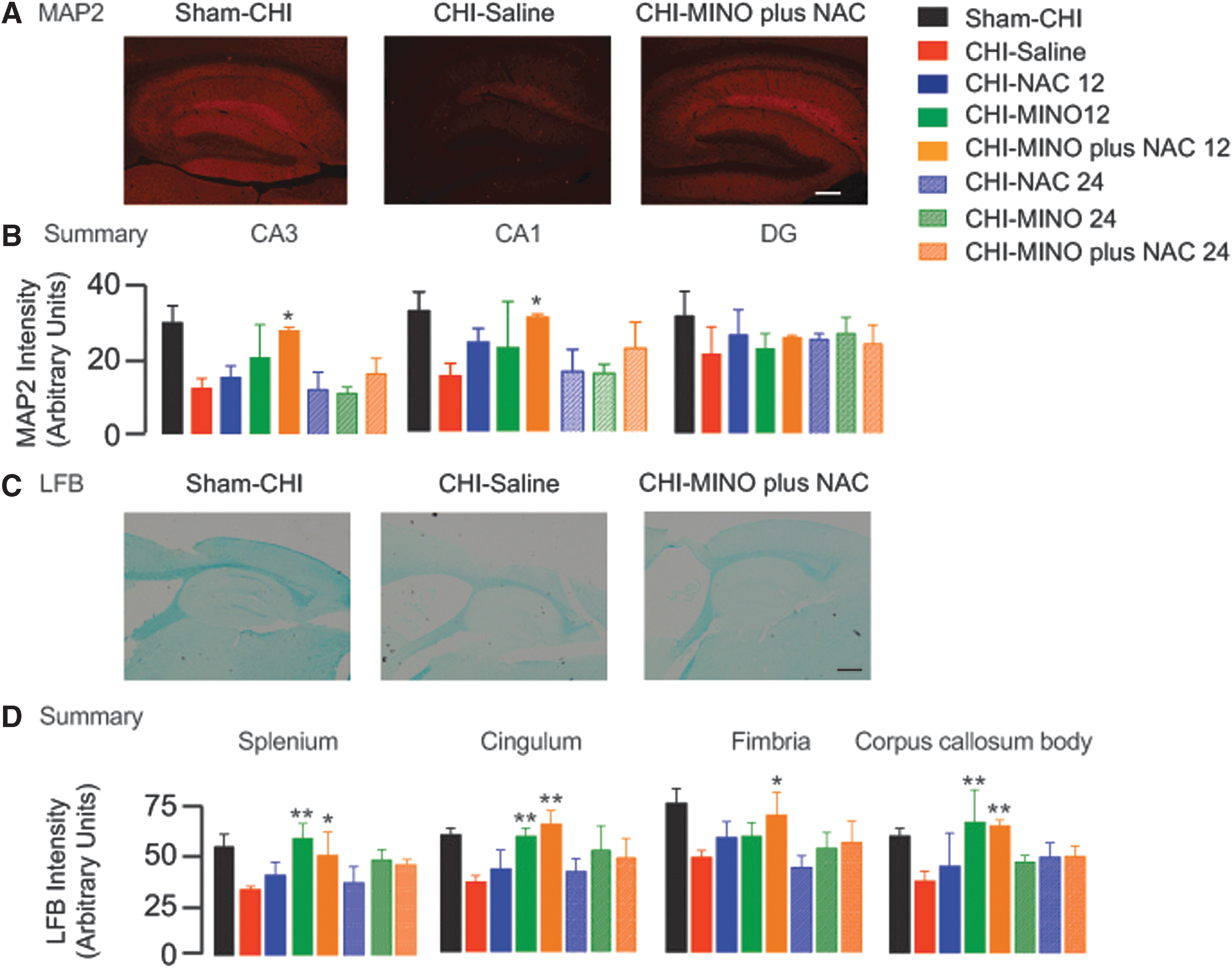
Assessment of gray and white matter injury in mice after closed head injury (CHI).
MAP2 immunoreactivity in the sham-CHI group was significantly reduced in the CHI-saline group (post hoc, p < 0.01) (Fig. 5B). MAP2 immunoreactivity was significantly increased in injured mice first dosed at 12 h PI MINO plus NAC compared with the CHI-saline group (post hoc, p < 0.05). This increase was observed in both CA3 and CA1 (Fig. 5B).
Myelin content was also assessed using LFB (Fig. 5C). LFB stain intensity was assessed in the body of the corpus callosum, fimbria, splenium, and cingulum; these are white matter regions that are frequently demyelinated in animal models of TBI. 36 All white matter regions showed a significant effect of treatment and time to first dose, except for a strong trend toward significance in the fimbria (treatment; splenium, F4,32 = 10.0, p < 0.0001; cingulum, F4,32 = 13.0, p < 0.0001; fimbria, F4,32 = 12.95, p < 0.0001; corpus callosum body, F4,32 = 6.94, p < 0.05; time to first dose, splenium, F4,32 = 6.25, p < 0.05; cingulum, F4,32 = 5.3, p < 0.05; fimbria, F4,32 = 3.26, p = 0.08; corpus callosum body, F4,32 = 6.94, p < 0.05). Treatment and time to first dose interacted significantly in all regions (splenium, F4,32 = 3.8, p < 0.05; cingulum, F4,32 = 5.3, p < 0.05; corpus callosum body) except for a strong trend toward significance in the fimbria, (F4,32 = 3.3, p = 0.08).
The sham-CHI saline had significantly higher LFB intensity in all regions assayed compared with the CHI-saline group (post hoc, p < 0.01) (Fig. 5D). LFB intensity was significantly increased in injured mice first treated with MINO plus NAC at 12 h PI in all regions assayed (post hoc, p < 0.05). LFB intensity also increased in injured mice first treated with MINO alone at 12 h PI in all regions except the fimbria (post hoc, p < 0.05)
Analysis of therapeutic time window in a rat CCI model
The rat CCI model of TBI was also used to test the therapeutic time window of MINO plus NAC, MINO, or NAC. Saline or drugs were first dosed at 6 h, 12 h, or 24 h PI. Sham-CCI rats were treated with saline. Seven days later, sham-CCI and CCI-injured rats treated with saline or drugs were tested on Barnes maze (Fig. 6). Path length, primary errors, and latency were measured. Latency had a significant effect of treatment (F10,54 = 14.81, p < 0.0001) and day (F10,54 = 23.82, p < 0.0002) with a significant interaction between treatment and day (F30,176 = 1.78, p < 0.05) (Fig. 6). The sham-CCI group had a significantly shorter latency that the CCI-saline group (post hoc, p < 0.005). A first dose of NAC, MINO, or MINO plus NAC at 6 h, 12 h, or 24 h PI significantly lowered primary latency compared with saline treatment (post hoc, p < 0.02). Primary errors also had significant effect of treatment (F10,54 = 3.89, p < 0.005) and day (F10,54 = 19.81, p < 0.0005) with a trend toward a significant interaction between treatment and day (F30,176 = 1.48, p = 0.08).
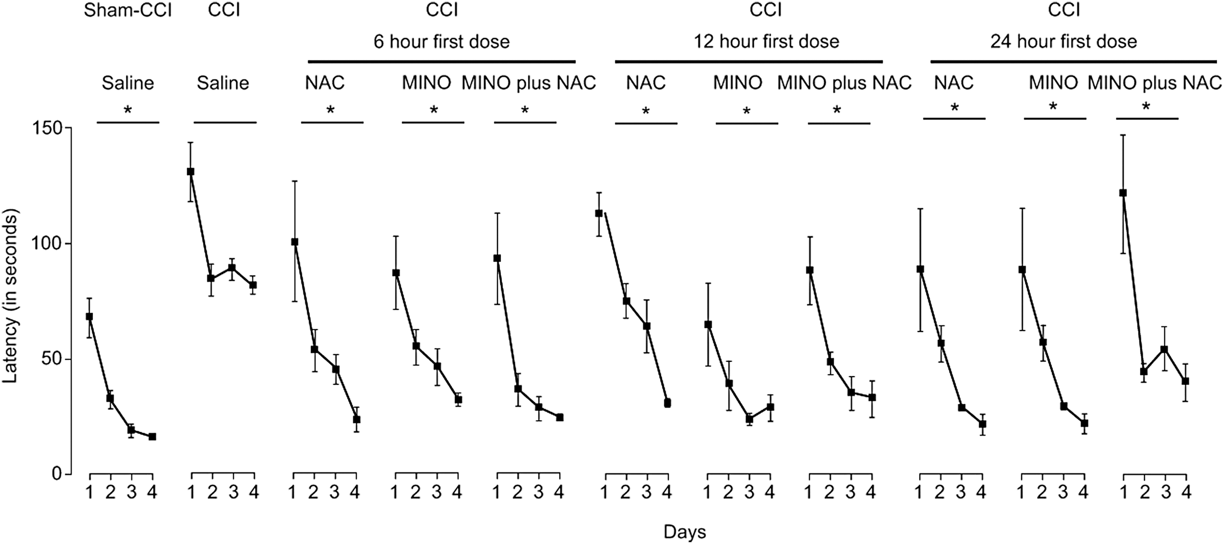
Assessment of drug efficacy on Barnes maze in rats after controlled cortical impact (CCI). When first dosed at 6 h, 12 h, or 24 h post-injury, all groups significantly lowered the time to find the escape hole except injured rats treated with saline (*p < 0.05). MINO, minocycline; NAC, N-acetylcysteine.
The sham-CCI saline group had significantly fewer errors than the CCI-saline group (Supplementary Table 4; see online supplementary material at
Eleven days after sham-CCI or CCI, rats were tested on open field, passive place avoidance, and APA (Fig. 2B). All groups performed equally in open field and passive avoidance, suggesting similar motor and sensory abilities (Supplementary Table 5; see online supplementary material at
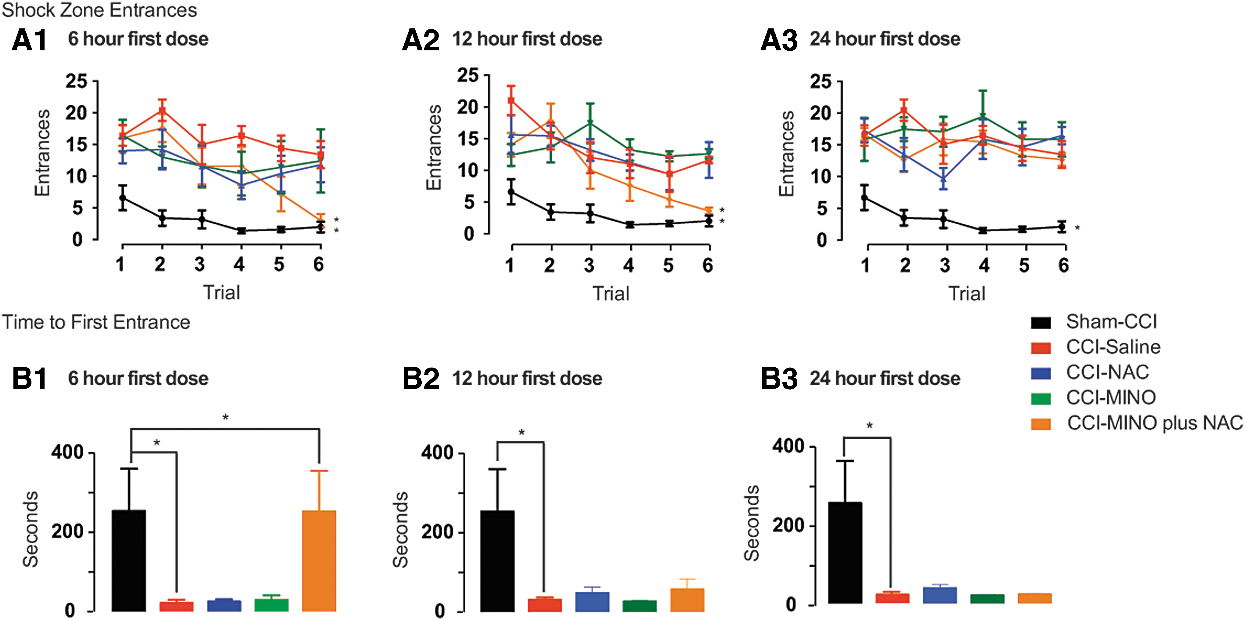
Assessment of drug efficacy on active place avoidance (APA) in rats after controlled cortical impact (CCI). (
Time to first entrance also showed a significant effect of treatment (F10,54 = 5.74, p < 0.0001). Both the sham-CCI group and the CCI-injured group treated with MINO plus NAC first dosed at 6 h increased time to first entrance. This increase was not seen in any of the other groups (post hoc, p < 0.05) (Fig. 7B1–3). These data suggest that a first dose of MINO and NAC at 6 h PI synergized to restore APA acquisition and retention while a 12 h first dose synergized to restore only APA acquisition.
At 14 days after sham-CCI or CCI, the amount of immunoreactivity of dendritic protein MAP2 was examined in the CA1, CA3, DG, and hilar regions of the dorsal hippocampus (Fig. 8A). All hippocampal regions showed a significant group effect (CA1, F9,40 = 25.56, p < 0.0001; CA3, F9,40 = 18.80, p > 0.0005; hilus, F9,39 = 21.65, p > 0.0001, DG, F9,39 = 21.00, p > 0.0001). The amount of immunoreactive MAP2 in the sham-CCI group was significantly lowered compared with the CCI saline group (post hoc, p < 0.0005) (Fig. 8B). This effect was observed in all regions assayed. Injured rats first dosed with MINO plus NAC at 6 h PI significantly increased MAP2 immunoreactivity in all hippocampal regions compared with the CCI saline group (p < 0.0001).
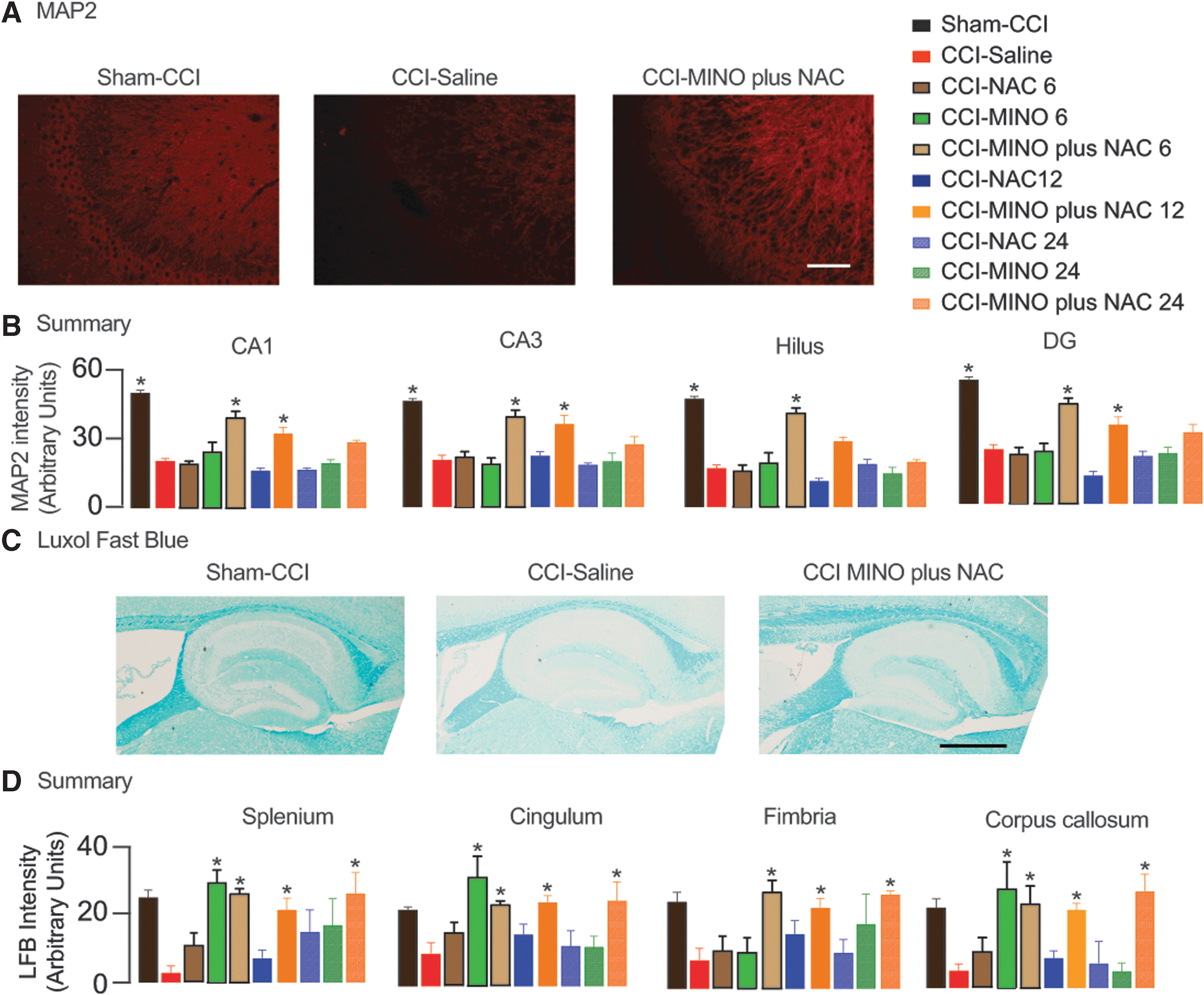
Assessment of gray and white matter injury in rats after controlled cortical impact (CCI). (
Increased MAP2 immunoreactivity was also seen in injured rats with a first dose of MINO plus NAC at 12 h PI. These effects were significant in CA1, CA3, and the DG (p < 0.05) and trended toward significance in the hilus (p = 0.07). MAP2 immunoreactivity was similar in the CCI -saline group and in groups treated with MINO or NAC alone. These data suggest that a first dose of MINO plus NAC at 12 h PI synergized to increase MAP2 immunoreactivity. Dosing of the drugs individually or in combination at 24 h PI had no effect on MAP2 immunoreactivity.
Myelin content was assessed using LFB in the rats previously analyzed for MAP2. LFB intensity was measured in the splenium, cingulum, fimbria, and body of the corpus callosum in the hemisphere ipsilateral to the impact site (Fig. 8C). All regions showed a significant treatment effect (splenium, F8,40 = 5.89, p < 0.0005; cingulum, F8,40 = 5.59, p < 0.0005; fimbria, F8,40 = 3.56, p < 0.005; corpus callosum, F8,40 = 6.30 p < 0.0001). LFB stain intensity in the sham-CCI group was significantly lower than in the CCI saline group (p < 0.005). Significantly higher LFB staining was seen in injured rats first dosed with MINO alone at 6 h or 12 h PI than injured rats treated with saline (Fig. 8D). These effects were seen in the splenium, cingulum, and corpus callosum (post hoc, p < 0.05). Injured rats receiving a first dose of MINO plus NAC at 6 h, 12 h, or 24 h PI increased LFB staining compared with CCI saline. These effects were seen in all white matter regions assayed except in the fimbria (post hoc, p < 0.05). Similar LFB staining was seen in injured rats first dosed with saline or NAC. These data suggest that a first dose of MINO plus NAC synergize to increase in LFB intensity when first dosed at 24 h PI.
Discussion
This study identified the most potent formulation of MINO plus NAC that improved behavioral outcomes (Fig. 1). This formulation of MINO plus NAC reduced behavioral deficits in both a spatial task (Fig. 3, 7) and a more challenging behavioral task involving sensory segregation (Fig. 4, 7). MINO plus NAC limited hippocampal gray matter injury (Fig. 5B, 8B) and myelin loss (Fig. 5C, 8C). Most importantly, these therapeutic effects were seen in both the CHI and CCI models with the drugs dosed at a clinically relevant time window.
MINO (22.5 mg/kg) plus NAC (75 mg/kg) was the most potent combination tested (Fig. 1). MINO plus NAC are drugs approved by the Food and Drug Administration (FDA), which increases the probability that the combination is safe. The safety of the combination has not been established in humans, yet the doses used in this proposal are close to what have been used clinically. The NAC dose (75 mg/kg) used in this study is two-fold lower than the maximum clinical dose. NAC (75 mg/kg) was also well tolerated in a small clinical study of TBI.
2
The MINO dose (22.5 mg/kg) used in the study is higher than the antimicrobial dose but is within the range of the dosage used to test MINO in a clinical trial to manage TBI (
The MINO plus NAC combination improved the ability of mice and rats to acquire and retain Barnes maze and APA (Fig. 3, 4, 6, 7). The therapeutic time window assessed by Barnes maze was larger than for APA. Increasing the therapeutic time window also decreased the protection against gray and white matter injury by MINO plus NAC. Barnes likely has a longer therapeutic time window because it requires fewer brain regions to function properly than for APA. Lesion studies support this hypothesis. Mice can acquire Barnes maze with one functional hippocampus or with lesions in the corpus callosum and ventral hippocampal commissure. 37,38 Acquisition of APA requires two hippocampi and the white matter tracts that connect them. 34,39 Recent experiments also suggest that Barnes maze, but not APA, can be acquired with localized demyelination of corpus callosum (E.N and P.J.B, unpublished results). These data suggest that severely injured rodents can acquire Barnes maze but not APA. Hylin and coworkers 40 also reported injured mice that acquired Barnes maze, but not APA. 40
Either MINO alone or MINO plus NAC prevented myelin loss (Fig. 5, 8). The mechanism used by the drugs first dosed 12 h or 24 h PI is not known. One hour dosing of MINO or MINO plus NAC repaired demyelinated white matter through remyelination. 6 MINO plus NAC induced remyelination through protection of resident oligodendrocytes. MINO promoted remyelination through proliferation and differentiation of oligodendrocyte precursor cells. Twelve or 24 h dosing of the drugs likely uses similar mechanisms.
The maximum therapeutic time window tested for MINO plus NAC was 24 h PI. When first dosed to mice at 24 h PI, MINO plus NAC restored acquisition of Barnes maze (Fig. 3). CHI-injured mice are impaired on Barnes maze, suggesting that both hippocampi lack function. Mice first treated with MINO plus NAC acquired Barnes maze even though the hippocampus ipsilateral to the injury site had substantial gray matter damage (Fig. 5B). The need for only one hippocampus to acquire Barnes maze suggests that a first dose of MINO plus NAC at 24 h PI may restore the acquisition of Barnes maze by acting on the contralateral hippocampus.
In the CHI model, MINO improved performance on APA (Fig. 3), Barnes maze (Fig. 4), and improved myelin content (Fig. 5D). NAC alone improved performance on Barnes maze (Fig. 4). MINO plus NAC previously showed synergy when dosed 1 h after rat CCI. 6 This synergy was retained when dosed up to 12 h (Fig. 6). MINO plus NAC also synergized to maintain MAP2 expression and myelin content (Fig. 5, 8).
There are limitations to the conclusions in this study. In addition, immunohistochemical analyses may not be quantitative. The large number of determinations in this study, however, provides evidence that MINO (22.5 mg/kg) plus NAC (75 mg/kg) not only partially restore cognition, but also provide histological improvements in the hippocampus ipsilateral to the impact site.
Conclusion
We demonstrated in two animal TBI models that a combination of two FDA-approved drugs, MINO and NAC, effectively restores cognition and memory while preserving white and gray matter. Most importantly, MINO plus NAC was effective when first dosed at a clinically relevant time window. The drug combination presents a very promising therapy to treat patients with TBI.
Footnotes
Acknowledgments
This work was supported by an award from the USAMRCC (09127004) to P.J.B.
Author Disclosure Statement
No competing financial interests exist.
References
Supplementary Material
Please find the following supplemental material available below.
For Open Access articles published under a Creative Commons License, all supplemental material carries the same license as the article it is associated with.
For non-Open Access articles published, all supplemental material carries a non-exclusive license, and permission requests for re-use of supplemental material or any part of supplemental material shall be sent directly to the copyright owner as specified in the copyright notice associated with the article.
