
Abstract
The aim of this study was to determine the current trends in magnetic resonance imaging (MRI)/computed tomography (CT) utilization for spine trauma in various clinical scenarios. We conducted a survey across six AO regions and preformed pair-wise comparisons between responses obtained from different AO regions. The survey was sent to 5813 surgeons and had a 9.6% response rate with the majority being orthopedic followed by neurosurgeons. In a neurologically intact patient, the predominant imaging modality for all AO regions was CT. For patients with spinal cord injury (SCI), the predominant choice for all AO regions was CT + MRI + x-ray except North America, which was CT + MRI; pair-wise comparisons revealed significant differences involving LATAM (Latin America) versus (Asia-Pacific [APAC], Europe [EU], and Middle East [MEA]) and APAC versus (LATAM and North America [NA]). In a patient with incomplete SCI (ISCI) who presented within 4 h and had CT, the predominant choice for all AO regions was “forgo MRI and proceed to operating room (OR).” Similar to ISCI, in a patient with complete SCI, the predominant option for all AO regions was the same as ISCI, but the range was lower. Pair-wise comparisons noted significant differences between MEA and APAC, with both exhibiting differences compare to NA, LATAM, and EU for complete and ISCI. Most AO regions obtained post-operative MRI only if there was a new deficit. In summary, decisions about the use of a particular imaging modality across AO regions appears to be influenced by the neurological status of the patient upon admission and the presence of neurological deficits post-surgery. Type of residency training and fellowship training did not have an influence on choosing the appropriate imaging modality for both intact and impaired patients. Further study is needed to determine whether accessibility to MRI would change surgeons' attitude toward obtaining MRI in patients with SCI.
Introduction
Imaging is a crucial component during the initial phase of management of the polytrauma patient in the clinical setting. The choice of imaging modality deployed as part of the institutional trauma protocol depends upon the anatomical structure(s) to be visualized with computed tomography (CT) and magnetic resonance imaging (MRI) utilized most commonly. Currently, the AOSpine guidelines suggest that MRI be performed in adult patients with acute spinal cord injury (SCI) before surgical intervention to help with clinical decision making and possibly after to help with predicting neurological outcomes. 1 However, current trends for the utilization of aforementioned imaging techniques by physicians across different geographical regions remain unchartered. This underscores a gap in knowledge, which AOSpine, in its commitment to enhancing the understanding of the role of imaging in the standard of care in spine trauma and spinal cord injury (SCI), attempts to answer. Consequently, the present report highlights the findings from an AO survey aimed at understanding the role of imaging in decision making by focusing on differences in the usage rates for CT and MRI among different AO regions.
Methods
To determine the utilization of CT and MRI among different geographical regions, a survey was conducted across the six global AO regions (Africa, Asia Pacific, Latin America, Middle East, Europe, and North America). The questions included in the survey were structured in a way to evaluate preferences for various imaging modalities (x-rays, CT, and MRI) in different clinical scenarios (Table 1). The questions for clinical scenarios were structured based on the location of neurological injury, type of imaging modality that was performed (i.e., x-ray vs. CT), and/or the presence of neurological deficit to evaluate the preferred imaging modality in each scenario. The next set of questions were structured to assess the importance of obtaining MRI in complete versus incomplete SCI in setting of having fracture on the CT.
Characteristics of AO Respondents
CSS, Cognitive Science Society; KF, Knowledge Forum; SCI, Spinal Cord Injury; NACTN, North American Clinical Trials Network®.
We also looked to determine whether type of residency (i.e., trauma, neurosurgery, and orthopedic surgery) and fellowship training has any impact on obtaining preferred imaging modality in each clinical scenario (Table 1). The data for each clinical case question were collected and analyzed using SPSS Statistics software (version 22.0; SPSS, Inc., Chicago, IL). Responses were tabulated with frequencies and percentages. Pair-wise comparisons regarding the distribution of responses between AO regions were performed with the Mann-Whitney U test for ordinal responses and with a chi-square test for nominal responses. Two-sided p < 0.05 was considered statistically significant.
Results
Table 1 provides an overview of all respondents. In total, 561 completed surveys were obtained. The survey was sent to total of 5813 members, and 561 completed surveys were received for a response rate of 9.6% (Table 2). Of the members that completed the survey, 165 were neurosurgeons, 351 were orthopedic surgeons, 28 were trauma surgeons, and 17 were other (emergency room physicians); 66% (370) surgeons had fellowship training in the spine (Table 2).
Survey Questions
Of note in cases 9 and 10: You are going to take him to the OR for a C5 vertebrectomy, cage reconstruction, and fusion and a subsequent posterior instrumented stabilization. The OR is available right not, but an MRI is ordered, and you are informed that the MRI will require time to transport the patient there, obtain the scan, and transport the patient back. Also, when the patient is finished with the MRI, the OR may not be available right away and you may need to wait before the next room is available.
CT, computed tomography; MRI, magnetic resonance imaging; SCI, spinal cord injury; OR, operating room.
Clinical scenarios
The description of each question of the survey (n = 15) is provided in Table 2. Figure 1 summarizes the responses regarding preferences for imaging modalities based on various case scenarios for each AO region, and Table 3 provides the pair-wise comparisons.
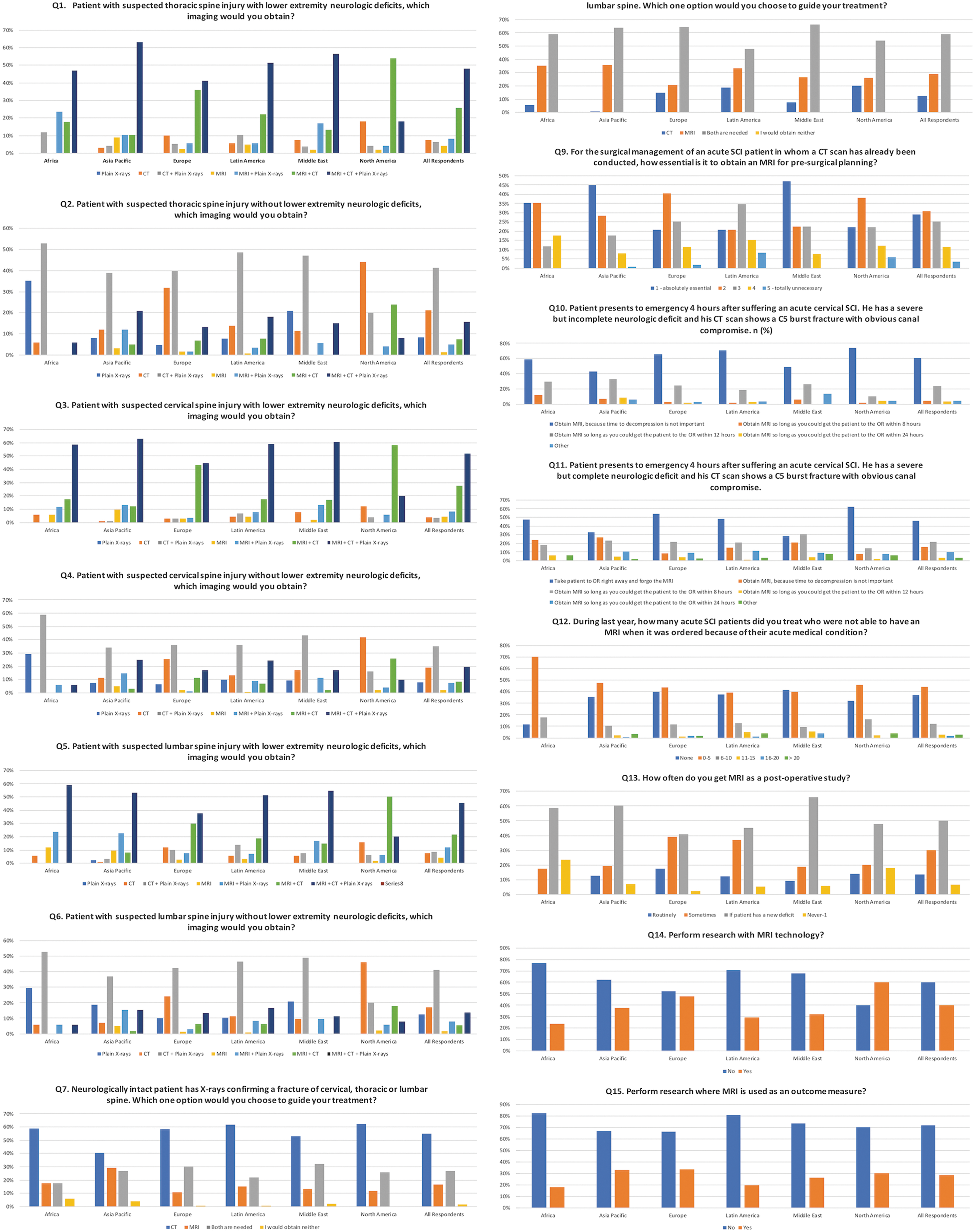
Bar graph showing the reponses from each of the six AO regions for the 15 questions included in the survey. Color image is available online.
Pair-Wise Comparisons within Table 3, with p Values
The decision to use a particular imaging modality was seen to be strongly influenced by the presence of neurological deficits. For clinical cases 1 (suspected thoracic spine injury with lower extremity deficits), 3 (suspected cervical spine injury with lower extremity deficits), and 5 (suspected lumbar spine injury with lower extremity deficits), the responses were significantly different between North America and most of the other AO regions (see Fig. 1). For North America, the predominant imaging combination in the presence of any neurological deficit was MRI + CT (at least 50% of respondents, depending on suspected spinal level); for all other AO regions, the predominant imaging combination was MRI + CT + x-rays (ranging from 41% to 62.9% of respondents, depending on suspected spinal level). On the other hand, in the absence of neurological deficits, such as clinical cases 2 (suspected thoracic spine injury), 4 (suspected cervical spine injury), and 6 (suspected lumbar spine injury), the responses from North America were not significantly different compared with other AO regions.
For the three clinical scenarios (cases 2, 4, and 6), pair-wise comparisons involving Africa were significant against Asia (cases 2 and 4) and Latin America (cases 2, 4, and 6), with Africa showing a greater percentage of respondents utilizing only plain x-rays (ranging from 29.4% to 35.3%, depending on suspected spinal level) compared with other regions.
The next set of questions centered around the influence of neurological exam findings on the decision for the next requested imaging modality in the setting of confirmation of a fracture on x-rays. For a neurologically intact patient (case 7), the predominant choice for all AO regions was CT (ranging from 40.3% to 62% of respondents within each region), with pair-wise comparisons revealing significant differences between Latin America and Asia Pacific. For a neurologically impaired patient (case 8), the predominant choice for all AO regions was CT + MRI (ranging from 47.9% to 66.0% within each region); pair-wise comparisons revealed significant differences between more AO regions, largely involving Latin America (vs. Asia Pacific, Europe, and Middle East) and Asia Pacific (vs. Latin America and North America).
The respondents were then queried about the role of MRI in the surgical management of an acute SCI patient in whom a CT scan has already been conducted (question 9). There were marked differences between AO regions on whether MRI was essential for pre-surgical planning, with regional responses of “absolutely essential” ranging between 20.8% and 47.2% and “totally unnecessary” ranging between 0% and 8.3%.
Questions that followed assessed the role of neurological deficits with regard to the request of an MRI—for example, given the specific clinical finding of a C5 burst fracture noted with CT scanning in a patient presenting 4 h after SCI. In a patient with incomplete SCI (case 9), the predominant choices for all AO regions were “Take patient to OR [operating room] right away and forgo MRI” (ranging between 42.7% and 74%) and “Obtain MRI so long as you could get patient to the OR within 8 h” (ranging between 10% and 33%). North America respondents were most likely to forgo MRI and proceed to the operating theater. For a complete SCI (case 10), the first predominant option for all AO regions remained the same as for case 9, but the ranges shifted lower (28.3–62%), whereas the option “Obtain MRI, because time to decompression is not important” became more prominent, ranging between 8% and 26.6%. Pair-wise comparisons noted numerous significant differences between AO regions (see Table 3). Notably, Middle East and Asia Pacific both exhibited differences compared to North America, Latin America, and Europe for both cases; Middle East and Asia Pacific were the two least likely to forgo MRI.
The remaining set of questions were focused on the use of MRI. The response to the question concerning the number of acute SCI patients treated by the physician last year in whom MRI was ordered but not performed because of their acute medical condition (question 12) exhibited no significant differences on pair-wise comparisons between AO regions (Table 4). When asked about the use of post-operative use of MRI (question 13), significant differences on pair-wise comparisons were observed (see Table 3), with the predominant choice for all AO regions being to conduct an MRI “if the patient has a new deficit,” ranging between 41% and 66%. Notably, for Africa, 0% obtained a routine MRI post-operatively whereas 23.5% never obtained an MRI post-operatively (even with new deficits). Another question inquiring about the use of MRI technology in performing research (question 14) revealed that only North America exhibited a majority of respondents (60%) who answered “yes,” with significant pair-wise comparisons to all other AO regions. Last, when asked about the use of MRI as an outcome measure in research (question 15), respondents from all AO regions largely answered in the negative about the use of MRI as an outcome measure, with “no” answers ranging from 66.5% to 82.4%.
Cases Analyzed with Multiple Answers by AO Region
CT, computed tomography; MRI, magnetic resonance imaging.
Table 4 highlights multiple-choice answers given the same case scenarios as above. Overall, for a suspected spinal injury with no neurological deficits (cases 2, 4, and 6), most respondents were less likely to obtain CT and/or MRI. Notably, the responses from Africa were the least likely to obtain an MRI scan (only 5.9–11.8%, depending on the spinal level).
Orthopedic, neuro, and trauma surgeons chose CT as a main imaging modality in neurologically intact patients. However, the “other” group had equal distribution among CT and CT + MRI. For patients with neurological deficit, CT + MRI was the main imaging modality across different surgeon groups. With respect to fellowship training, no differences were noted for either intact or impaired patients for obtaining the relevant imaging modality, with CT being more preferred in intact and CT + MRI in impaired patients.
Discussion
The finding of differences in the utilization of imaging modalities across AO regions in the present study highlights the influence of neurological status of the patient on clinical management. A deeper dive of the implications of the differences observed for individual questions is attempted below.
The first six clinical scenarios (cases 1–6) highlights the preferences for imaging modalities across AO regions, with all AO regions predominantly utilizing multi-modal imaging in the presence of a neurological deficit and suspected spine injury. For North America, the predominant imaging combination in the presence of any neurological deficit is MRI + CT (at least 50% of respondents, depending on suspected spinal level); for all other AO regions, the predominant imaging combination is MRI + CT + x-rays (ranging from 41% to 62.9% of respondents, depending on suspected spinal level). On the other hand, with no deficits, Africa appears to utilize only plain x-rays (ranging from 29.4% to 35.3%, depending on suspected spinal level) more so than other regions, likely reflecting restricted imaging resources, overcapacities issues, and limited access to specialist. 2 The preference for plain x-rays along with MRI + CT in the face of neurological deficits by all regions except North America is unclear. We suspect this observed finding could be attributed to differences in clinical mentality to evaluation of spine trauma and access to imaging facilities and/or personnel. 3 –5
Accordingly, even though a large percentage of surgeons that responded to the survey might be able to obtain CT scan within 4 h, when dealing with a patient with neurological deficits this time duration might be too long, and some might be inclined to obtain additional x-ray imaging. Further, results from recent U.S. studies noting increasing CT utilization in emergency departments for trauma patients, even when controlling for injury severity, hint to the possible influence of physicians' approach to trauma evaluation on imaging utilization. 6,7
The present analysis explored the clinical dilemma surrounding the role of MRI in a patient with SCI. The AOSpine guidelines suggest MRI to be performed in adult patients with acute SCI before surgical intervention to help with clinical decision making, but different practices were noted across the AO regions. 1 Given a CT scan in a patient with an acute SCI (question 9), marked differences were observed between AO regions whether an MRI was essential for pre-surgical planning, ranging from 20.8% to 47.2% for “absolutely essential” and 0% to 8.3% for “totally unnecessary.” Even though all AO regions seemed be in agreement that an MRI might be beneficial, Europe, Latin America, and North America were less adamant about the “absolute necessity compared to the other AO regions, namely, Middle East and Asia Pacific.” This might appear at odds with the response received from North American surgeons when inquired about their imaging preferences in the presence of a neurological deficit and suspected spine injury, with over half picking MRI + CT.
However, the tendency toward obtaining MRI + CT for patients with acute SCI needs to be seen in the context of the time interval elapsed between injury and presentation to the hospital. Based on the response to a question inquiring whether the surgeon would obtain an MRI when a CT scan had already been obtained, a large percentage of surgeons indicated that they would forgo MRI if a CT scan had been obtained in patients and more than 4 h had passed between time of injury and admission to the hospital. So, we believe that the urgency to treat a patient with neurological deficits might supersede the likelihood of obtaining an MRI. Oddly, respondents from Africa believed that an MRI is essential, but the region probably has the least access to radiology units. The overall agreement noted in favor of obtaining an MRI stems from the many noted advantages associated with this imaging modality, including a better visualization of soft tissues. Studies have also noted that MRI is more sensitive for detection of ongoing spinal cord compression compared to CT. 8,9 In addition, the modality is advantageous for assessment of comatose patients or unexplained neurological deficits. 10
In cases of thoracolumbar spinal trauma, MRI augments the detection of fractures and soft tissue damage, refining the overall trauma classification compared to CT alone. 9,11 However, a recent article involving the classification of 30 thoracolumbar fractures by 41 spine surgeons noted that although MRI was more sensitive, the decision to operate was not affected by MRI data. 11
For cases 9 and 10, preferences regarding MRI imaging after CT-confirmed burst fracture/canal compromise were broached. The scenarios allowed for surgery within 24 h in the following clinical context: “You are going to take him to the OR for a C5 vertebrectomy, cage reconstruction, and fusion, and a subsequent posterior instrumented stabilization. The OR is available right now, but MRI is ordered, and you are informed that the MRI will require time to transport the patient there, obtain the scan and transport the patient back. Also, when the patient is finished the MRI, the OR may not be available right away and you may need to wait before the next room is available.” Notably, Middle East and Asia Pacific both exhibited differences compared to North America, Latin America, and Europe for both cases; Middle East and Asia Pacific were the two least likely to forgo MRI. These findings appear to correlate with question 9, given that the top response for both regions was that an MRI was “absolutely essential” for pre-surgical planning. However, there was a pre-defined surgical approach, so a different clinical mentality may be at work.
With an incomplete injury, 2 (11.8%) of respondents from Africa believed that time to decompression is not important, compared to the next highest at 7.3% for Asia Pacific. With a complete injury, lower proportions from all AO regions wanted to operate immediately, forgoing MRI; moreover, high proportions believed that timing is not important compared to an incomplete injury.
The perceptions around the need for MRI in the context of acute SCI interestingly highlight differences in opinion about the timing of surgical intervention. Though timing of surgery remains controversial, the recent trends with data have leaned toward earlier surgical intervention. 12 In 2012, the Surgical Timing in Acute Spinal Cord Injury Study (STASCIS) compared early (<24 h) to late (>24 h) surgical intervention for cervical trauma (American Spinal Injury Association [ASIA] A–D). At 6 months follow-up, the former group demonstrated 2.8 times more likelihood for ≥2 ASIA grade improvement. 13 This conclusion was reaffirmed in subsequent studies regarding improved motor scores. 14,15 Early decompression has also been associated with shorter hospitalizations. 15
A recent systematic review concluded that results of earlier studies have been variable, but underscores that evidence supports early surgery for acute cervical SCI, but not for other spinal levels. 16 The results from the AO survey align with a recent Netherlands survey, which found that surgery was less urgent in complete SCI compared to incomplete SCI; notably, 43% patients with complete SCI were not operated within the recommended 24-h guideline. 17 In addition to timing of the surgery, Aarabi and colleagues recently discussed the importance of complete canal decompression for improved recovery in patients with SCI. They used pre-operative MRI to better visualize the extent of injury to help with better surgical planning and used post-operative MRI to confirm the complete extent of decompression to maximize neurological outcomes. 18
The present analysis contains a number of limitations. The survey was mainly sent to AOSpine members, which could be a source of bias given that AOSpine members, despite spanning multiple continents, might not be as a valid proxy for all the spine surgeons throughout the world. The present study was carried out with the intention of obtaining a high response rate, but the exploratory nature of the study, coupled with the goal of covering several geographical regions with many different questions concerning the accessibility to MRI and management of patients with acute SCI in different scenarios across trauma, orthopedic, and neurosurgeons, does not allow us to comment with certainty on the adequacy of the response rate. Consequently, a power analysis was not attempted for the present analysis.
Conclusions
The presence of differences regarding preferences for various imaging modalities shows that decisions about the use of a particular imaging modality across various AO regions appear to be influenced by the neurological status of the patient upon admission and the presence of neurological deficits post-surgery. Whether this is a consequence related to the availability of resources remains unanswered and requires further study to determine whether the accessibility to MRI would change surgeons' attitude toward obtaining MRI in patients with SCI.
Footnotes
Acknowledgments
This study was organized and funded by AOSpine through the AOSpine Knowledge Forum Spinal Cord Injury, a focused group of international spinal cord injury experts as well as Bryon Riesch Paralysis Foundation. AOSpine is a clinical division of the AO Foundation, which is an independent, medically guided, not-for-profit organization. Study support was provided directly through the AOSpine Research Department.
Author Disclosure Statement
No competing financial interests exist.