




Abstract
Chronic subdural hematoma (CSDH) is an angiogenic disease that is involved with many inflammatory mediators. Tie2 is predominantly expressed in the embryonic endothelium and plays an important role in the maturation and stabilization of the vasculature. Angiopoietin (Ang)1 and Ang2 are well-known ligands of the Tie2 receptor. We examined the expression of Ang1 and Ang2 in CSDH fluid and the expression of Tie-2 receptor and components of the angiogenic signaling pathways in the outer membrane of CSDH. Twenty-five samples of CSDH fluid and eight samples of outer membrane of CSDH were included. The concentrations of Ang1 and Ang2 in the CSDH fluid were measured using enzyme-linked immunosorbent assay (ELISA) kits. The expression of Tie2, phosphoinositide 3-kinase (PI3K), protein kinase B (Akt) mechanistic target of rapamycin (mTOR), GβL, 70 kDa ribosomal protein S6 kinase (p70S6K), eukaryotic initiation factor 4E (eIF-4E), and β-actin was examined by a Western blot analysis. The expression of Tie2, Akt, and mTOR was also examined by immunohistochemistry. The concentration of Ang2 in CSDH fluid was significantly higher than that in the serum or cerebrospinal fluid (CSF), and also higher than that of Ang1 in CSDH fluid. Tie2, PI3K, Akt, mTOR, GβL, p70S6K, and eIF-4E were detected in all cases. In addition, Tie2, Akt, and mTOR were localized in the endothelial cells of vessels in the CSDH outer membrane. Our data suggest that Ang2, although not Ang1, in CSDH fluid promotes angiogenesis in endothelial cells through the Tie2 receptor. The Ang2/Tie2 signaling pathway might therefore be a useful therapeutic target for treating the growth of intractable CSDH.
Introduction
Chronic subdural hematomas (CSDHs) occur in elderly people who have sustained mild head trauma. Patients with CSDH present with symptoms such as headache, hemiparesis, gait disturbance, and a decreased cognitive function. The treatment for symptomatic CSDH is trepanation surgery, even in elderly people. However, recurrence of CSDH after the surgery has been observed at a rate of 5–17%. 1,2 The precise mechanism underlying the development and recurrence of CSDH still remains to be elucidated.
Vascular endothelial growth factor (VEGF) is a potent angiogenic factor, and high concentrations of VEGF in CSDH fluid have been reported, 3 –5 suggesting that VEGF might be involved in the angiogenesis of the CSDH outer membrane. VEGF in CSDH fluid activates the nuclear factor kappa-light-chain-enhancer of activated B cells (NF-κB) and Ras/ mitogen-activated protein kinase (MAPK) extracellular signal-regulated kinase (ERK) (MEK)/ERK pathways in endothelial cells, resulting in angiogenesis in the outer membrane of CSDH. 6,7 The inflammatory cytokine interleukin (IL)-6) has been reported to be elevated in CSDH fluids and significantly correlated with recurrence. 8 IL-6 activates the janus kinase (JAK)/signal transducer and activator of transcription (STAT) signaling pathway in fibroblasts and endothelial cells in CSDH outer membranes and plays a role in the growth of CSDH. 9,10 These data suggest that angiogenesis and inflammation play key roles in the development of CSDH. 11
Angiopoietins are another family of growth factors that are involved in blood vessel formation during developmental and pathological angiogenesis. Ang1-deficient mice die between E11.5 and E12.5 because of dilated vessels and a decreased complexity of the vascular network. 12 A previous study showed that mRNA species encoding Ang1, Ang2, and Tie-2 were detected in outer membrane samples, suggesting that angiogenic mechanisms induced by angiopoietins play an important role in the pathophysiology of CSDH. 13 However, the protein levels of these molecules are still unknown, as the expression may differ between mRNA and protein.
Therefore, we explored whether or not angiopoietins exist in CSDH fluid, and investigated the angiogenic signaling pathway in the outer membrane of CSDH using immunoblotting and immunohistochemical analyses.
Methods
This study was approved by Certified Clinical Research Review Board of Aichi Medical University Hospital (17-H047). Informed consent was obtained from each patient or the patient's family. Burr hole irrigation and drainage surgery were performed on all patients under local anesthesia at Aichi University Hospital.
Materials
All chemicals were obtained from Sigma Chemical (St. Louis, MO) unless otherwise specified.
Analysis of angiopoietins in CSDH fluid
Fluid from 25 consecutive CSDHs were obtained during trepanation surgery. As a control, CSF samples were obtained from 10 patients undergoing neck clipping for unruptured cerebral aneurysm, and serum samples were obtained from 6 patients with CSDH. After collection, all samples were immediately centrifuged, and the supernatant was stored at -80°C until the analysis.
We measured the concentrations of Ang1 and Ang2 using enzyme-linked immunosorbent assay (ELISA) kits (R&D Systems, Minneapolis, MN), according to the manufacturer's instructions. The mean minimum detectable doses of these assays were 3.5 pg/mL for Ang1 and 8.3 pg/mL for Ang2.
Western blotting analyses
Eight samples of the outer membrane of the CSDH were obtained during surgery. The membranes were homogenized in 75 μL of homogenization buffer containing 50 mM Tris base/HCL (pH 7.5), 0.1 mM dithiothreitol, 0.2 mM ethylenediaminetetraacetate, 0.2 mM ethylene glycol bis(aminoethyl ether)tetraacetate, 0.2 mM phenylmethylsulfonyl fluoride, 1.25 μg/mL of pepstatin A, 0.2 μg/mL of aprotinin, 1 mM sodium orthovanadate, 50 nM sodium fluoride, 2 mM sodium pyrophosphate, and 1% Nonidet™ P-40. The homogenates were centrifuged at 12000g for 10 min at 4°C. The protein concentrations of the supernatants were separated using 7.5% or 10% sodium dodecyl sulfate (SDS)-polyacrylamide gel electrophoresis.
The proteins in the gel were transferred to polyvinylidene difluoride membrane and incubated with primary polyclonal antibodies against Tie2 (R&D Systems), phosphoinositide 3-kinase (PI3 kinase, Cell Signaling Technology, Danvers, MA), protein kinase B (Akt; Cell Signaling Technology), mechanistic target of rapamycin (mTOR; Cell Signaling Technology), GβL (Cell Signaling Technology), 70 kDa ribosomal protein S6 kinase (p70S6K; Cell Signaling Technology), eukaryotic initiation factor 4E (eIF4E; R&D Systems) and β-actin (Sigma). Akt and mTOR antibody were diluted 1:1,000, and all the others were used at a 1:500 dilution and incubated overnight at 4°C. After unbound antibodies were washed away, the membranes were incubated in secondary antibodies with horseradish peroxidase (Sigma) at a 1:3000 dilution for 30 min at room temperature. The reactions were developed with ECL Plus (GE Healthcare, Buckinghamshire, UK). Positive controls were Jurkat whole cell lysate (Cell Signaling Technology) and NIH/3T3 whole cell lysate (Santa Cruz Biotechnology, Dallas, TX).
Histological examinations
For the analysis of the cellular localization of Tie2, Akt, and mTOR, immunohistochemical staining was performed on samples from three patients at room temperature using the avidin-biotinylated peroxidase complex (ABC) technique. To preserve the outer membrane of the CSDH samples, the membranes were incubated in 10 mL of ice-cold 4% paraformaldehyde in 0.1 M phosphate buffer (pH 7.4) for 3 h and then embedded in paraffin.
In this study, 10-μm-thick sections were prepared on a microtome and mounted onto MAS-coated glass slides (Matsunami Glass, Kishiwada, Japan). The sections were deparaffinized in xylene, immersed in decreasing concentrations of ethanol, and rehydrated in water. Endogenous peroxidase activity was blocked with 0.3% H2O2 in 100% methanol for 20 min. All sections for immunostaining were processed for microwave-enhanced antigen retrieval. Slide-mounted sections immersed in 0.01 M sodium citrate buffer (pH 6.0) were placed for 15 min in a 700 W microwave oven at maximum power.
Non-specific immunoreactivity was blocked by incubation with goat or donkey serum for 30 min, depending on the primary antibody. The samples were treated with primary antibodies against Tie2 (R&D Systems) at a dilution of 1:150, Akt (Cell Signaling Technology) at a dilution of 1:300, and mTOR (Cell Signaling Technology) at a dilution of 1:100 over 2 nights at 4°C. After several rinses in phosphate buffered saline (PBS), the samples were incubated with secondary biotinylated antibodies (anti-goat IgG 1:200, anti-rabbit IgG 1:200; Santa Cruz Biotechnology) at room temperature for 2 h. After several more rinses in PBS, they were incubated with the Vectastain ABC reagent (Vectastain ABC Kit; Vector Laboratories, Burlingame, CA) for 1 h. After several more rinses in PBS, the bound peroxidase was visualized by incubating the sections with a solution containing 0.05% 3,3'-diaminobenzidine tetrahydrochloride (Sigma Aldrich) and 0.01% H2O2 in 0.05 M Tris-HCl (pH 7.4) for 10 min. After several rinses in water, the immunostained sections were dehydrated and cover-slipped with Entellan New® rapid mounting medium (Merk, Kenilworth, NJ).
Statistical analysis
Data are expressed as the mean ± standard error. Significant differences between groups were assessed using a one-way analysis of variance (ANOVA) followed by Bonferroni's/Dunn's tests for multiple comparisons. The Mann–Whitney U test was used for the analysis between two groups. Significance was indicated when p < 0.05.
Results
Clinical data
Clinical data are presented in Table 1. There were 18 men and 7 women in this study, ranging in age from 51 to 92 years, with an average age of 78.1 years. In total, 25 CSDH fluid samples were obtained and included two recurrent cases. One patient (No. 16) died from a pancreatic cancer as the comorbidity. Hematoma type of computed tomographic (CT) images are divided according to the classification into four types grouped by Nakaguchi and colleagues. 1 There were four homogenous types, seven laminar types, six separated types and eight trabecular types.
Characteristics of 25 Patients with Chronic Subdural Hematoma
No. 7 and 11 are recurrent cases. The size refers to the largest extent measured in pre-operative computed tomography (CT) slices. Shift refers to the midline sift.
M, man; W, woman, lt, left; rt, right; H, homogeneous type; L, laminar type; S, separated type; T, trabecular type; GCS, Glasgow Coma Scale at admission; GOS, Glasgow Outcome Scale at discharge.
Concentrations of Ang1 and Ang2 in CSDH fluid
The concentration of Ang1 in serum (29,327.3 ± 5282.6 pg/mL) was significantly higher than that in CSDH fluid or CSF (1614.9 ± 184.3 pg/mL and 294.3 ± 37.7 pg/mL, respectively) according to a one-way ANOVA followed by Bonferroni's/Dunn's test (p < 0.05, Fig. 1A). In contrast, the concentration of Ang2 in CSDH fluid (97,523.8 ± 14,455.5 pg/mL) was significantly higher than that in the serum or CSF (1203.2 ± 162.2 pg/mL and 1596.3 ± 272.1 pg/mL, respectively) according to a one-way ANOVA followed by Bonferroni's/Dunn's test (p < 0.05, Fig. 1B). The concentration of Ang2 in CSDH fluid was significantly much higher than that of Ang1in the CSDH fluid, according to a Mann–Whitney U test (p < 0.01, Fig. 1C).

Concentrations of angiopoietin-1 (Ang1)
Western blotting analyses of Tie2 and the angiogenic signaling pathway
Figure 2 shows the results of Western blotting analyses of Tie2 and the angiogenic signaling pathway. Nearly constant β-actin levels were detected in CSDH outer membrane samples. Tie2, PI3K, Akt, mTOR, GβL, p70S6 kinase, and eIF-4E were detected in almost all samples; however, in some cases, the signals were weak. Positive controls revealed that these molecules had been correctly detected.
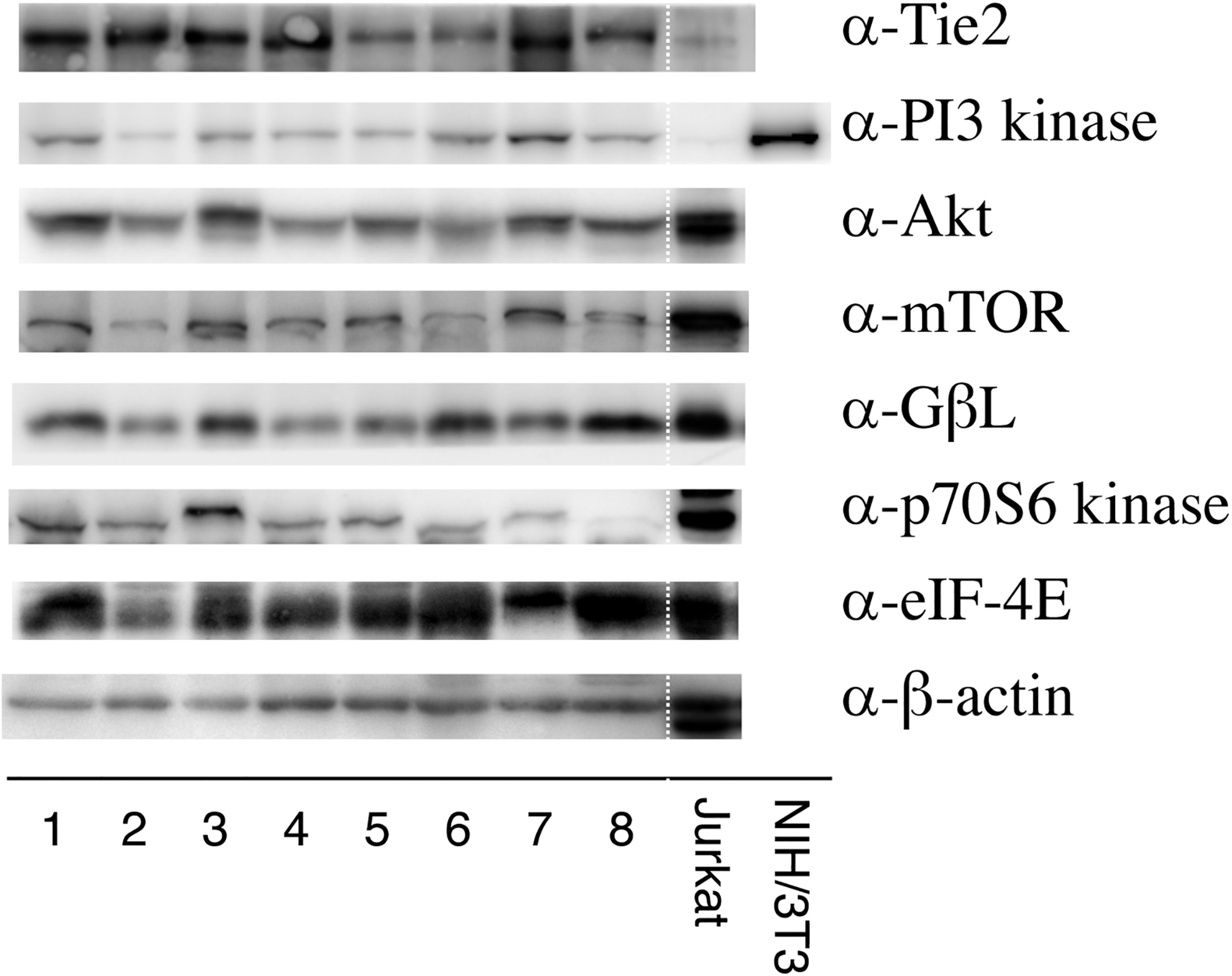
Western blots showing the expression of angiogenic pathway molecules in the outer membrane of chronic subdural hematomas from eight patients. Tie2, phosphoinositide 3-kinase (PI3 kinase), protein kinase B (Akt), mechanistic target of rapamycin (mTOR), GβL, 70 kDa ribosomal protein S6 kinase (p70S6K), and eukaryotic initiation factor 4E (eIF4E) were detected in almost all cases. Positive controls are shown in the right two lanes and suggest that these molecules were correctly detected. Jurkat, Jurkat whole cell lysate; 3T3/NIH, 3T3/NIH whole cell lysate
Historical observations
Tie2 (Fig. 3A and B), Akt (Fig. 3C and D), and mTOR (Fig. 3E and F) were localized in the endothelial cells of vessels within the outer membrane. Note that vascular longitudinal cross-sections in 10 μm consecutive slices at a higher magnification clearly demonstrate that these molecules are expressed in the endothelium (Fig. 3B, D, and F). In the negative controls examined without primary antibodies, the endothelial cells were consistently negative for the markers listed (Fig. 3G).
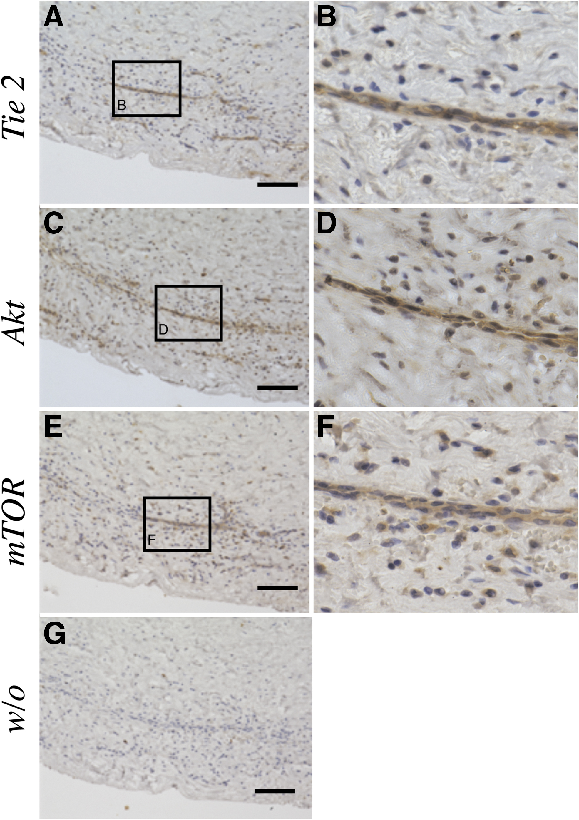
Ten-micrometer consecutive slices were immunostained with polyclonal antibodies against Tie 2 (
Discussion
The expression of Ang2 in CSDH fluid was significantly higher than that of Ang1 in CSDH fluid. Molecules of the angiogenic pathway, including Tie2, were detected in the outer membrane of CSDH. Tie2, Akt, and mTOR were expressed in the endothelium of vessels of the CSDH outer membrane.
Tie2 was identified as a receptor tyrosine kinase expressed principally on the vascular endothelium. 10 Ang1 phosphorylates Tie2 and induces vessel maturation. Ang1 mediates the migration, adhesion, and survival of endothelial cells. Both Ang1 and Ang2 bind to the Tie2 receptor. In general, Ang2 antagonizes Ang1 by keeping Ang1 from binding to Tie2 receptors. Ang2 disrupts the connections between the endothelium and perivascular cells, resulting in vascular regression. 14,15 However, some reports have shown that Ang2 is a Tie2 agonist, suggesting that the action of Ang2 as a Tie2 agonist or antagonist is context dependent. 16 The increased expression of Ang2 results in increased tumor vascular density, and the increased expression of Ang2 relative to Ang1 in tumors correlates with a poor prognosis. 17 Glioblastomas are characterized by prominent vascularization. Ang1 mRNA was expressed in tumor cells, whereas Ang2 mRNA was confirmed in endothelial cells of glioblastoma blood vessels, especially in small capillaries with few periendothelial support cells. 18,19 Hepatocellular carcinoma (HCC) is also a tumor frequently associated with increased vascularity. Ang2 expression may play an important role in vascular remodeling and angiogenesis in HCC tumors. 20,21 In systemic sclerosis patients, the serum levels of Ang1 were shown to be significantly lower and those of Ang2 were shown to be significantly higher than in the control group. 22 Altered Ang1/Ang2 levels might underlie the aberrant angiogenesis in systemic sclerosis. These data are agreement with our own finding that the level of Ang1 in CSDH fluids was lower whereas that of Ang2 was higher than the relevant serum levels in CSDH patients. A previous study revealed that the mean ratio of the Ang1/Ang2 mRNA expression in CSDH membrane was 0.48, 13 whereas the mean ratio of the Ang1/Ang2 protein level in CSDH fluid was 0.02 in our data. Ang2 can act as an agonist of the Tie2 receptor in the absence of Ang1. 16 Recently, Ang2 was shown to be upregulated in multiple inflammatory diseases and was implicated in the mediation of inflammatory processes as well. 23 Taken together, these findings suggest that the high concentration of Ang2 in CSDH fluid might be involved in the regulation of angiogenesis and inflammation within CSDH.
The expression of PI3K/Akt signaling molecules was confirmed by Western blot analyses. PI3K/AkT constitutes an important pathway regulating the signaling of multiple biological processes, such as apoptosis, metabolism, and cell proliferation. Dysregulation of the PI3/Akt pathway leads to diseases, such as cancer, diabetic mellitus, and cardiovascular disease. 24 Ang2 induced the phosphorylation of Tie2, PI3K, and Akt in human umbilical vein endothelial cells, resulting in the endothelial cell survival. 25 A previous study showed that activated Akt was expressed in the endothelial cells of vessels in CSDH, which is in agreement with our immunohistochemical result. 26 mTOR, which is a downstream kinase of the PI3K/Akt signaling pathway, plays an important regulatory role in cell survival, vascular permeability, migration, and proliferation, as well as in angiogenesis. 27,28 mTOR exists in two protein complexes: mTOR complex 1 (mTORC1) and mTOR complex 2 (mTORC2). mTORC1 consists of mTOR, raptor, and GβL. Activated mTORC1 catalyzes p70S6K and eukaryotic initiation factor 4E-binding protein-1 (4E-BP1) phosphorylation. 29,30 p70S6K plays an important role in regulating tumor angiogenesis in endothelial cells, 31 whereas 4E-BP1 is a translational repressor. The phosphorylation of 4E-BP1 leads to the release of eIF-4E from the 4E-BP1/eIF-4E complex. eIF-4E is a regulatory protein that binds to the mRNA cap-binding 32 and induces tumor vascularization in vivo. 33 Hypoxia-inducible factor (HIF)1 is a heterodimeric transcriptional factor composed of α and β subunits. Accumulated HIF1α translocates to the nucleus and dimerises with HIF1β to form a functional transcription factor capable of DNA binding at hypoxia response elements (HREs) and activating the transcription of target genes (Fig. 4). 34 HIF1 plays a central role in tumor progression and angiogenesis in vivo. The PI3K/Akt signaling pathway mediates the upregulation of HIF1α by increasing HIF1α synthesis or HIF1α stability, resulting in the transcriptional activity of HIF1. 35 HIF1α was confirmed to be positive in the outer membrane of CSDH and induced the excessive development of fragile microvessels through VEGF. 3 Both p70S6K and HIF1 are essential for tumor growth and angiogenesis. 31 Vanillic acid, an active compound isolated from green tea, inhibits angiogenesis in human colon cancer cells by suppressing the HIF-1α expression and mTOR/p70S6K/4E-BP1 pathway. 36 Given our findings that Tie2, Akt, and mTOR were expressed in endothelial cells, the PI3K/Akt/mTOR/eIF-4E signaling pathway activated by Ang2/Tie2 receptor may play an important role in angiogenesis in the CSDH outer membrane.
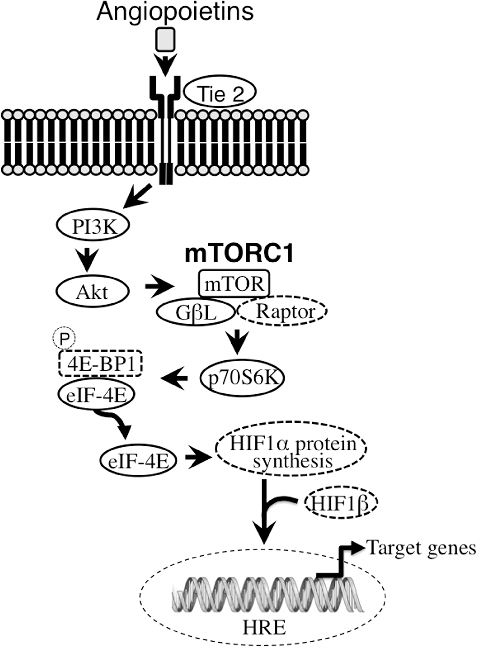
The signal transduction of angiopoietin in the outer membrane of chronic subdural hematoma (CSDH). Angiopoietins binds to Tie 2 receptor. Mechanistic target of rapamycin (mTOR), which is a downstream kinase of the phosphoinositide 3-kinase (PI3K)/protein kinase B (Akt) signaling pathway, plays an important regulatory role in the cell survival, vascular permeability, migration, and proliferation. mTORC1 consists of mTOR, Raptor, and GβL. Activated mTORC1 catalyzes 70 kDa ribosomal protein S6 kinase (p70S6K) and eukaryotic initiation factor 4E-binding protein-1 (4E-BP1) phosphorylation, resulting in the release of eukaryotic initiation factor 4E (eIf-4E) from the 4E-BP1/eIF-4E complex. eIF-4E is a regulatory protein of hypoxia-inducible factor (HIF)-1α synthesis that dimerizes with HIF-1α and HIF-1β and translocates into the nucleus, where it regulates transcription factors. The expression of molecules bordered by a solid line was confirmed by Western blot analyses. HRE, hypoxia response elements.
Several limitations associated with the present study warrant mention. First, from our limited case number, we were unable to detect the correlation among the level of angiopoietin in CSDH fluid, the data from Western blot analyses, and the growth stage of CSDH. Further studies, including more patients, will be needed in order to clarify this point. Second, we only detected the existence of these angiogenic signaling molecules in the outer membrane of CSDH. Whether or not this cascade is activated remains to be elucidated. We also need control cases, such as cases of chronic subdural effusion, in order to explore whether or not this Ang/Tie2 signaling pathway plays an important role in angiogenesis of CSDH.
Conclusion
In the present study, we detected the expression of Tie2 and the PI3K/Akt/mTOR angiogenic signaling pathway in the CSDH outer membrane for the first time. High concentrations of Ang2 in CSDH fluid might play an important role in angiogenesis and inflammation in CSDH, resulting in the growth of the hematoma. This Ang2/Tie2 signaling pathway might be a therapeutically attractive target for the treatment of intractable CSDH.
Footnotes
Funding Information
This work was supported in part by a Japanese Grant-in-Aid for Scientific Research (C), grant number 17K10853 (K.O.).
Author Disclosure Statement
No competing financial interests exist.