
Abstract
Intracranial pressure (ICP) monitoring is necessary for managing patients with traumatic brain injury (TBI). Although gold-standard methods include intraventricular or intraparenchymal transducers, these systems cannot be used in patients with coagulopathies or in those who are at high risk of catheter-related infections, nor can they be used in resource-constrained settings. Therefore, a non-invasive modality that is more widely available, cost effective, and safe would have tremendous impact. Among such non-invasive choices, transcranial Doppler (TCD) provides indirect ICP estimates through waveform analysis of cerebral hemodynamic changes. The objective of this scoping review is to describe the existing evidence for the use of TCD-derived methods in estimating ICP in adult TBI patients as compared with gold-standard invasive methods. This review was conducted in accordance with the Joanna Briggs Institute methodology for scoping reviews, with a main search of PubMed and Embase. The search was limited to studies conducted in adult TBI patients published in any language between 2012 and 2022. Twenty-two studies were included for analysis, with most being prospective studies conducted in high-income countries. TCD-derived non-invasive ICP (nICP) methods are either mathematical or non-mathematical, with the former having slightly better correlation with invasive methods, especially when using time-trending ICP dynamics over one-time estimated values. Nevertheless, mathematical methods are associated with greater cost and complexity in their application. Formula-based methods showed promise in excluding elevated ICP, exhibiting a high negative predictive value. Therefore, TCD-derived methods could be useful in assessing ICP changes instead of absolute ICP values for high-risk patients, especially in low-resource settings.
Introduction
Intracranial pressure (ICP) monitoring is often recommended in the management of patients with severe traumatic brain injury (TBI). 1 Such monitoring allows for the titration of specific medical therapies, the assessment of response to said therapies, and the timing of necessary surgical interventions such as decompressive craniectomies. 2 In fact, intracranial hypertension is independently associated with increased morbidity and mortality in TBI, although there is still a lack of level one evidence about the role of ICP monitoring in patient outcomes. 3,4 In addition, ICP monitoring is one of the main variables necessary for obtaining more advanced functional data such as cerebral autoregulation (e.g., estimates of pressure-reactivity index [Prx], and optimal cerebral perfusion pressure [CPP]) and brain compliance (e.g., the RAP index, cerebral compensatory reserve index, and mean ICP pulse amplitude), among others. 5 –7
Currently, the gold-standard technique for estimating ICP requires invasive methods, particularly intraventricular and intraparenchymal transducers. 1,8 Because of its invasive nature, invasive ICP monitoring may face challenges in its widespread use because of several factors such as patient comorbidities (e.g., coagulopathies in the early post-TBI phase), device availability in resource-limited settings such as low- or middle-income countries (LMICs), physician experience with the necessary devices, the risk of catheter-related infections (particularly with prolonged use, with an incidence of 9.2% for intraventricular and 0.8% for intraparenchymal)52, and controversies surrounding proper indications for ICP monitoring, placement, and catheter withdrawal timing. 9 –11 Given the aforementioned drawbacks of invasive ICP monitoring, non-invasive modalities may be a key alternative, especially in low-resource contexts, in which cost is of paramount concern. Such modalities are indeed cost effective, and are also safe, as similarly deployable at bedside as invasive methods are, useful in emergent settings, and available for patients for whom there is uncertainty about contraindications for invasive ICP monitoring. 12 The literature describes many different methods for non-invasive ICP (nICP) estimation, including optic nerve sheath diameter (ONSD) measurement assessed by ultrasound, computed tomography (CT) or magnetic resonance imaging (MRI); transcranial doppler (TCD)-derived indices (e.g., pulsatility index [PI] and flow velocities [FV]), and the measurement of pupil size along with other dynamic pupillary variables, such as neurological pupilar index (NPi), latency, constriction velocity, and dilation velocity. 13 –16 This article, however, focuses exclusively on characterizing the evidence regarding TCD's ability to estimate nICP.
Review Questions
The objective of this scoping review is to describe the extent and type of evidence regarding non-invasive methods for ICP monitoring in traumatic brain injury (TBI) using TCD as compared with standard, invasive methods in the adult population. Applying the population, concept, and context (PCC) framework, the following specific questions were formulated.
Which methods are available for non-invasive ICP monitoring using TCD?
What evidence exists for the accuracy and comparison of non-invasive ICP monitoring using TCD versus invasive monitoring (IPCi) for ICP estimation?
Methods
This scoping review was conducted in accordance with the Joanna Briggs Institute (JBI) methodology for scoping reviews.
Inclusion criteria, participants.
This scoping review considered studies including patients >18 years old across the entire TBI-severity spectrum, who underwent non-invasive ICP monitoring using TCD and also required diagnostic invasive ICP monitoring for ICP estimation. All studies in the pediatric population (< 18 years) were excluded.
Concept
The concept of this scoping review was to review studies that investigated nICP monitoring by TCD in adult patients with all degrees of TBI (mild, moderate, and severe) as compared with the analysis derived from gold-standard invasive methods. Topics in this concept include but are not limited to device features, methodological details, variables derived from said methods, the diagnostic accuracy of each method in detecting intracranial hypertension, the reliability of these methods, and the sensitivity and specificity of a specific TCD method in the diagnosis of intracranial hypertension.
Context
This scoping review did not consider the specific race, gender, or geographic location of participants in the selected studies. Given that the anatomy and pathophysiology of TBI within the pediatric population differ substantially from those of their adult counterparts, exclusion was determined solely by participant age, with only studies conducted in the adult population (> 18 years of age) being included.
Types of sources
The present scoping review assessed both experimental and quasi-experimental study designs including randomized controlled trials, non-randomized controlled trials, before-and-after studies, and interrupted time-series studies. In addition, analytical observational studies including prospective and retrospective cohort studies, case-control studies, and analytical cross-sectional studies were considered for inclusion. This review also considered descriptive observational study designs including case series, individual case reports, and descriptive cross-sectional studies for inclusion. Qualitative studies that focus on qualitative data were also considered, including, but not limited to, designs such as phenomenology, grounded theory, ethnography, qualitative description, action research, and feminist research. In addition, systematic reviews that met the inclusion criteria were also considered, depending on the research question.
Search strategy
An initial search in EMBASE and PubMed was undertaken, aimed at locating published studies in the adult population between January 2012 and June 2022 so as to obtain the most updated evidence and technological advances on the subject. Additionally, studies published in any language were included, as the available and useful literature is in a variety of languages. Studies that contained non-invasive monitoring with techniques other than TCD were excluded. Studies containing invasive or non-invasive ICP monitoring for the diagnosis of intracranial hypertension from etiologies other than TBI were also excluded. A detailed search strategy for both databases is contained in Supplementary Table S1.
Source of evidence screening/selection
The initial Embase and PubMed search yielded 608 studies. All identified citations were collated and uploaded into Covidence, and 35 duplicated studies were removed. Studies were screened by two independent researchers (K.M. and S.V.) and one collaborator (O.A.F.). After examining 573 titles and abstracts for inclusion, 520 irrelevant studies were removed, 53 full-text studies were assessed for eligibility, and 31 studies were excluded for reasons described in Figure 1. The results of the search are reported using the Preferred Reporting Items for Systematic Reviews and Meta-Analyses Extension for Scoping Reviews (PRISMA-ScR) checklist. 21

Extraction methodology
Results
After reviewing and applying inclusion and exclusion criteria, 22 studies were included for final analysis. Table 1 provides the characteristics of the included publications. Supplementary Table 2 provides the extracted information based upon the formulated research questions.
Characteristics of Included Publications
TCD Methods for ICP Estimation and Their Correlation With Invasive ICP
ABP: arterial blood pressure (in those studies, mean arterial pressure); anICP, signal transformation-estimated ICP; AUC, area under the curve; CPPe, non-invasively-estimated cerebral perfusión pressure; CI, confidence interval; CRA, central retinal artery; cnICP, matrix method ICP estimation; DIR ,signal transformation; eICP, estimated formula-based non-invasive intracranial pressure; EDV, end-diastolic velocity; FV, flow velocity; ICP, intracranial pressure; ICPi, invasive ICP; ICP_TCD, transcranial doppler-estimated intracranial pressure, nICP, non-invasive intracranial pressure, nICP_BB, “black box” method; nICP_CrCP, critical closing pressure method; FVdia (FVd), diastolic flow velocity; FVm, mean flow velocity; FVs, systolic flow velocity; FVsv, straight sinus systolic flow velocity; nICP, non-invasive intracranial pressure; MAP, mean arterial pressure; MCA, middle cerebral artery; MFV, mean flow velocity; PCA, posterior cerebral artery; PCiA, posterior ciliary artery; PSV, peak systolic velocity; PI, pulsatility index; ROC, receiver operating characteristic; SS, straight sinus; TCD, transcranial Doppler; VA, vertebral artery; Vmax, maximal velocity.
Non-invasive ICP monitoring using TCD-derived methods
Among the studies analyzed, the reported technical settings were generally similar in terms of probes, frequencies, insonation windows, and vessels chosen for insonation. In general, a 2 MHz transducer was used with a transtemporal window to insonate the middle cerebral artery (MCA) (Table 2). Five studies included other windows and vessels in addition to the aforementioned. Two used transorbital windows and the central retinal and posterior ciliary arteries, 32,33 one used basilar artery (BA) insonation, 34 and two included venous TCD methods (with a transforaminal window for straight sinus flow velocities and indices). 19,47 A few studies reported complementary data, such as insonation depth, 19,35 transducer site positioning (injury vs. side of ICP monitoring), 35,36 and, for bilateral measurements, a final values estimation. 19 With the exception of that by Robba and coworkers, no study reported patient positioning when describing the technical aspects of TCD measurement (Table 2). 19 In addition, there were scarce descriptions of probe placement (band or frame-holding vs. in-place holding by sonographer), and, for non-simultaneous bilateral measurements, there were no inter-measurement time descriptions. 17 Although detailed description and analysis of the different kinds of biases is outside of the scope of this review, it is interesting to mention that, given the operator and technology-dependent nature of ultrasound devices, Singer and coworkers used two different machine brands for TCD studies, with different variables and results for each one of them (Table 2). 38
According to the extracted information, nICP estimation using TCD-derived methods can be divided into mathematical techniques and non-mathematical ones. Non-mathematical methods use TCD variables in isolation, such as the PI, resistance index (RI), or flow velocities (systolic, diastolic, and mean), which are often automatically estimated by the device. Conversely, mathematical methods extract those TCD variables (using standardized software) and associate them with pre-established or newly designed algorithms derived from complex operational models, allowing for the simultaneous, real-time integration and analysis of these variables among themselves and with other systemic (e.g., mean arterial pressure) and/or multimodal monitoring-derived (e.g., ICP signals) variables. 15
In this review, the identified mathematical methods were  the critical closing pressure-based model (
nICP by TCD versus ICPi for IPC estimation
The reviewed studies included TBI patients with both high ICP values (≥ 20 mm Hg at minimum) and lower ICP values (≤ 15 mm Hg). The term “elevated ICP” (defined as ICP >20 mm Hg for at least 5 min, >22 mm Hg for at least 10 min, or otherwise not specified) was interchangeably used with the term “intracranial hypertension” across the studies. The vast majority of the articles described the (ICPi) monitoring method comparator (either intraventricular and/or intraparenchymal) (Table 2); however, only two of them made any distinction regarding the direct comparison of TCD with either invasive method. Additionally, these same two studies were the only two to disclose the site of the injury and the site of the ICP probe placement on the side indicated for insonation. 35,36 There were no specifications about the type of injury (e.g., contusion vs. subdural hematoma) for ICPi and nICP estimation, and one of them did not distinguish between traumatic and non-traumatic diseases (e.g., ischemic stroke). 36 Most of the studies were conducted in Europe (especially in the United Kingdom) and the United States, with a low proportion of studies performed in LMICs (Table 1).
Kashif and coworkers
39
performed a diagnostic test accuracy study in 37 patients using an nICP calculation based on FV and arterial blood pressure (ABP) in an electric circuit analog model (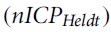 . In this study, sensitivity was reported as 83% and specificity was reported as 70%, and there was a high correlation (r = 0.90) for ICPi values >20 mm Hg.
Kim and coworkers 41 used a semi-supervised machine learning model showing a high predictive ability (area under the curve [AUC] = 0.92) and correlation (r = 0.55) for ICPi >30 mm Hg, outperforming a PI-based method studied before by the same group.
Cardim and coworkers,
42
on behalf of Cambridge's Neurophysics Research Group, performed a prospective study in 40 TBI patients with four different nICP methods, establishing different correlations with global ICPi, with the time domain, and in ICPi variations (change in ICP >7 mm Hg) (Table 2). In general, all methods showed significant but not strong correlation with invasive ICP. However,
Cardim and coworkers,
15
in a cohort study of 27 patients assessing the relationship between ICPi plateau waves and TCD using the same four methods described, found that the best correlation with ICPi >35 mm Hg was with
Given the theoretical relationship between hemodynamic changes in the venous circulation and global ICP, Robba and coworkers
19
developed a prospective cohort trial in 64 patients (45 with TBI) aimed at comparing arterial (MCA) and venous (straight sinus) FV, both measured by TCD and ICPi methods. The straight sinus FV (
In a cohort study, Zhou and coworkers 47 assessed both arterial and venous TCD for nICP estimation in 73 TBI patients. This work identified that straight sinus MFV seems to be the most highly correlated with Invasive intracranial pressure (ICPi) measurements, even over other arterial TCD variables (r = 0.9298, p < 0.0001).
Rasulo and coworkers,
35
in a cohort study of 38 patients (20 with TBI), used a validated nICP formula (Table 2), and found that
Schmidt and coworkers 36 conducted a prospective study including 41 patients (20 with TBI) using a “black box-like” model with different calibration methods (Table 2). The cnICP calibration method showed the best results, with a significantly decreased delta ICP compared with ICPi. Nevertheless, it was not specified if the craniotomy patients had TBIs or other pathologies (e.g., ischemic stroke), which limits the interpretation in this population regarding each method.
Cardim and coworkers,
17
using a prospective study of 100 TBI patients in the United Kingdom, identified that there was no correlation between
Regarding isolated PI correlation with ICPi, Martin and coworkers 48 conducted a prospective cohort study in 54 patients with TBI and found a weak correlation between these two variables (ROC = 0.67), despite a sensitivity and negative predictive value (NPV) of 100%. The maximal PI cutoff was found to be 0.9.
Sari and coworkers, 37 in a cohort study of 52 TBI patients, determined that both PI and maximal flow velocity from the MCA were poorly correlated with ICPi measurements; however, subgroup analysis was not performed on patients with decompressive craniectomy (16 patients), and PI values were considered abnormal in absolute numerical terms (PI >1).
Sawicki and coworkers, 43 in another study assessing PI correlation with ICPi using a commercial software and a mathematically obtained index (the DI) in 96 patients with TBI, showed that both variables (DI and PI) were correlated with ICP plateau waves (defined as a sudden rise in ICP >40 mm Hg that lasted at least 5 min) (p < 0.0001).
Zweifel and coworkers 46 performed a prospective cohort trial in 290 TBI patients assessing the reliability of the PI for ICP estimation. This study found a poor association between PI and ICP (r = 0.31; p < 0.001). The predictive value of PI was indeed relatively high, with AUC values >0.75; however, this was only the case in highly elevated ICP values (> 35 mm Hg).
De Riva and coworkers, 49 in another prospective cohort trial performed in a small sample (20 TBI patients), found a moderate correlation between PI and ICPi (r = 0.7) and a linear model 95% prediction margin for ICP of 21 mm Hg. This needs to be interpreted in context with other variables that have a significant influence on absolute values of PI (Table 2).
Glenn and coworkers 34 analyzed flow velocities and indices from anterior and posterior circulation vessels in 99 patients in a prospective cohort study, concluding that the right MCA PI had the strongest Pearson correlation with ICPi (r = 0.45) in comparison with other vessels and variables. Nevertheless, this correlation was ultimately very weak in absolute terms for predicting elevated ICPi.
Singer and coworkers, 38 in a prospective study of 135 patients (66 with TBI), analyzed flow velocities and PI using two different brands of ultrasound machines. They concluded that the MCA peak systolic velocity (PSV) had a better correlation with ICPi than PI (with some differences between each ultrasound machine). In all, however, correlations were weak overall.
Shrestha and coworkers 50 studied MCA flow velocities and their correlation with ICPi in a cohort of 26 patients in an LMIC. Here, they noted a good MCA PSV correlation with ICPi (r = 0.715, p < 0.000) and a PSV threshold of 39.6 cm/sec with 82.1% sensitivity and 84.4% specificity for ICP values >15 cm H2O (>11 mm Hg)
Lucinskas and coworkers, 32 in a randomized experimental study, considered a mathematical model to establish a BFF derived from indices of internal and external segments of the ophthalmic artery. This model had a strong positive correlation (r = 0.94) with ICPi and low measured differences between the two methods (Table 2).
Youm and coworkers, 33 similarly to Lucinskas and coworkers, 32 but without a mathematical model, assessed TCD variables in the central retinal artery (CRA) and posterior ciliary artery (PCA) in a cohort study in 50 TBI patients. They noted a poor correlation for each parameter with ICPi measurements.
Finally, Rasulo and coworkers 44 published the results of the most recently published study analyzed, the IMPRESSIT-2, a prospective multi-center, international diagnostic test accuracy study in 100 patients (20 with TBI), that used a validated formula employed in other studies. 17,35,42,45 They established a correlation with three different ICPi values and three different times, with sensitivity, specificity, NPV, and AUC values increasing with ICPi. This shows that TCD is a reliable non-invasive method for excluding ICP >20 mmHg and that is has better detection capacity for even higher pressures (e.g., >25 mmHg) (Table 2).
Discussion
TCD is a reliable method for estimating nICP, with some mathematical and formula-based methods showing slightly greater correlations with ICPi than isolated, non-mathematical ones. This is particularly true in cases of increased ICP and when considering a serial assessment (trends) rather than obtaining one-time absolute values. Although more and larger controlled studies are needed, some studies analyzed here suggest that TCD could be a useful tool mainly for excluding elevated ICP and detecting ICP variations (so-called “delta ICP”) in patients with TBI, particularly in scenarios in which invasive monitoring is infeasible or contraindicated.
TCD uses a device composed of a 2-MHz handheld transducer that is placed on the surface of the scalp. Arterial information is gleaned via the placement of a probe with a range-gated ultrasound Doppler instrument. 30 An ultrasonic beam transmitted by the probe crosses the skull at pre-specified locations and is reflected back from flowing erythrocytes in the blood vessels. These erythrocytes move at different speeds, and the resultant Doppler signal obtained is a combination of different frequency components. Insonation of cerebral arteries can be achieved using four different windows: temporal, orbital, foraminal, or submandibular. Spectral analysis then presents three-dimensional Doppler data in a two-dimensional format. 51 The time vector is represented on the horizontal scale while velocity (frequency shift) is displayed on the vertical scale. Color brightness represents the signal intensity translated into variables, such as mean cerebral blood flow velocity (CBFV), PI, PSV, RI, and end-diastolic velocity (EDV), which allow for the assessment of cerebral hemodynamic changes from different etiologies (e.g., TBI, vasospasm, and aneurysmal subarachnoid hemorrhage [aSAH]), among others. 31 Measuring these changes in relation to cerebral blood flow (CBF) provides, through different models, an indirect estimation of ICP.
The rationale behind the use of TCD variables for nICP estimation rests in its ability to reflect hemodynamic changes that take place in the cerebrovascular circuitry in response to local changes and/or changes in other components, such as the cerebrospinal fluid (CSF), brain parenchyma, or venous system. These changes subsequently influence ICP. 22 As such, CPP estimation is in fact a “driving force” for CBF and has a physiological basis in assessing ICP changes indirectly, given their close relationship (as in complex vasodilation and vasoconstriction cascades). 22 –24 A detailed explanation of ICP waveform physiology is outside the scope of this review, but it is well known that ICP pulse amplitude and waveform are shaped by (1) changes in the pulsatile component of the cerebral blood volume in the amplitude (and in P2 waveform), (2) synchronic systolic arterial pressure peak (P1), and (3) synchronic venous blood outflow (P3). 25,26 As a consequence, methods that consider either the calculation of an indirect CPP via TCD or the transformation of ABP and TCD signals may represent a physiological correlation with ICPi. 15 Some of these methods deserve to be analyzed deeply in response to this scoping review's questions.
Mathematical methods have the advantage of considering different variables for nICP estimation that are calculated and depicted both instantly and continuously, allowing for detection of ICP fluctuations in specified time frames.
39,42,43
On the other hand, their complexity in terms of model design as well as the need to interpret algebraic and physical formulas and invest in software, computational resources, and expertise to do so limit their widespread use in the neurocritical care setting.
32,40
–42
This may even reverse the cost effectiveness in LMICs touted at the outset. In addition, because of the numerous variables considered in each model, some may individually represent different components of ICP. An example of this scenario is exhibited in a study using four different methods, in which
With respect to the non-mathematical methods for nICP estimation, it must be stated that the classical and mistaken concept of the PI as an isolated marker of intracranial hypertension (even with consideration of flow velocity changes) needs to be abandoned. Although some studies showed correlations of PI with ICPi, they seem to be weak, because of the complex relationship that PI itself has with multiple hemodynamic variables such as heart rate, cerebral arterial compliance, and CPP. 34,48 Therefore, PI is not simply an indirect marker of distal cerebrovascular resistance. 49 Lastly, the methodological aspects of two studies need to be considered. The first is related to the estimation units of ICP (cm H2O instead of mm Hg) in the study by Shrestha and coworkers, which was not specified by the authors, even when mentioning that “invasive intracranial pressure monitoring” was used. 50 The second is related to different TCD machines (and, by consequence, variables) that were employed in the study by Singer and coworkers 38 which, although not typically an objective of a scoping review such as theirs, may condition biases related to accuracy and performance of a given device.
As a final point, the results of the recently published trial by Rasulo and coworkers using a validated and easily applicable nICP formula exhibited a very high value for real-world daily clinical practice, a setting in which crucial decisions must be made in ICP screening, ICPi monitoring insertion, and early detection of potentially severe TBI patients. 20,44 However, only time and the dissemination of the results of this work will be able to validate this assertion. Future studies should consider the complex issues behind TCD analysis and interpretation, specific trial designs (namely randomized control trials), the implications of comparing nICP with either intraventricular or intraparenchymal probes, proper definitions of intracranial hypertension (in physiological parameters rather than by absolute ICP values), and the relationship of TCD variables with “numerically exclusive” ICP properties that are not “numerically exclusive,” such as ICP waveform changes.
Limitations
This scoping review has several limitations. First, only studies in patients ≥18 years of age were included, potentially leaving out valuable information from patients ≥16 years who are also considered part of the “adult population”. Second, as a matter of a scoping review design and according to the PRISMA-ScR recommendations, 21 in-depth statistical analyses or risk-of-bias assessments (usually performed in systematic reviews) were not conducted in our study. This represents a significant limitation for data interpretation in terms of diagnostic accuracy and nICP-ICPi correlation comparison between studies. Third, narrative reviews, which may contain valuable information from other sources in a clear format and consider expert opinions in both technical and data interpretation from TCD studies, were not included in our search. Fourth, during the review process, we did not consider studies related to indirect markers of ICP changes (e.g., TCD-derived autoregulation indices) because of their huge implications in other pathophysiological processes. Undoubtedly, we believe that they deserve a detailed analysis in the context of nICP estimation in TBI patients in future studies. Finally, we only included studies in TBI patients. As such, the analyses and conclusions derived from this work cannot be extrapolated to other neurocritical care patient populations (e.g., those with aSAH, ischemic stroke, or intracerebral hemorrhage).
Conclusion
This review serves as an informative tool for understanding the different methods for bedside ICP monitoring derived from TCD in patients with TBI. We hope that this allows for the consideration of this non-invasive technique as a proven means of assessing ICP changes rather than ICP values, with the most utility being in settings where invasive devices are not available and/or are considered to be of high risk because of patient factors.
Transparency, Rigor, and Reproducibility Summary
This scoping review was conducted in accordance with the JBI methodology for scoping reviews. 21 Although a protocol was written before beginning the review, the protocol was not published because of project time constraints. A total of two databases (PubMed and Embase) were searched, with initial search strategy formation being undertaken in PubMed. The final searches were completed in June of 2022, yielding 608 studies for review. All identified citations were collated and uploaded into Covidence, and 35 duplicated studies were removed. Studies were screened by two independent researchers (K.M. and S.V.) and one collaborator (O.A.F.). After examining 573 titles and abstracts for inclusion, 520 irrelevant studies were removed, 53 full-text studies were assessed for eligibility, and 31 studies were excluded for reasons described in Figure 1. Any conflicts that arose in the selection process were resolved by a tie-breaking vote from the third member who did not participate in the initial vote. The results of the search are reported using the PRISMA-ScR checklist. 21 Data were extracted from included studies by all three researchers using a data extraction tool developed in the protocol, and modified after initial review of the articles, to present data in the clearest manner (Table 2). After two researchers completed extraction, consensus was achieved by mediation between the two researchers involved. In accordance with JBI methodology for scoping reviews specifically, included studies were not reviewed for quality or risk of bias.
Footnotes
Acknowledgments
We thank the non-invasive ICP monitoring international consensus participants for their comments on this article based on their experience in the field:
Yasser Abulhassan, Amos Adeleye, Amelia Ain, Marcel Aries, Rafael Badenes R, Pierre Bouzat, Sérgio Brasil, Luis Bustamante, Danilo Cardim D, Marek Czosnyka, Thomas Geeraerts, Daniel Godoy, Mohammad Hirzallah, Bhagavatula Indiradevi, Manuel Jibaja, Pier G. Lochner, Julio Mijangos Mendez, David Menon, Thangaraj Munusamy, Wellingson Paiva, Juan Pinedo, Frank Rasulo, Chiara Robba, Andrés Rubiano, Diana Sanchez, Aarti Sarwal A, Hamisi Shabani, Gentle Shrestha, Dhaval P Shukla, Gene Sung, Fabio Silvio Taccone, Abenezer Tirsit, Franly Vasquez, Sebastián Vásquez, Yu Lin Wong and Getaw Worku.
Authors' Contributions
Karol Martínez-Palacios and Sebastián Vásquez-García were equally responsible for methodology and writing – original draft preparation. Olubunmi A. Fariyike was responsible for software, visualization, writing – reviewing and editing. Chiara Robba was responsible for writing – reviewing and editing. Andrés M. Rubiano was responsible for conceptualization, supervision, writing – reviewing and editing.
Funding Information
There are no sources of funding to report.
Author Disclosure Statement
No competing financial interests exist.
Supplementary Material
Supplementary Table S1
Supplementary Table S2
References
Supplementary Material
Please find the following supplemental material available below.
For Open Access articles published under a Creative Commons License, all supplemental material carries the same license as the article it is associated with.
For non-Open Access articles published, all supplemental material carries a non-exclusive license, and permission requests for re-use of supplemental material or any part of supplemental material shall be sent directly to the copyright owner as specified in the copyright notice associated with the article.