
Abstract
Abstract
Alzheimer's disease (AD) is a progressive neurodegenerative disorder and the most common form of dementia. However, biomarkers that require testing in the brain tissue pose a formidable practical barrier to AD diagnostic innovation. MicroRNAs (miRNAs) are responsible for control of gene expression at the posttranscriptional level and are essential for the function of neuronal networks and neuronal survival. miRNA expression can impact the regulation of APP (amyloid beta A4 precursor protein), PSEN1 (presenilin 1), PSEN2 (presenilin 2), and BACE1 (beta-secretase 1) genes in the brain that were previously implicated in AD pathophysiology. Little is known, however, on the extent to which peripheral tissue (e.g., whole blood) miRNA variation might offer clinical predictive value for AD. Moreover, few studies have examined multiple peripheral miRNA expression data at the same time. We report here, to the best of our knowledge, the first whole-blood-based and parallel study of seven miRNAs (hsa-miR-9-5p, hsa-miR-29a-3p, hsa-miR-106a-5p, hsa-miR-106b-5p, hsa-miR-107, hsa-miR-125a-3p, and hsa-miR-125b-5p) in relation to AD susceptibility. Notably, these miRNAs are situated “upstream” to the genes implicated in AD. We measured the whole-blood miRNA expression by a real-time polymerase chain reaction in a large study sample (n = 281), comprising patients with AD (n = 172) and healthy controls (n = 109). A reduction in whole-blood expression of hsa-miR-9-5p, hsa-miR-106a-5p, hsa-miR-106b-5p, and hsa-miR-107 was significantly associated with an increased risk of AD (p < 0.05). Notably, after receiver operating characteristics curve analyses, hsa-miR-106a-5p displayed, as a predictor variable, 93% specificity and 68% sensitivity. On the other hand, the expression of hsa-miR-29a-3p, hsa-miR-125a-3p, and hsa-miR-125b-5p was not significantly different between patients and controls (p > 0.05). In conclusion, these observations warrant replication in larger samples while making a contribution to translational research, precision medicine, and biomarker literatures, by expanding the current efforts for AD diagnostic innovation to the realm of epigenomic pathways such as miRNA expression variation among patients.
Introduction
A
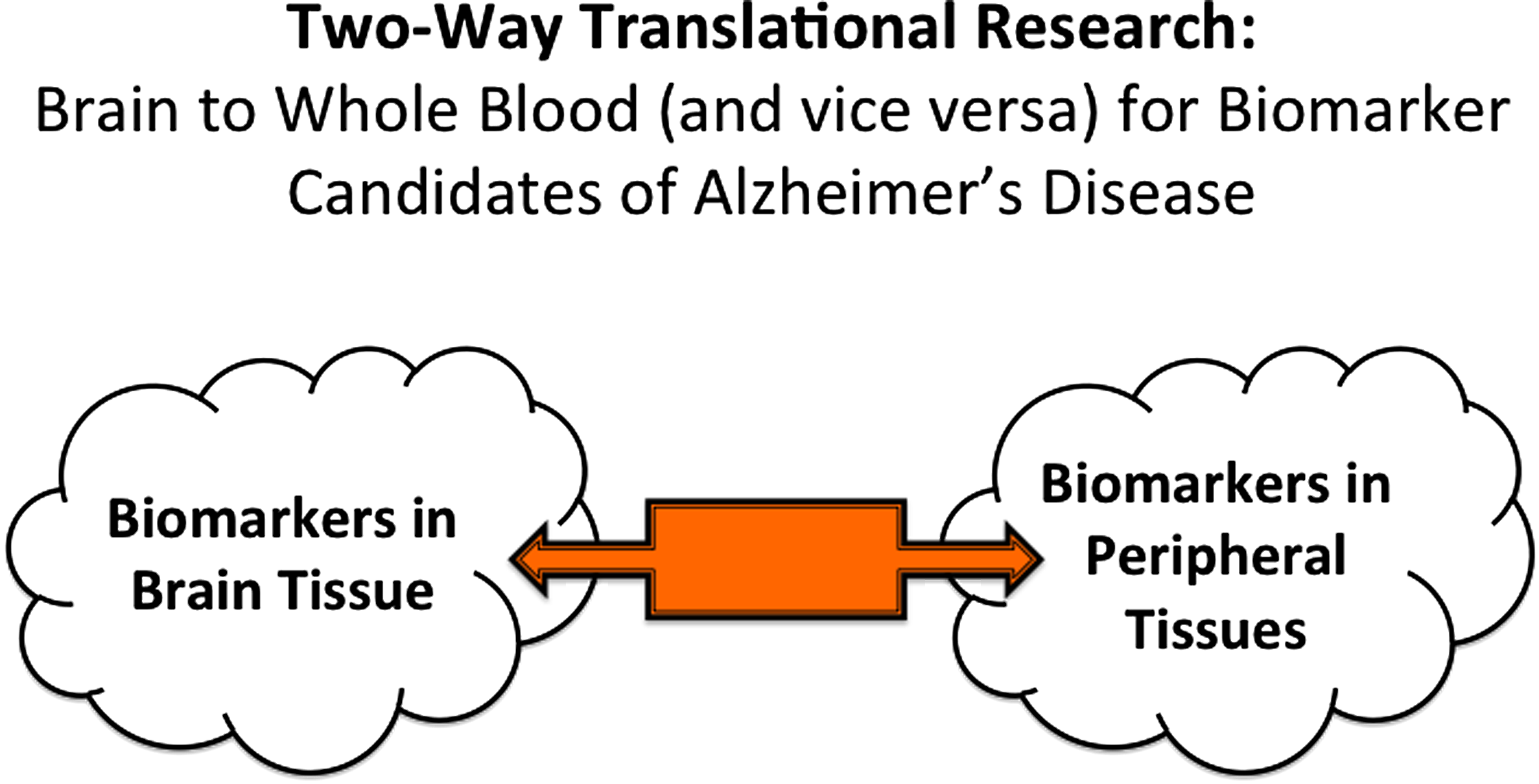
Underscoring the need for “bi-directional translational research” so as to move brain-derived and peripheral tissue-based biomarker candidates toward robust clinical diagnostic applications.
MicroRNAs (miRNA) are a group of endogenous, small, noncoding RNA molecules consisting of nearly 22 nucleotides and are responsible for control of gene expression at the posttranscriptional level by binding to the 3′ untranslated region of target mRNAs (Ambros, 2004). miRNAs are essential for the function of neuronal networks and neuronal survival (Esteller, 2011). For example, miRNA expression can impact the regulation of APP (amyloid beta A4 precursor protein), PSEN1 (presenilin 1), PSEN2 (presenilin 2), and BACE1 (beta-secretase 1) genes in the brain that were previously implicated in AD pathophysiology (De Strooper and Christen, 2010). Little is known, however, on the extent to which whole-blood miRNA variation targeting such AD-relevant candidate genes might offer clinical predictive value.
We report here, to the best of our knowledge, the first whole-blood-based and parallel study of seven miRNAs (hsa-miR-9-5p, hsa-miR-29a-3p, hsa-miR-106a-5p, hsa-miR-106b-5p, hsa-miR-107, hsa-miR-125a-3p, and hsa-miR-125b-5p) in the same study sample, in relation to AD susceptibility. Notably, these miRNAs are situated “upstream” to the genes implicated in AD (Table 1).
miRNA, MicroRNAs.
Materials and Methods
Study sample
The participants were recruited from the Neurology Department of the Medical Faculty of Mersin University in Turkey. The total study sample (n = 281) comprised AD patients (n = 172) and healthy controls (n = 109). Both study groups had a Turkish descent and were aged 60 years and older. The patients with AD were diagnosed according to the DSM-IV-TR (American Psychiatric Association, 2000) and the ICD-10 Classification of Mental and Behavioral Disorders (World Health Organization, 1992). The patients were late-onset AD type (except four individuals). The healthy control subjects had no history of AD, were evaluated with the DSM-IV-TR and ICD-10, and were to be ascertained as control subjects. Importantly, co-morbid complex disorders such as hypertension, diabetes, and vascular disease were neither among the cases nor among the controls, as determined by detailed medical history, thus lending confidence for the present AD peripheral biomarker association study. The data on age and gender were recorded and accounted for in multivariate statistical analyses between the cases and the controls. The present study sample comprised, by and large, the late-onset type of AD, except four patients. The study was approved by the Institutional Research Ethics Committee of the Faculty of Medicine at Mersin University. A written informed consent was obtained from all subjects. A 10 mL whole-blood sample was drawn from each subject into a coded and heparinized tube for miRNA expression analysis.
Selection of miRNA genes for expression
We selected the miRNAs targeting the APP, BACE1, PSEN1, and PSEN2 genes that had been previously implicated in relation to AD-related phenotypes. These miRNAs were predicted using MicroCosm Targets, TargetScan, miRNAMap, microInspector, PicTar, miRWalk, mirbase, mirdb, Patrocles, Diana lab, and Human MiRNAs & Diseases databases. Subsequently, we specified and corroborated the normal blood tissue expression of the candidate miRNAs that were identified earlier using Ferrolab (Ferrolab, the Bioinformatics and Data Mining Group, 2010) and Miracle (Zotos et al., 2012) databases. Finally, to demonstrate the efficacy values (matching scores) of the identified miRNAs' match with their target genes, we used the microRNA database (Wang, 2008).
Synthesis of primers and probes
We used the pdC and fluorogenic Zip Nucleic Acids (ZNA™) probe technologies for synthesis of the probes. Substitution of C-5 propynyl-dC (pdC) for dC is an effective strategy to enhance base pairing. Using these base substitutions, duplex stability and melting temperatures are raised by C-5 propynyl-C 2.8° per substitution. ZNA probes provide broad flexibility in assay design and represent an effective alternative to minor groove binder and locked nucleic acid-containing oligonucleotides (Paris et al., 2010). The specific stem–loop primers, reverse transcriptase–polymerase chain reaction (RT-PCR) primers, and probes for the miRNAs were designed using Primer Express 3.0 software provided by Applied Biosystems (Table 2) (Yılmaz, 2013).
F, forward; PR, probe; R, reverse; RT, reverse transcriptase.
Reverse-transcriptase PCR
RNA was extracted from whole blood using the method of acide guanidinium–phenol chloroform (Chomczynski and Sacchi, 1987). Reverse-transcriptase reactions contained 10 ng total RNA, 50 nM stem–loop reverse transcriptase (RT) primer (Metabion International AG), 5 × RT buffer, 10 mM dNTPs, 0,25 mM MultiScribe Reverse Transcriptase (Thermo Scientific), 0,1 mM RiboLock RNase inhibitor (Thermo Scientific), and nuclease-free water to a total reaction volume of 15 μL. The reaction was performed on an automated Thermal Cycler (Techne Flexigene). RT-PCR was performed for 30 min at 16°C, 30 min at 42°C, and 5 min at 85°C; then, cDNA samples were held at −20°C.
Quantitative-comparative CT (ΔΔCT) real-time PCR
Quantitative-comparative CT (ΔΔCT) real-time PCR was performed in an ABI Prism 7500 Real-Time PCR System (Applied Biosystems) using the SDS 2.0.6 software. The hsa-miR-26b-5p (Applied Biosystems application note cms 044972) was used as an endogenous control. We used a reference RNA sample (AM7155; Life Technologies). The 25 μL PCR included 5 μL RT-PCR product, 2 × TaqMan Universal PCR Master Mix (Applied Biosystems), 900 nM of each primer (Primer F and Universal Primer R; Metabion International AG), and 200 nM TaqMan® probe. The reactions were incubated in a 96-well plate of preincubation at 52°C for 2 min, and at 94°C for 10 min, followed by 50 cycles at 94°C for 15 sec, and at 60°C for 90 sec. All reactions were run in triplicate. ΔΔCT values were used and calculated of 2 −ΔΔC T values (Livak and Schmittgen, 2001).
Statistical analyses
The data were processed and analyzed using the statistical package IBM SPSS-V22.0 (IBM Corp.) for Windows. The normality assumption of the 2 −ΔΔC T values was checked by the Shapiro–Wilk test. Since the assumption of normality was not met, the 2 −ΔΔC T values were expressed as median (50th percentile), first quartile (25th percentile), and third quartile (75th percentile), and the comparison between groups was performed using the Mann–Whitney U test. Univariate and multivariate logistic regression analyses were used to obtain odds ratio of variables that were significantly related with disease. The chi-squared test was used for categorical variables (gender), and the t-test was used for continuous variables (age) between AD patients and controls. Differences (two-tailed p) less than 0.05 were regarded as significant. A receiver operating characteristics (ROC) curve was performed, and the area under the curve was assessed for the specificity and sensitivity of AD prediction for the whole-blood miRNAs.
Results
The study sample comprised 281 individuals: 172 patients with AD (111 men and 61 women) and 109 (56 men and 53 women) controls. The age distribution for the patients and controls was 73.92 ± 1.06 and 69.42 ± 1.10, respectively. Age and gender were statistically controlled in univariate and multivariate analyses, as noted earlier.
We found that a decrease in whole-blood expression of hsa-miR-9-5p, hsa-miR-106a-5p, hsa-miR-106b-5p, and hsa-miR-107 was significantly associated with an increased risk of AD (p < 0.05) (Tables 3 and 4). In contrast, the expression of hsa-miR-29a-3p, hsa-miR-125a-3p, and hsa-miR-125b-5p was not significantly different between patients and controls (p > 0.05).
Median (50th percentile), first quartile (25th percentile), and third quartile (75th percentile) are reported here. n = Numbers of individuals included in the statistical analyses for the patient and the control groups; the numbers noted for each miRNA association analysis are the numbers of patients and controls who could be characterized technically for the given miRNA. Note that in general, miRNA expression levels were lower in patients than in controls.
p < 0.05 is considered statistically significant, and after adjustment for age and gender. Note that in the case of miRNAs that were significantly associated with AD, they appear to offer a protective role against AD, in that their higher expression confers a lower risk for AD.
AD, Alzheimer's disease.
Out of the seven miRNAs whose expression was analyzed in the whole blood, four turned out as being significantly associated with AD. Hence, we conducted further joint analysis of these four miRNA variables together as composite predictors of AD (Table 5). Additionally, after ROC curve analyses, hsa-miR-106a-5p displayed, as a predictor variable, 93% specificity and 68% sensitivity (Table 6).
p < 0.05 is considered statistically significant, and after adjustment for age and gender. p-Values and the odds ratios are derived after composite analyses of the four miRNAs in statistical analysis.
AUC, area under the curve; CI, confidence interval.
Discussion
AD is a common form of dementia, with a paucity of robust clinical diagnostics (Hallock and Thomas, 2012; Yılmaz et al., 2016). Moreover, it is essential that future diagnostics also consider the feasibility of practical applications in the clinic, not to mention in both developed and developing countries (Evans et al., 2015; Reddy et al., 2015; Wonkam and Hurst, 2014). Blood contains circulating miRNAs and could serve as a source of novel noninvasive biomarkers for the diagnosis of common complex neurodegenerative disorders such as AD (Kumar and Reddy, 2016). With the availability of biotechnologies targeted for biomarker discovery, much research has been conducted on AD and its putative diagnostics in the brain tissue and peripheral samples such as the whole blood. For these two sets of biomarker literature to be meaningful, we need to examine the emerging findings from both the brain biomarker research and peripheral diagnostics research in a context of AD pathophysiology. Only through such two-pronged approaches and insights, we will be able to decipher and translate biomarker data between the brain and the periphery in a context of AD (Fig. 1).
In the case of the seven miRNAs examined in the present study, they are reportedly all expressed in various brain regions (Table 1), and were associated in some studies with AD, when measured in the brain and/or peripheral tissues (Kumar and Reddy, 2016). On the other hand, and to the best of our knowledge, no previous study has examined these seven miRNAs in the same study sample, with a view to their potential for future clinical diagnostics applications. Moreover, no previous whole-blood-based study of miR-106a-5p and miR-106b-5p was reported in clinical association studies of AD.
We underscore that the miRNA expression levels were, by and large, lower in patients than in healthy controls. Additionally, hsa-miR-9-5p, hsa-miR-106a-5p, hsa-miR-106b-5p, and hsa-miR-107, which were significantly associated with AD in the present study (Tables 3 and 5), seem to confer protection against AD, since controls had displayed a higher expression of these miRNAs in the whole blood. From a biological plausibility standpoint, this makes sense, because the observed significantly lower expression of hsa-miR-9-5p, hsa-miR-106a-5p, hsa-miR-106b-5p, and hsa-miR-107 in patients would mean that the corresponding target genes for these miRNAs (BACE1, APP, and PSEN1) would be expressed in higher abundance owing to the release of the traditionally, inhibitory effect of miRNAs on the target genes regulated by them.
Taken together, these observations bring about new possibilities for future clinical development of miRNAs, hsa-miR-9-5p, hsa-miR-106a-5p, hsa-miR-106b-5p, and hsa-miR-107, as potential peripheral diagnostics for AD. We wish to underscore that considering the ROC analyses reported in Table 6, hsa-miR-106a-5p, in particular, warrants future research attention as a putative regulatory or epigenomic biomarker of AD susceptibility.
Conclusions
In sum, we report here, to the best of our knowledge, the first whole-blood-based and parallel study of seven miRNAs (hsa-miR-9-5p, hsa-miR-29a-3p, hsa-miR-106a-5p, hsa-miR-106b-5p, hsa-miR-107, hsa-miR-125a-3p, and hsa-miR-125b-5p) situated “upstream” to the genes targeting AD. These observations require replication in larger samples while making a contribution to translational research, precision medicine, and biomarker literatures, by expanding the current efforts for AD diagnostics to the realm of epigenomic regulatory mechanisms such as miRNA expression variation among patients.
Footnotes
Acknowledgments
This study was supported by the intramural research fund of the Mersin University (BAP-SBE TB [Ş.G.Y.] 2010-4). This article is based on the work done in the first author's PhD thesis (Ş.G.Y.); it has never been published as a journal article, and it represents original data and research. The authors thank the research participants who volunteered their time for this study.
Author Disclosure Statement
The authors declare that no conflicting financial interests exist.