
Abstract
Idiopathic acute eosinophilic pneumonia (IAEP) is characterized by febrile hypoxic respiratory failure with diffuse radiographic infiltrates and peripheral and pulmonary eosinophilia in a previously healthy child. Diagnosis is by exclusion, but promptness is imperative, as IAEP can lead to life-threatening acute lung injury and acute respiratory distress syndrome. Prognosis is usually good after steroid treatment with total recovery and absence of relapse in reported cases. We report a case of a previously healthy 14-year-old boy with this diagnosis who presented with acute hypoxia and characteristic radiographic and bronchoalveolar lavage findings. He had full recovery after corticosteroid treatment. This case report highlights the diagnostic approach to a patient with IAEP, an uncommon diagnosis, and one of the exclusions, in children.
Introduction
Case Report
A 14-year-old boy with unremarkable medical and family histories was admitted for respiratory distress. He presented with a history of nausea and loss of appetite 3 weeks before admission. He was seen by his pediatrician, and at that time, had normal physical examination and laboratory findings; cell blood count (CBC) showed white blood cells (WBC) of 6.0×10³ with a normal differential count. A week later, he developed facial urticarial rash managed with oral antihistamine. He then developed dry cough progressing to difficulty breathing on the day of admission.
On admission, he was found to be in respiratory distress. He was tachypneic with a respiratory rate of 32 breaths/min; oxygen saturation in room air was 90%. He was febrile with a temperature of 39.5°C. Pertinent physical examination findings revealed chest retractions, poor air entry with decreased breath sounds in all lung fields with crackles in both bases, and urticarial rash in the trunk and face. Chest X-ray showed bilateral alveolointerstitial infiltrates (Fig. 1). His condition worsened within the next 12 h with progressive hypoxemia despite provision of supplemental oxygen via a non-rebreather mask. He became anxious and drowsy necessitating admission to the Pediatric Intensive Care Unit (ICU) with an initial diagnosis of acute pneumonia. Upon admission to the ICU, venous blood gas showed pH 7.37, pO2 26 mmHg, pCO2 37 mmHg, and bicarbonate 26 mmol/L. Laboratory tests revealed elevated WBC of 22.4×10³ cells/mm3 (normal: 6–17.5) and eosinophilia of 8.5×10³ cells/mm3 (normal: ≤0.3). C reactive protein (CRP) was also elevated at 51 mg/L (normal: <6), while procalcitonin was normal at 0.18 ng/mL (normal <0.3). Serum electrolytes, BUN, creatinine, and liver function tests were normal. Noninvasive ventilation in the form of nasal continuous positive airway pressure (CPAP) was initiated. Antibiotics were given. He underwent bronchoscopy with bronchoalveolar lavage (BAL) and was intubated for this procedure. There was a note of abundant yellowish secretions emanating from all bronchi. The BAL cell count showed 70% eosinophils, 10% leukocytes, 7% neutrophils, and 13% macrophages. Microbiologic cultures were negative.
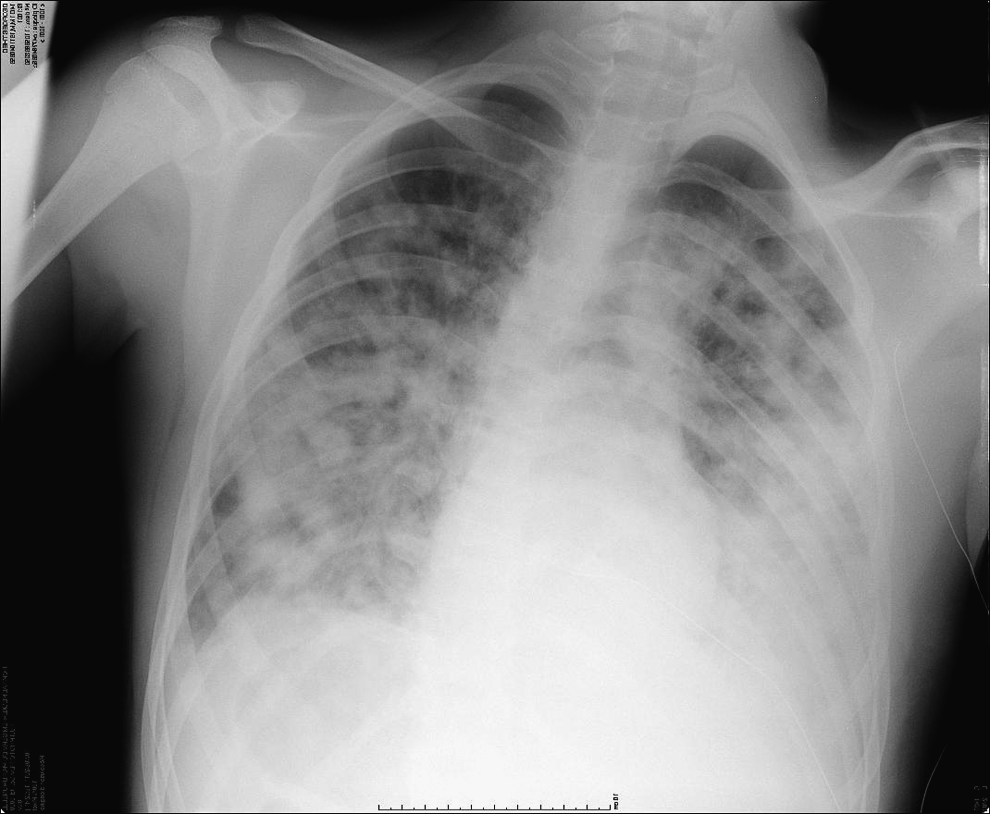
Chest X-ray postero anterior (PA) view on admission showing bilateral alveolointerstitial infiltrates.
Given the findings of peripheral and pulmonary eosinophilia, acute eosinophilic pneumonia was considered, and further workup was done. The IgE level was normal at 20.2 kU/L (normal: <114); anti-neutrophil cytoplasm antibodies (ANCA) and anti-nuclear antibodies (ANA) were negative. Stool examination and serology for roundworm, tapeworms, and flukes were negative; no recent travel history to endemic areas was ascertained. No drug reaction was suspected, as the patient and his parents declared no history of recent drug use or toxic inhalation. Myeloproliferative hypereosinophilic syndrome was included in the differential diagnosis. Bone marrow aspirate with cytogenetic study, lymphocyte immunophenotyping, and molecular studies to detect T-cell disorder were carried out. Bone marrow aspiration showed an increased number of eosinophils up to 27% of the total cell count, but otherwise normal results.
Treatment was then initiated on the second hospital day while awaiting laboratory results for acute eosinophilic pneumonia using intravenous methylprednisolone at 10 mg/kg/day for 3 days and then decreased to 2 mg/kg/day. Antihistamine was added to prevent the secondary effect of eosinophil lysis.
Patient improved rapidly and was extubated to nasal CPAP by the third day. He was maintained on CPAP for another 24 h and weaned to room air thereafter and then transferred to the general ward. Repeat chest X-ray on the fourth day showed improvement in previously noted diffuse infiltrates (Fig. 2). Similarly, there was a note of significant decline in peripheral eosinophilia to 0.8×10³ by the fifth day (Fig. 3). Skin rashes resolved as well. He was discharged after 7 days to continue systemic steroids in the form of oral methylprednisolone at 40 mg/day followed by a tapering regimen for 6 weeks. On follow-up 3 months after discharge, physical examination, chest X-ray, and pulmonary function remained normal.

Chest X-ray PA on hospital day 4 showing a marked improvement of diffuse infiltrates.

Evolution of peripheral eosinophil count showing dramatic decline after 24 h of steroid treatment.
Discussion
Our case is a classical representation of IAEP. The diagnostic criteria were defined in a retrospective study by Pope-Harman and coworkers 5 and consisted of (1) acute onset (onset of symptoms such as fever within 7 days), (2) bilateral infiltrates in chest radiographs, (3) severe hypoxemia (pO2 on room air <60 mmHg, oxygen saturation on room air <90%, or A-a gradient >40), (4) pulmonary eosinophilia (BAL differential count with at least 25% eosinophils), (5) negative history of exposure to drug associated with eosinophilic lung disease, negative history or laboratory evidence of infection, and no other known cause of acute eosinophilic lung disease. From these criteria, pulmonary and peripheral eosinophilia and bilateral infiltrates on chest radiography resulting in hypoxemia remain the most important. The urticarial rash as found in our patient may be ascribed to release of histamine from lysis of eosinophils; the response to antihistamine was documented.
IAEP is a diagnosis of exclusion. A thorough history to rule-out the exposures with the known association with eosinophilic lung disease such as to drugs and toxins is important (Table 1). 6 Secondary eosinophilia associated with intestinal parasitism, allergy, or certain forms of leukemia and types of myeloid disorders have to be ruled out. 7 The most frequent cause of secondary eosinophilia worldwide is tissue-invasive parasitism. The pulmonary eosinophilia occurs in almost all metazoan infections. Most common infections are caused by Strongyloides, Ascaris, Toxocara, and Ancylostoma species. 7 Eliciting a history of recent travel to endemic areas, stool exams, and serology allow ascertainment of parasitic infection. Pulmonary eosinophilia secondary to allergy and autoimmune disease usually complicates long-standing asthma or atopic disease and is uncommon in a previously healthy patient. Diagnosis is established by documenting increased levels of IgE and autoimmunity markers such as ANCA and ANA. The diagnosis of clonal eosinophilia requires the demonstration of either a cytogenetic/molecular marker of clonality or bone marrow histological features that are consistent with an otherwise-classified myeloid malignancy. Several types of myeloid disorder can be accompanied by clonal eosinophilia such as acute lymphoblastic leukemia or acute myeloblastic leukaemia. 8 Peripheral blood and bone marrow aspirate show associated abnormalities such as macrocytosis, monocytosis, left-shift granulocytosis, presence of circulating blasts, thrombocytosis, multilineage myeloproliferation, dysmyelopoiesis, and reticulin fibrosis. The more recently described forms of hypereosinophilic syndrome must also be investigated and include the myeloid variant associated with the FIP1L1-PDGFRA fusion gene and the lymphocytic variant associated with an abnormal and often clonal IL5-secreting lymphocyte subset. 9
Reprinted with permission from Knutsen et al. 6
Chest X-ray is a characteristic of the disease, and radiographic findings in IAEP were well described in a series of 12 patients. 5 Diffuse bilateral alveolar–interstitial involvement, with or without pleural effusion, is often seen. The chest X-ray findings dramatically improve within 24 to 48 h of initiation of corticosteroid treatment, which is unlikely for infectious etiologies, pulmonary infection being an important differential diagnosis. The dose and duration of corticosteroid treatment are not clearly established, but initiation of treatment with intravenous administration is the recommendation. Initial dosage ranged from 60 mg/day to 125 mg every 6 h in an adult series and was followed by oral prednisone for a total duration of several months.2,10 We recommended an initial dose of intravenous methylprednisolone at 10 mg/kg/day for the first 3 days, followed by oral prednisolone at a dose of 1 mg/kg/day slowly tapered over 3 months. The prompt and complete response to steroids further confirms the diagnosis of IAEP. No relapses have been reported, and total recovery is the rule [2].
Conclusion
We report a case of IAEP in a 14-year-old boy with prompt and complete recovery after steroid therapy. The diagnosis is one of the exclusions. A thorough history and examination coupled with review of radiographic findings and laboratory testing to rule out secondary etiologies, notably infectious processes, are important. Diagnostic investigations for secondary causes such as from infectious, including parasitic, allergic, or clonal need to be done expeditiously, since IAEP can be life-threatening, and early initiation of systemic steroids provides a dramatic response. Peripheral and pulmonary eosinophilia is key to diagnosis. Prognosis is usually good with corticosteroid treatment with a complete recovery and absence of relapse.
Footnotes
Author Disclosure Statement
Authors declare no potential, perceived, or real conflict of interest. Authors declare no study sponsor. Jean Bergounioux wrote the first draft of the manuscript and no honorarium, grant, or other form of payment was given to anyone to produce the manuscript.