
Abstract
Introduction
5-Aminolevulinic acid (ALA) is used to diagnose and treat neoplastic tissues. Although ALA is not a photosensitizer, it acts as a precursor of an endogenous porphyrin in the heme biosynthesis pathway. 1 When patients are given exogenous ALA, the photosensitizer protoporphyrin IX (PpIX) is produced by mitochondria because the negative feedback control of heme is bypassed. 4,5 Since PpIX mainly accumulates in mitochondria, these organelles may be the specific target of the cytotoxic photochemical reaction induced by PDT with ALA. Several clinical studies have shown that the local application of PDT with ALA successfully treats various superficial diseases, such as basal cell carcinoma, Bowen disease, and early squamous cell carcinoma. 6 However, since ALA is administered locally and has low permeability, PpIX production is limited to the skin. Consequently, this treatment regimen is considered to be ineffective for deep-seated tumors. 7
Hyperthermia (HT) has also been used to treat tumors. HT at 42°C or higher directly damages cells by injuring the DNA, destroying the plasma membrane, inhibiting protein synthesis and energy metabolism, and inducing mitochondrial damage. 8 –11 Consequently, HT is used, alone or with radiotherapy and chemotherapy, to treat malignant tumors.
In order to reach deep-seated tumors, such as osteosarcomas, and to broaden the treatment options that are available for neoplastic disease, including malignant tumors, we examined the efficacy of simultaneous application of PDT with ALA hexyl ester (hALA) and HT using a human-derived osteosarcoma tumor model. 12
hALA induces much lower PpIX synthesis in tumor cells compared to ALA because retention of hALA in peripheral tissues, such as the blood vessels or capillaries of the tumor, is much higher, although intracellular PpIX content is increased when various tumor cells are treated with hALA in vitro. 13 However, a higher concentration of hALA in the peripheral tissues around a tumor would increase tumor PpIX synthesis in the tumor, since hALA not only freely diffuses into cells but also binds to cell membranes with low affinity. 13 This is why we previously employed local injection to administer hALA to deep-seated tumors. We also remodeled a linear-polarized near-infrared hyperthermic irradiator to enable simultaneous irradiation of cells with red light for PpIX excitation and heating with infrared light, which allowed PDT and HT treatment to be performed simultaneously. By using this approach, we found that PDT with hALA (hALA-PDT) and HT acted synergistically to inhibit the tumor growth of human-derived osteosarcomas. 12
We also examined the effect of combination therapy of hALA-PDT and HT (PDT + HT) on various cell lines in vitro. 14 During that study, we observed that two osteosarcoma cell lines derived from the same tumors, namely HOSM-1 and HOSM-2, showed marked differences in their sensitivities to hALA-PDT. The aim of the present study was to investigate why these two closely related lines show such disparities in their hALA-PDT sensitivities. We also wished to determine whether hALA-PDT phototoxicity in the less hALA-PDT–sensitive line could be elevated by a simultaneous application of HT.
Materials and Methods
Cells and cell culture
The two human osteosarcoma cell lines HOSM-1 15 and HOSM-2, 16 which were derived from a tumor that developed in the mandible of a 40-y-old Japanese man, were cultured in 25 cm2 flasks (Nalge Nunc International Co., Tokyo, Japan) in RPMI/FBS (RPMI-1640 [Gibco, Grand Island, NY, USA] supplemented with 10% fetal bovine serum [FBS] [Invitrogen Co., Carlsbad, CA, USA]) at 37°C in a humidified atmosphere containing 5% CO2 and 95% air atmosphere. The cells were diluted to a density of 1 × 10⁵/mL and harvested in the exponential phase of growth after a week.
Chemicals
5-Aminolevulinic acid hexylester hydrochloride (hALA; Cosmo Bio Co., Tokyo, Japan) was used. hALA was dissolved in phosphate-buffered saline (PBS) and added to RPMI-1640 cells supplemented with 10% FBS (1:1 v/v) to a final concentration of 0.2 mM. hALA alone did not show a significant cytocidal effect in this culture medium.
Hyperthermic conditions
HT was achieved by using a hot plate. The temperature in the well was closely monitored by placing a digital microprobe thermometer in the bottom of an adjoining well, which was maintained under the same conditions. By using a hot plate, the temperature in the well reached 43.5°C within 2 min after the start of heating, and was maintained at 43.5 ± 0.5°C during treatment.
Light source
A linearly polarized near-infrared hyperthermic irradiator with a halogen lamp as a light source (model HA-550: Tokyo Iken, Inc., Tokyo, Japan) was used. The irradiator was remodeled by installing two light filters for ALA-PDT. The original specifications of the irradiator were wavelength, 600–1600 nm; power output, 1800 mW; weight, 24 kg; dimensions, 370 × 510 × 990 mm. The attachment of two light filters allows the irradiator to generate wavelengths in the range of 580 to 740 nm, which includes the appropriate wavelength for PpIX. 14 The output in the 700–740 nm wavelength region was too small to affect the temperature, as confirmed by using a digital thermometer.
Measurement of the intracellular accumulation of PpIX
Each well of a 24-well flat-bottomed plate was filled with 5 × 10⁴ cells in 500 μL RPMI/FBS containing 0.2 mM hALA. Cells in RPMI/FBS without hALA served as controls. After incubation for 1, 3, 6, 9, 12, and 24 h, the cells were trypsinized, centrifuged, and resuspended in 1 mL PBS. PpIX fluorescence was detected by using the FL-3 channel (650 nm long pass) of a FACSCalibur flow cytometer (Becton Dickinson, NJ, USA). The fluorescence intensity of 2 × 10⁴ cells was recorded and the intracellular accumulation of PpIX was evaluated from the mean fluorescence intensity (MFI). 17
Measurement of the cytotoxic effects of various treatments
The cytotoxic effects of HT alone, PDT alone, and PDT + HT were assessed. Cells were prepared as described in the previous section. For HT alone, the treatment was performed by using the hot plate for 4, 8, 16, 24, and 32 min, after which the medium was removed and 250 μL of fresh hALA-free medium was added. For PDT alone, the prepared cells were incubated for 6 h with 500 μL of RPMI/FBS containing 0.2 mM hALA. After removal of the medium, 250 μL of fresh hALA-free medium was added and the wells were irradiated with a light dose of 10, 20, 40, 60, or 80 J/cm2 delivered at a fluence rate of 42.4 mW/cm2. For PDT + HT, the cells were treated as for PDT alone but at a temperature of 43.5°C during light irradiation. There were five treatment combinations: 10 J/cm2 + 4 min, 20 J/cm2 + 8 min, 40 J/cm2 + 16 min, 60 J/cm2 + 24 min, and 80 J/cm2 + 32 min. After each treatment, the cells were rinsed with PBS, 500 μL of fresh hALA-free RPMI/FBS was added, and the cells were incubated for a further 24 h. Cell survival was then assessed by using the 3-(4,5-dimethylthiazoyl-2yl)-2,5-diphenyl-2H-tetrazolium bromide assay (MTT assay). To determine whether HT and PDT in combination acted synergistically, a synergism index [SI = (survival rate with HT alone × survival rate with PDT alone)/survival rate with PDT + HT] was calculated. 18
Mitochondrial membrane potential
As an indicator of the mitochondrial membrane potential, 5,5′,6,6′-tetrachloro-1,1′,3,3′-tetraethylbenzimidazolylcarbo-cyanine iodide (JC-1; Molecular Probes, Eugene, OR, USA) was used. JC-1 is a J aggregate–forming dye that accumulates in mitochondria where it can result in multimer formation depending on the potential across the inner mitochondrial membrane. It is generally used as an indicator of mitochondrial activity. 17 Fluorescence emission of the JC-1 aggregates after 488 nm laser excitation is shifted to the red and can be easily distinguished from monomer fluorescence (emission maximum 527 nm, detected through channel FL1, 530/30 nm bandpass) on a FACSCalibur flow cytometer. Mitochondrial depolarization is indicated by the fluorescence intensity ratio. A stock solution of 500 μg/mL JC-1 in dimethylsulfoxide was prepared. Cells were seeded onto 24-well culture dishes and treated as described above. After trypsinization and centrifugation at 200 g, the supernatants were removed and the cells were resuspended in 1 mL of PBS, and 20 μL of the JC-1 stock solution was added to yield a final JC-1 concentration of 10 μg/mL. After a 10 min incubation at 37°C, the fluorescence of the cell suspension was measured on a FACSCalibur flow cytometer. 17
Morphological alterations after treatments
Morphological alterations were examined 3 h after each treatment by phase contrast microscopy.
Statistical analysis
All experiments were performed in triplicate or more, and the data were statistically analyzed by using the Student t test. Significance was defined as a calculated p value < 0.05.
Results
PpIX intracellular accumulation in HOSM-1 and HOSM-2
When HOSM-1 and HOSM-2 cells were incubated with hALA, the amount of intracellular PpIX accumulated as a function of the time of incubation. In HOSM-1 cells, PpIX levels peaked 9 h (MFI = 119.40) and remained almost constant thereafter until at least 24 h. In HOSM-2 cells, PpIX levels peaked between 9 to 12 h (MFI = 220.75) and decreased thereafter (the MFI at 24 h was 59.82). At 6 h after administration, HOSM-2 cells had about 1.53-fold more PpIX than HOSM-1 cells (Fig. 1).

Intracellular accumulation of protoporphyrin IX (PpIX) by the two osteosarcoma cell lines HOSM-1 and HOSM-2. HOSM-1 (circles) and HOSM-2 (squares) cells were incubated with 0.2 mM aminolevulinic acid hexyl ester (hALA) for 3, 6, 9, 12, or 24 h. Each data point represents the mean ± SD of three experiments. MFI, mean fluorescence intensity.
Cytotoxic effects of PDT and/or HT treatments
HT alone, PDT alone, and PDT + HT increased HOSM-1 and HOSM-2 cell cytotoxicity in a dose-dependent manner. The survival rate after HT alone at the maximal dose (32 min) was high at 88.4% and 76.3% for HOSM-1 and HOSM-2 cells, respectively. The survival rate with PDT alone was much lower, with the maximal dose (80 J/cm2 irradiation) resulting in 35.7% and 10.9% survival for HOSM-1 and HOSM-2 cells, respectively. The survival rates of the two cell types differed significantly at all PDT treatment doses (p < 0.05). The survival rate after PDT + HT was low for both cell types: after 40 J/cm2 + 16 min, the survival rates for HOSM-1 and HOSM-2 cells were 21.2% and 13.3%, respectively. Similarly, after 80 J/cm2 + 32 min, the survival rates for HOSM-1 and HOSM-2 cells were 14.1% and 10.7%, respectively. The two cells types differed significantly in their responses to 10 J/cm2 + 4 min and 20 J/cm2 + 8 min but not to 40 J/cm2 +16 min, 60 J/cm2 + 24 min, or 80 J/cm2 + 32 min (Fig. 2).
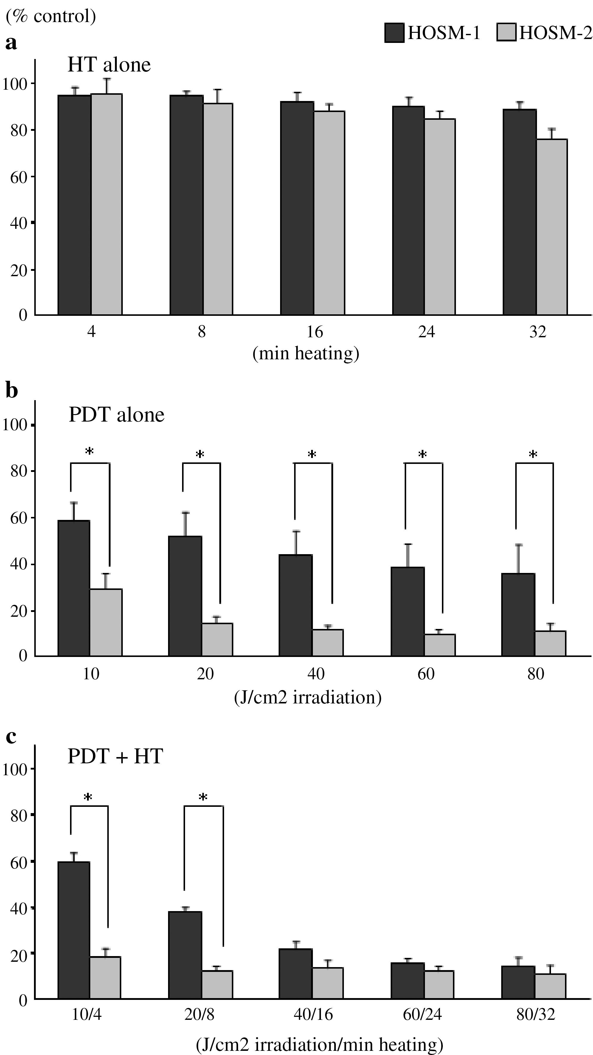
Effect on HOSM-1 and HOSM-2 cell survival of (
The SI of HOSM-1 cells treated with PDT + HT at all treatment doses except for 10 J/cm2 + 4 min was 1.3–2.2, which reveals that the two treatments acted synergistically. A similar synergistic interaction was observed for HOSM-2 cells treated with 10 J/cm2 + 4 min (Table 1).
Mitochondrial membrane potential
When we examined the effect of each treatment on mitochondrial membrane potential, we found that PDT alone and PDT + HT, but not HT alone, increased the FL1 values of HOSM-1 and HOSM-2 cells. However, PDT alone and PDT + HT had a greater effect on HOSM-2 cells than on HOSM-1 cells. The FL1 values for untreated HOSM-1 and HOSM-2 cells were 7.30 and 14.27, respectively. After treatment, the FL1 value was higher in both cell types. After PDT alone, the FL1 values for HOSM-1 and HOSM-2 cells were 22.89 and 48.96, respectively. Similarly, after PDT + HT, the FL1 values for HOSM-1 and HOSM-2 cells were 27.12 and 80.88, respectively (Fig. 3). Consequently, mitochondrial membrane potential decreased in the order PDT + HT > PDT alone > HT alone for both cell types.

Effect on HOSM-1 and HOSM-2 cell mitochondrial membrane potential of HT alone, PDT alone, and PDT + HT. The cells were treated as described in the legend to Fig. 2 with 0.2 mM hALA and 40 J/cm2 irradiation (42.4 mW/cm2) alone or with heating at 43.5°C for 16 min, or with heating alone. Their mitochondrial membrane potentials were then measured immediately by using JC-1 and flow cytometry. The figure shows representative data of three experiments.
Morphological alterations
Some HOSM-1 cells became swollen 3 h after treatment with HT alone (24-min heating) (Fig. 4b). Similarly, many swollen HOSM-1 cells with a round shape as well as some dead cells with blebbing were observed 3 h after treatment with PDT alone (60 J/cm2 irradiation; Fig. 4c). The percentage of atrophied cells increased further 3 h after PDT + HT treatment (60 J/cm2 + 24 min), and many of the cells were dead (Fig. 4d). HOSM-2 cells were mostly swollen 3 h after treatment with HT alone (24 min heating) (Fig. 4f). Some swollen cells with a round shape as well as some atrophied cells were also observed after a 3 h treatment with PDT alone (60 J/cm2 irradiation) (Fig. 3g). However, the number of atrophied and dead HOSM-2 cells markedly increased 3 h after treatment with PDT + HT (60 J/cm2 + 24 min) (Fig. 4h).

Effect of HT alone, PDT alone, and PDT + HT on phase contrast microscopic findings. (
Discussion
ALA is metabolized into the photosensitizer PpIX in mitochondria by the heme biosynthesis pathway. 1,5,17 In ALA-PDT, ALA is applied to the area to be irradiated and subsequent irradiation induces cytotoxicity, mainly by targeting mitochondria. ALA-PDT can be used to treat tumor cells because PpIX accumulates at higher rates in tumor cells than in normal cells. 4,5 ALA delivery can be improved by using ALA derivatives, in particular ALA esterified with aliphatic alcohols. The lipophilic ALA derivative, hALA, is taken up at a higher rate by cells and produces more PpIX than ALA. 13
In this study, two osteosarcoma cell lines derived from the same tumor, HOSM-2 and HOSM-1, differed significantly in their susceptibility to phototoxicity induced by hALA and PDT. The greater susceptibility of the HOSM-2cells could be due, at least partly, to the 1.53-fold higher amount of PpIX in this cell line than in the HOSM-1 cell line after 6 h incubation with hALA. This difference in PpIX production may reflect disparities in the activities of enzymes involved in the heme biosynthesis pathway and/or the amount of PpIX that leaks from the cell. In support of this conjecture, HOSM-2 cells have been suggested to accumulate more PpIX than HOSM-1 cells based on the results of a study which suggested that HOSM-2 have more ferrochelatase, an enzyme involved in the final step of heme biosynthesis. 19
The intracellular localization of PpIX determines the degree of ALA-PDT phototoxicity. 20 –22 Since the singlet oxygen generated by PpIX has a short diffusion distance and lifetime, ALA-PDT will only be phototoxic when a large amount of PpIX localizes to mitochondria. 19,22 In the present study, both cell lines evinced mitochondrial membrane depolarization after hALA-PDT, with HOSM-2 cells showing more depolarization than HOSM-1 cells. Thus, hALA-PDT induces more mitochondrial damage in HOSM-2 cells than in HOSM-1 cells. While we did not examine the intracellular localization of PpIX in the two cell lines, it is likely that HOSM-2 cell mitochondria accumulated more PpIX than HOSM-1 cell mitochondria (see above).
Several studies have reported that ALA-PDT induces apoptosis. 23 –25 Apoptosis is induced upon mitochondrial membrane depolarization. 26,27 Given that hALA-PDT caused mitochondrial membrane depolarization, we expected that it would kill HOSM-1 and HOSM-2 cells by promoting apoptosis. However, Hoechst 33342 staining 3 h after hALA-PDT revealed that apoptotic cells accounted for only 3% of the dead cells in both cell lines (data not shown). Instead, microscopic analysis revealed a great deal of cell swelling and plasma membrane damage 3 h after treatment. This suggests that hALA-PDT induces rapid cell necrosis and cell killing before apoptosis can be implemented. It is possible that the necrosis arises because of diffusion of PpIX into the cytoplasm, which would damage the cell membrane and other organelles.
It is well known that HT damages mitochondria, the plasma membrane, and DNA and inhibits protein synthesis and energy metabolism. 8 –11 For both cell lines, HT was poorly cytotoxic and did not induce mitochondrial membrane depolarization. This is largely consistent with previous observations of tumor cells treated with HT. 8,9
We observed that 20 J/cm2 + 8 min, 40 J/cm2 + 16 min, 60 J/cm2 + 24 min, and 80 J/cm2 + 32 min irradiations acted synergistically with HT to kill HOSM-1 cells, and that 10 J/cm2 + 4 min acted synergistically with HT to kill HOSM-2 cells. Significant synergism was not observed for the remaining combinations, namely 40 J/cm2 + 16 min, 60 J/cm2 + 24 min, and 80 J/cm2 + 32 min in HOSM-2 cells. A comparison of PDT + HT and PDT at the same dose revealed that HOSM-1 cells were killed more efficiently by PDT + HT than by PDT at all treatment doses except for 10 J/cm2 + 4 min. Thus, cytotoxicity decreased in the following order; PDT +HT > PDT alone > HT alone. For HOSM-2 cells, however, the order was PDT + HT = PDT alone > HT alone as there were no significant differences between the survival rates after PDT alone and those after PDT + HT for all treatment doses except 10 J/cm2 + 4 min.
Several reports have shown that HT immediately after PDT enhances the effect of PDT. 28,29 For example, synergism with HT was observed in a study using photofrin II, hypericin, or aluminum phthalocyanine as a photosensitizer. This may be because PDT inhibits the cellular repair system for thermal inactivation and/or because HT inhibits the cellular repair system required to counteract the damage caused by PDT. Thus, synergistic interactions between PDT and HT may arise because PDT induces mitochondrial damage while HT damages organelles, such as the nucleus and cell membrane, which are not injured by the singlet oxygen produced in mitochondria. This would also explain why only low PDT-HT doses acted synergistically in HOSM-2 cells: at higher doses (i.e., > 20 J/cm2 + 8 min), PpIX would diffuse into the cytoplasm and damage other cellular organelles, which would make HT-induced damage redundant.
In conclusion, although the osteosarcoma cell lines HOSM-1 and HOSM-2 were derived from the same tumor, they differed significantly in terms of PpIX accumulation and hALA-PDT–induced phototoxicity. However, when HT was applied simultaneously with hALA-PDT, these differences became insignificant, as both cell lines showed a survival rate of about 10%. Thus, hALA-PDT + HT can be highly cytotoxic, which suggests that this treatment combination could be very useful for treating tumors containing tumor cells that are insensitive to hALA-PDT.
Footnotes
Disclosure Statement
No competing financial interests exist.