
Abstract
Introduction
Materials and Methods
Since 1995, the 1st Department of Urology of Łódź Medical University (former Clinic of Urology of the Military Medical Academy) has been treating BPH with Nd:YAG lasers manufactured by Trimedyne (Lake Forest, CA) with Urolase® (Bard, Covington, GA) fibers terminating with a 7.5 F gold tip, whose reflective surface allows deflection of the laser beam in the desired direction. The tip of the fiber deflects the laser radiation beam at a 90° angle with dispersion to 30° (a noncontact probe emitting a free laser beam is used).
VLAP technique
A gold-tipped laser fiber is introduced into the urethral lumen by means of a cystoscope, so as to be able to direct the laser beam towards the prostatic adenoma tissue at any site, with two to four exposures of the right lobe at the 8:00 and 10:00 o'clock positions, and then two to four exposures of the left lobe at the 2:00 and 4:00 o'clock positions. Depending on the middle lobe size, one exposure at 6:00 o'clock or two at 5:00 and 7:00 o'clock positions are applied; in cases of considerable middle lobe enlargement, three exposures are made at 5:00, 6:00, and 7:00 o'clock. The time of exposure to the laser beam ranges from 60 to 90 sec with 40–60 W energy used. Appropriate application of the laser beam leads to necrosis of adenoma tissue, with its subsequent demarcation and positive effect of treatment.
Nine Urolase fibers damaged during VLAP and, for comparison, one new fiber not used for any surgical procedure were analyzed. The surfaces and cross-sections of gold fiber tips were observed under a Hitachi S 3000N scanning electron microscope (Hitachi, Tokyo, Japan). In order to improve imaging quality, reflective fiber tip cross-sections embedded in electrically conductive resin [PolyFast; Struers (Warsaw, Poland)] were analyzed.
Longitudinal sections and microsections were prepared using waterproof abrasive papers with gradually decreasing grain diameter with a Labo Pol-5 grinder (Struers). In addition to observations under a scanning electron microscope, the chemical composition of the fiber tip was determined using an X-ray chemical microanalysis method with energy dispersion. The analyses were carried out using a Noran Instruments (Middleton, WI) system coupled with the scanning electron microscope, consisting of a Pioneer detector and Ventage software. This method involves generation of characteristic X-ray spectra (characterized by specific energy or wavelength) in the investigated microscopic area. Analysis of these parameters allows the elements present in the composition of the investigated sample to be determined. Surface distribution of the particular elements was obtained by scanning the sample surface with an electron beam and analyzing the characteristic X-ray spectra obtained for the particular sites of the scanned area.
Results
For appropriate interpretation of changes taking place on the investigated reflective surfaces of Nd:YAG laser Urolase fibers, an intact fiber not applied in any ablation procedure was used as a standard. Intact surface integrity is clearly visible on the gold-coated tip of the Urolase fiber (Fig. 1a). The laser beam transmission canal is very clearly visible, and the meniscus-shaped reflective surface is ideally smooth (Fig. 1b). The cross-sectional view shows separate, well-delineated components of the tip made of different metals: gold, chromium, nickel, and iron (Fig. 1c).
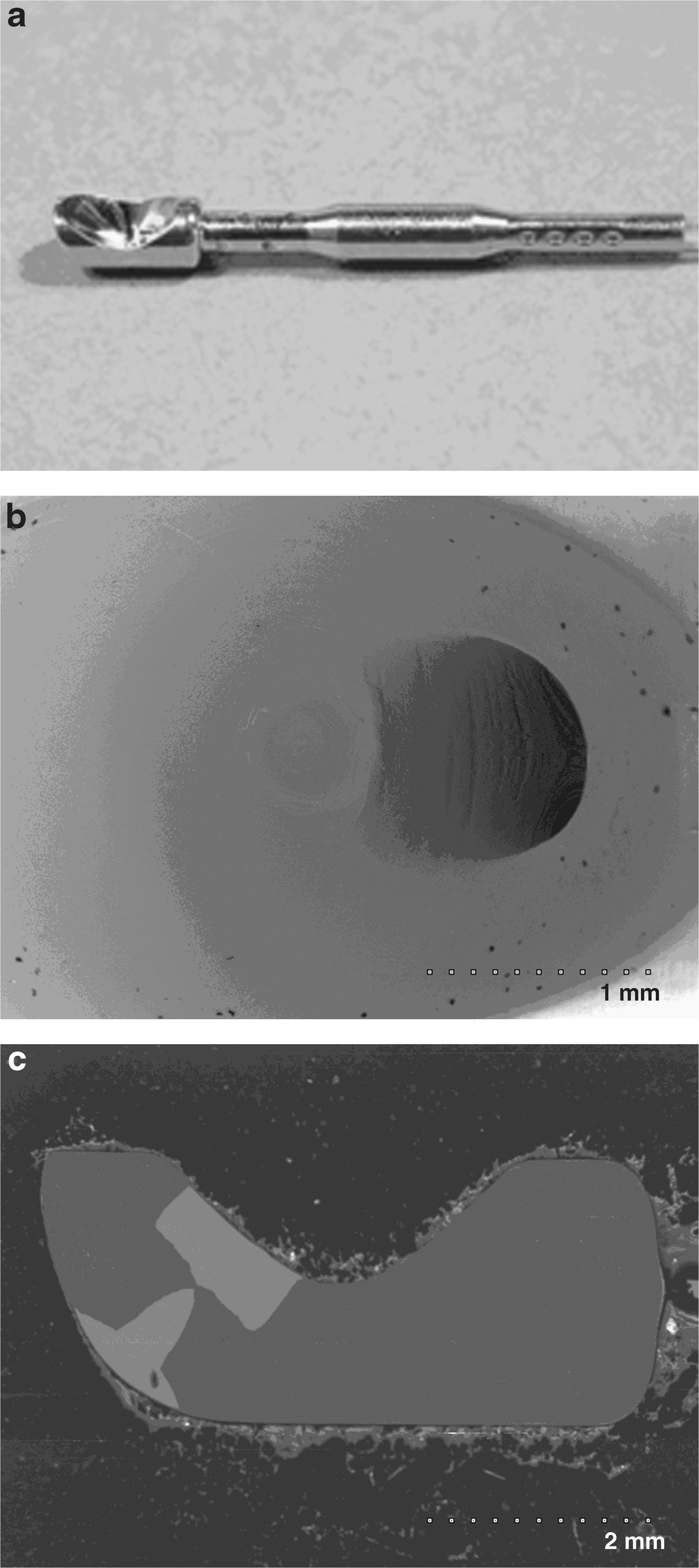
An intact gold reflective tip of the laser fiber. (
Incorrect handling of the laser fiber involving positioning the gold reflective surface of the tip too close to the adenoma tissue and the resultant disturbance of the correct laser beam course causes rapid damage to the surface. The meniscus of the reflective surface becomes covered with a black deposit (Fig. 2a), which affects the quality of fiber function. Damage to the smooth reflective surface and laser beam transmission canal is visible in the scanning microscopy images (Fig. 2b), and the cross-sectional view exhibits slight edge deformations of the parts made of different metals (Fig. 2c).
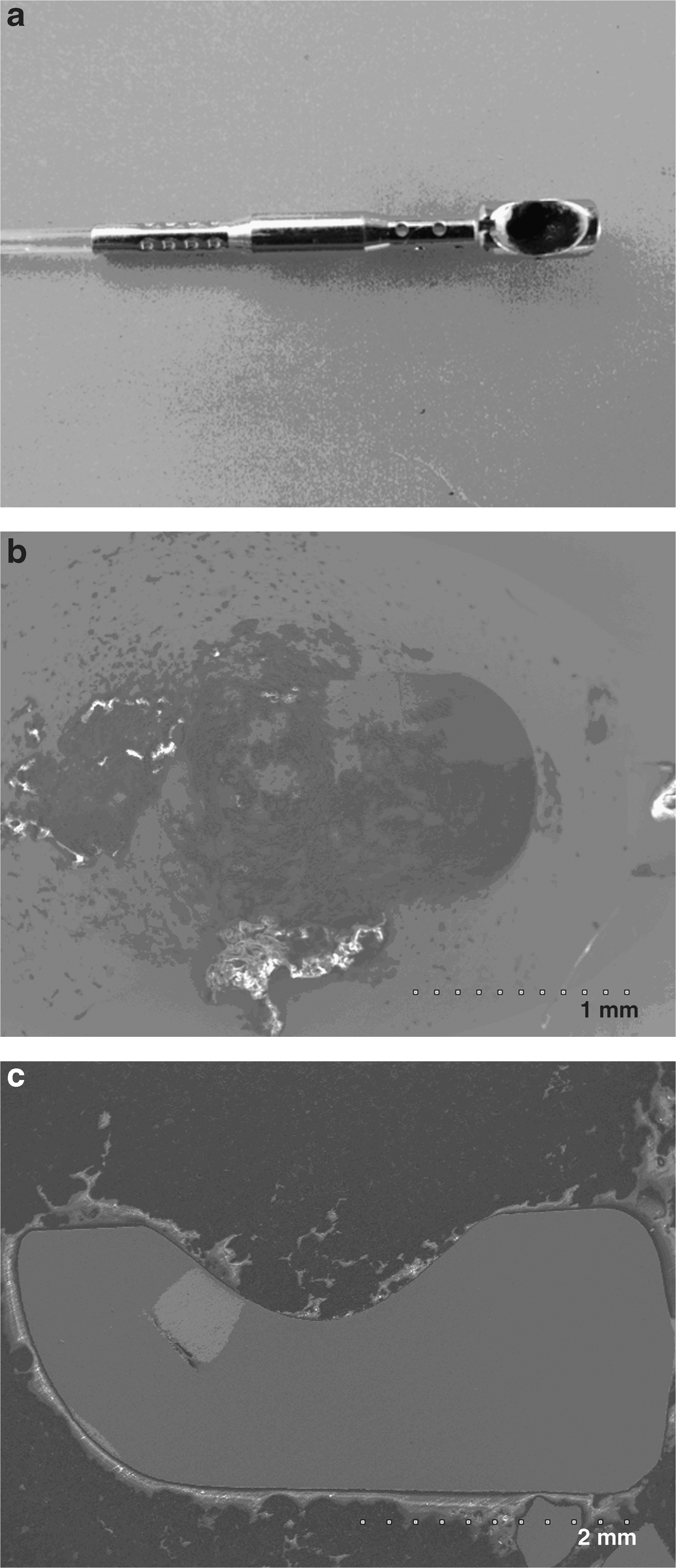
Reflective surface of a laser fiber tip damaged by 40 W energy applied for 90 sec. (
In case of prolonged direct contact between the gold fiber tip and adenoma tissue, and use of high energy approximating, for example, 60 W for 60 sec, the gold reflective surface of Urolase fiber rapidly undergoes complete destruction. The surface becomes black and considerably deformed (Fig. 3a). Scanning microscopy shows the complete destruction of the meniscus and the laser beam transmission canal (Fig. 3b). The cross-sectional view also demonstrates the complete destruction and deformation of the fiber tip elements, which develop irregular contours and become much different in shape from the structures shown in cross-sectional view of a new fiber tip (Fig. 3c).
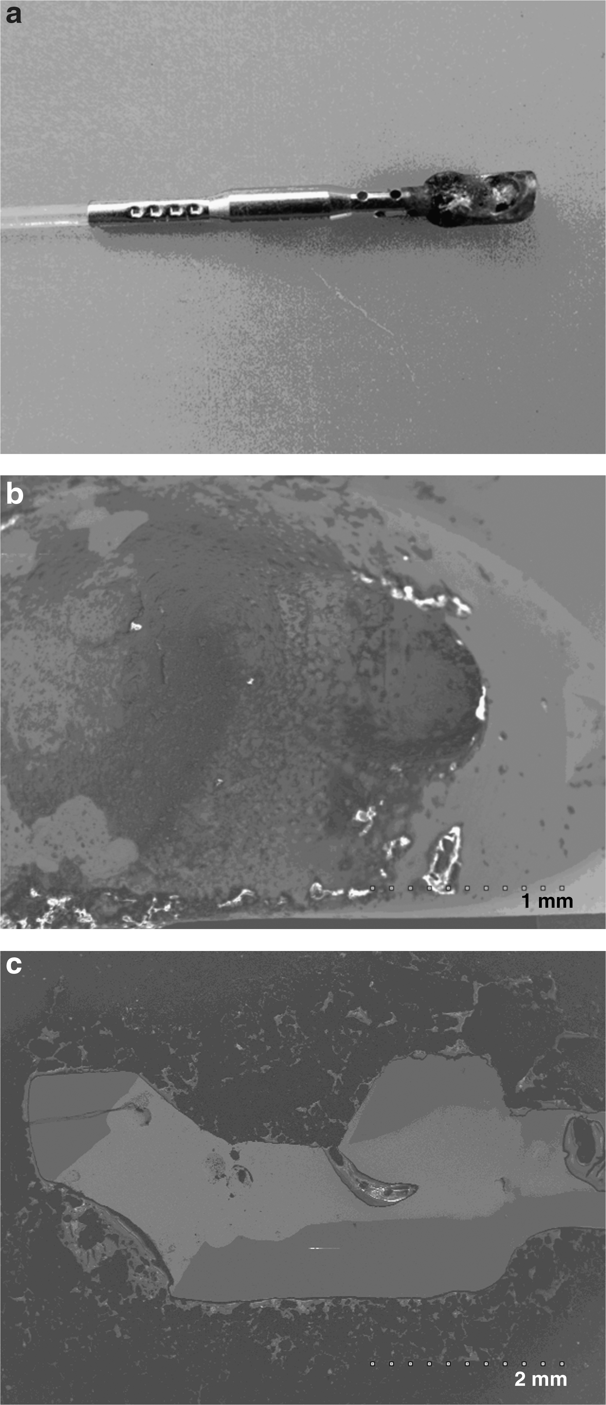
Reflective surface of a laser fiber tip damaged by 60 W energy applied for 60 sec. (
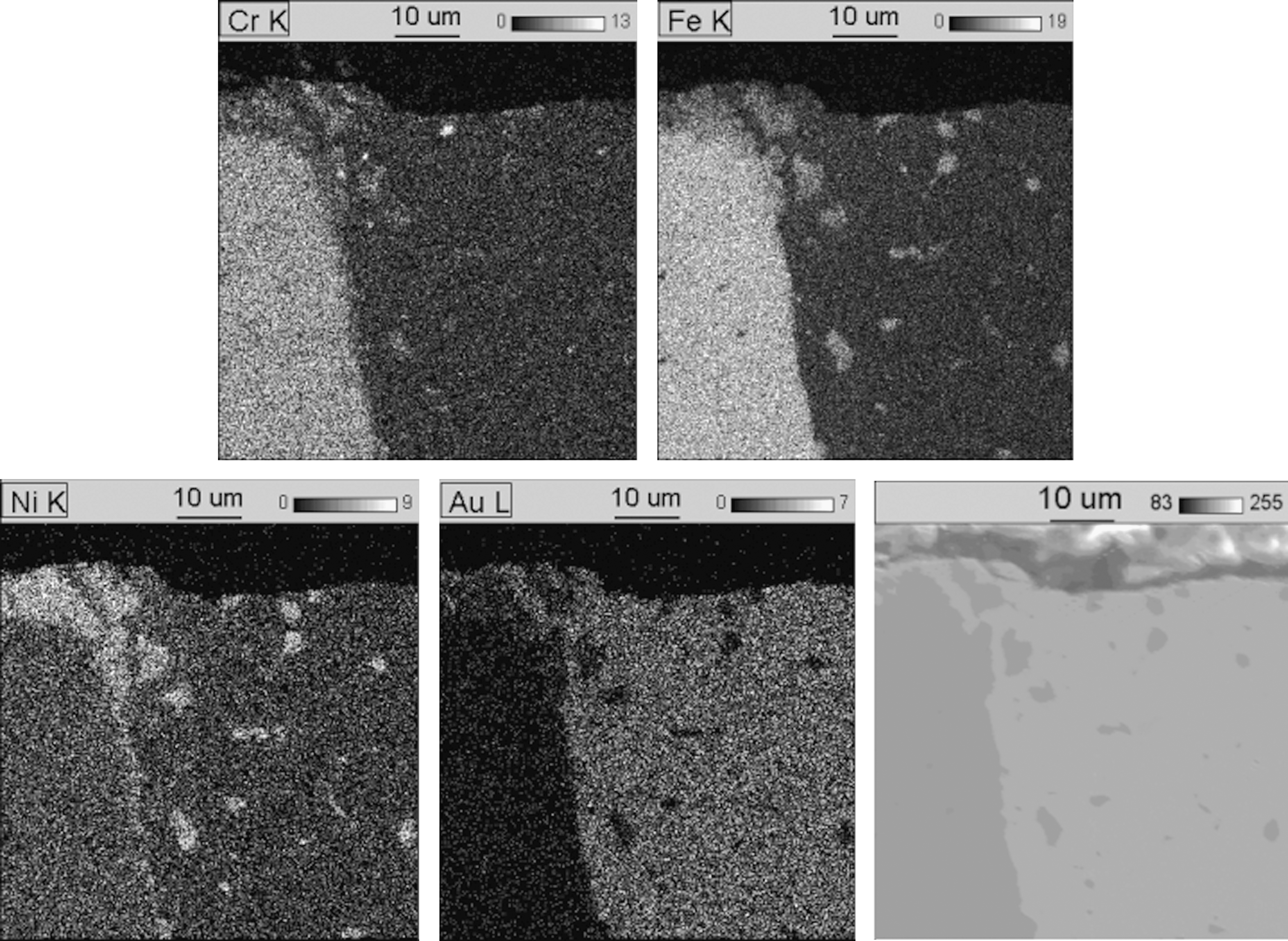
To confirm the aforementioned changes, the distribution of chemical elements making up the particular components of the gold-coated tip of the Urolase fiber was analyzed by atomic mapping. The Nd:YAG laser fiber tip is made up of gold and acid-resistant steel, containing chromium, nickel, and iron. The elements of the fiber tip made of steel are clearly separated from those made of gold. The individual chemical elements are distributed regularly, and their saturation intensity is homogeneous in all the parts of the fiber tip (Fig. 4).

Elemental map of the reflective surface of the intact fiber tip not used in visual laser ablation of the prostate.
If the fiber tip is positioned too close to the surface of prostatic tissue, with low rinsing fluid flow rate and laser work parameters of 40 W for 90 sec, changes appear in the elemental distribution maps. The obtained maps record the relative position changes of atoms within the gold-coated fiber tip. Destruction of the superficial layer of pure gold coating the fiber tip and partial disturbance of purity of the underlying structures is visible (Fig. 5).
Elemental map of the reflective surface of a fiber tip used in VLAP, performed with 40 W energy applied for 90 sec.
With 60 W energy applied for 60 sec, positioning the gold fiber tip too close to the prostatic tissue leads to the processes taking place on the surface and inside the tip, resulting in the complete destruction of its structure and mixing of the chemical elements, as well as its visible deformation. The superficial gold coating is destroyed, and gold fuses with the components of steel forming a homogeneous mass (Fig. 6).
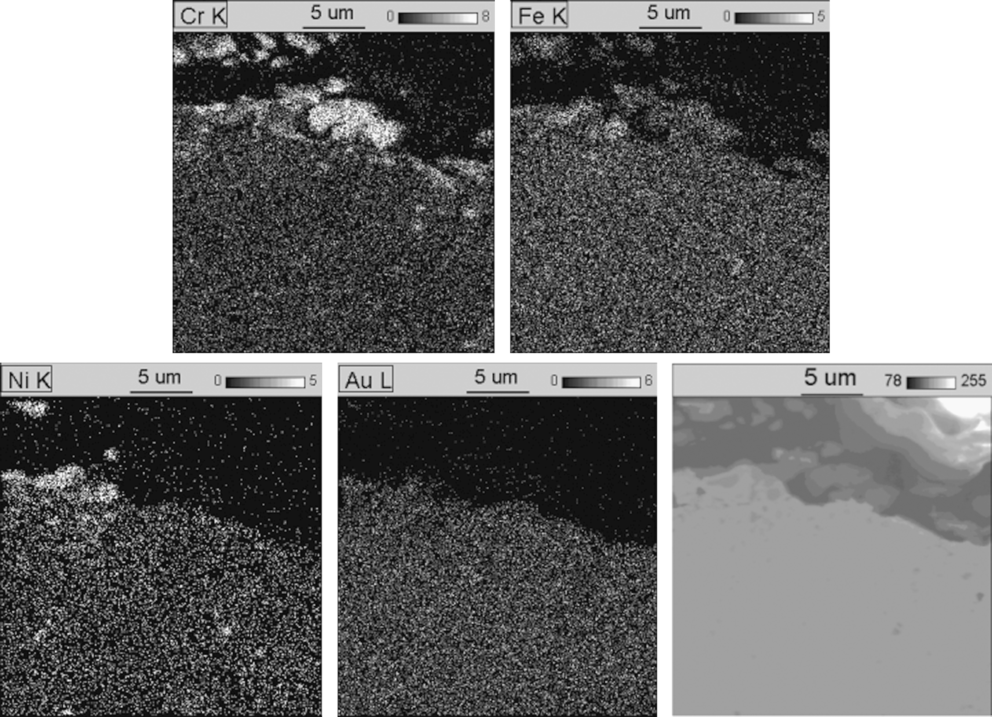
Elemental map of the reflective surface of a fiber tip used in visual laser ablation of the prostate, performed with 60 W energy applied for 60 sec.
It seems that in such a case much of the energy, instead of being transferred to the tissue, accumulates in the laser fiber tip or is dispersed in various directions. The amount of heat accumulated and released in this process is so high that it causes local melting of the reflective surface of the fiber tip. The extent of the molten area and the convection phenomena taking place there depend on the magnitude of energy used. At 40 W, evaporation of the superficial gold layer up to a few micrometers thick is observed (Fig. 5), whereas increasing the energy to 60 W causes the metal to melt to a much greater depth. Figure 6 shows total destruction of the superficial gold layer and intermediate nickel layer, as well as displacement of particles of the reflective fiber tip surface. In the gold-coated area, sites with markedly increased iron, chromium, and nickel content are clearly visible. The depth at which they are located shows the very high intensity of convection processes. Because the exposure times were relatively short (several dozen seconds) the observed processes of material displacement to such great distances could not be attributed to diffusion, but must have occurred in the liquid phase; therefore, the material must have melted at least to that depth.
The hypothesis concerning melting is confirmed by the presence of a black deposit on the surface of the gold coating layer, visible in Fig. 3a. It was formed most probably as a result of carbonization of the tissues that came into contact with the laser fiber tip. The organic origin of these deposits is indicated by microanalysis of their chemical composition (Fig. 7), which demonstrated the presence of carbon and oxygen. Because those elements are not present in new fiber tips, they must have originated from the external environment, i.e., the surrounding tissues. The lack of signals of elements comprising the underlying material indicates that the analyzed layer is at least several micrometers thick. In addition to deposits made up mainly of carbon, deposits containing considerable amounts of calcium and phosphorus as well as low amounts of magnesium and sodium can be observed. These elements may come from urine or from the rinsing fluid used during the procedure. The deposition of this layer markedly deteriorates the reflective properties of the gold-coated area and may cause accumulation of the laser energy in the fiber tip. Such a situation may result in complete destruction of the gold laser fiber tip (Figs. 2, 3).
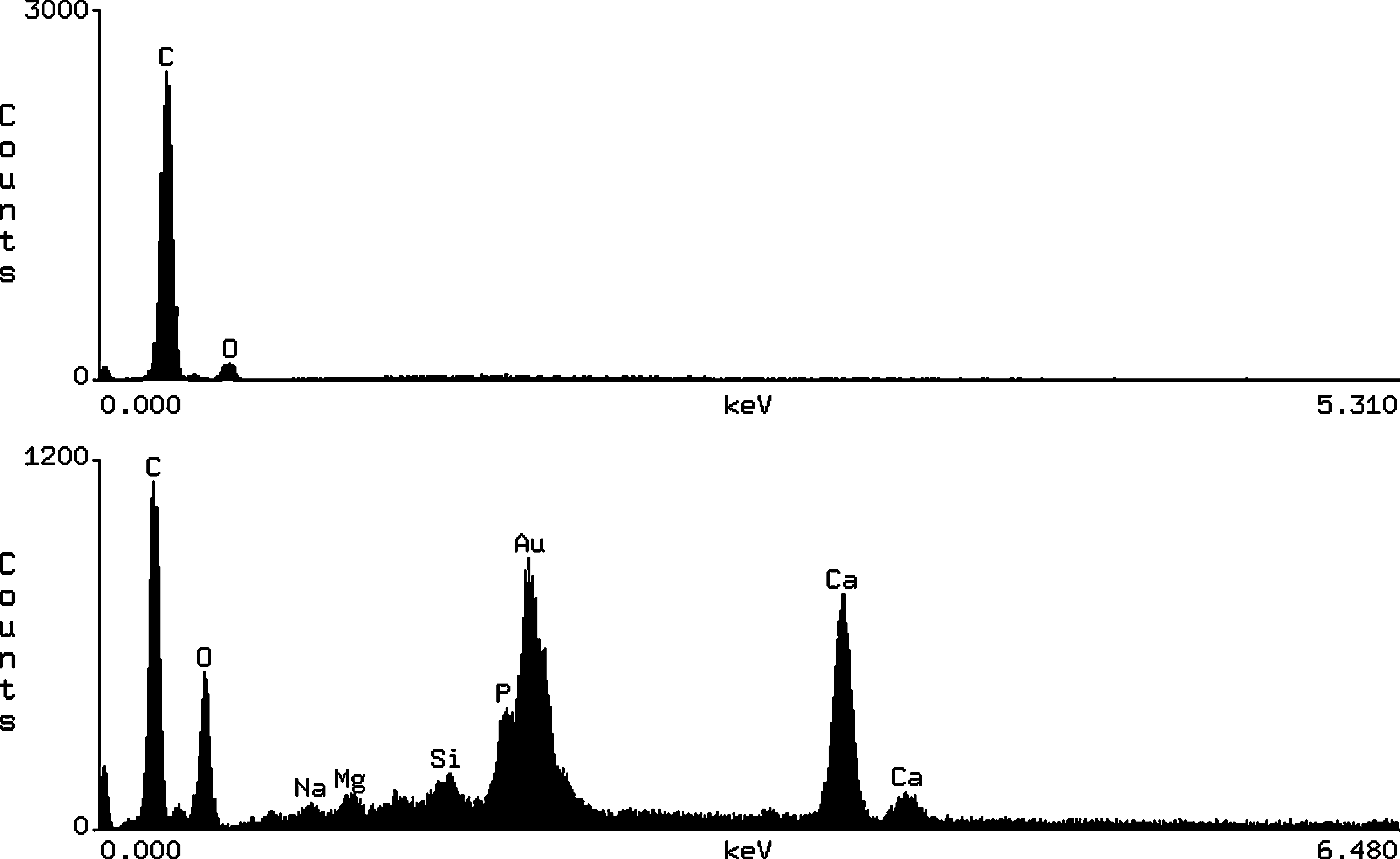
Characteristic X-ray spectra obtained from two different sites of the deposit present on the reflective surface of the fiber tip working with 60 W energy applied.
Discussion
White light is disordered, made up of different wavelength components, and propagates in various directions. Laser radiation is characterized by axial orientation of the beam and coherence of waves (all of them have the same phase, time, and space of radiation) and is monochromatic (the same wavelength and color). The aforementioned properties of laser radiation make it possible to focus its beam and direct it precisely onto the tissue that is to be operated on during a surgical procedure. However, some factors may interfere with the transmission of the laser beam through a laser fiber. Nazif et al. 11 and Knudsen et al., 12 in explaining the principles of laser beam transmission by flexible optic fibers, describe accumulation of energy on fiber curves and at the bending sites of flexible ureterorenoscopes. At such sites the fiber transmitting the laser beam, and in extreme cases, also the ureterorenoscope, can be damaged or even destroyed.
This problem concerns all fiber types, both straight- and angle-firing ones. With angle-firing laser fibers, an additional site where the laser energy can accumulate and cause fiber damage is its tip with a reflective surface causing deflection of the laser beam. The problem of tear and wear of laser fiber tips concern both the old and the most modern types of laser devices. 13
Nd:YAG laser has been used for treatment of BPH in the First Department of Urology of Łódź Medical University for 11 years. 9,10 This experience with VLAP allows objective assessment not only of the effects of treatment, but also of the device itself, and in particular fibers and their tips used during the procedure. Using Nd:YAG laser in ablation of prostatic adenoma tissue we observed the changes taking place on the surface of gold-coated fiber tips that direct the laser beam onto the adenoma tissue. The changes were most notable when a fiber tip with a gold-coated reflective surface came too near the adenoma tissue, or when the surface came into direct contact with the tissue under low water flow rate. The observed black deposit on the reflective surface formed as a result of reactions taking place within the fiber tip, accompanied by very high temperatures, which has been confirmed by atomic mapping images of the damaged tips with displacement and mixing of the atoms of metals making up the reflective surface elements. Atoms of gold, iron, nickel, and chromium become intermingled (Figs. 5, 6). On the macro scale, this corresponds to destruction of the fiber tip and its reflective surface, visible in Figs. 2 and 3, which causes incorrect transmission of the laser beam or even its complete fading. On the basis of observation of changes within the gold-coated Urolase laser fiber tip under a scanning microscope, it can be supposed that the reflective surface, if correct transmission of the deflected laser beam is impossible, becomes its receiver and undergoes self-destruction. The amount of energy accumulated in the fiber tip causes it to melt and in this situation it can become dangerous for the surrounding tissue and accidentally damage the urinary bladder wall or the urethra.
Conclusion
The gold reflective surface tip of a Urolase fiber, which directs a laser beam onto prostatic adenoma tissue, becomes damaged with inappropriate handling during visual laser ablation procedures. The amount of energy that accumulates in the fiber tip leads to a considerable temperature increase, which can be dangerous for living tissue and may lead to accidental damage of the urinary bladder wall or the urethra.
Footnotes
Disclosure Statement
No competing financial interests exist.