
Abstract
Introduction
A number of authors have described the effect of laser treatments of the external surfaces of teeth on intrapulpal temperature. 2,3 For endodontics, a significant limiting factor for the use of these lasers within the root canal is the extent of heat conduction to the supporting apparatus of the tooth. Several factors, including wavelength, energy, exposure time, and thickness of the radicular dentine, influence temperature conduction from the root canal through the radicular dentine to the periodontal apparatus. The use of coolants such as air and water during lasing can also influence the amount of heat conducted to the periodontal apparatus.
In an earlier study, 4 we showed that optically modified long cone fibers could remove smear layer better than conventional plain fiber because of their ability to better transfer energy to the walls of the root canal. This ability to deliver energy more efficiently to the walls of the root canal could also contribute to higher temperatures at the root surface, although the use of irrigants between lasing cycles could attenuate such an effect. Accordingly, the present study was undertaken to compare root surface temperatures using laterally emitting conical tips, versus conventional plain tipped quartz fiber optics, with Er:YAG and Er,Cr:YSGG lasers.
Materials and Methods
Sample selection and preparation
Single-rooted extracted teeth (n = 64) that had been stored in distilled water with 1% thymol were radiographed (Sopro digital X-ray, Satelec, Italy) in two planes. Only teeth that were free of preexisting root canal fillings and unusual root canal anatomy were chosen.
All teeth were then prepared 1 mm short of working length using rotary nickel–titanium ProTaper files, to an apical size corresponding to an F5 ProTaper instrument (Dentsply Tulsa Dental, Tulsa, OK). A 0.5-mm-diameter bead-type thermocouple was affixed to the external root surface with heat conductive silicon thermal compound in the apical third 2 mm from the apex; molding wax was used to cover the thermocouple and thereby maintain constant contact with the tooth surface (Fig. 1). The surfaces of the thermocouple that were not in contact with the tooth were sealed with a thick layer of sticky wax to remove the effects of the external environment. The thermocouple was connected to a digital multimeter (Protech 506, Dick Smith Electronics, North Ryde, Sydney, Australia) that had a computer interface for data capture. The thermocouple had a useful range of −17°C to 1200°C (0–2000°F), with an accuracy of ±3%, with a separate measurement every 0.01 s. All of the teeth were carefully mounted onto an individual grid, and a rubber dam and clamp were placed. The grid allowed the root to remain stationary while the tip of the optical fiber was moved. The teeth with their attached grids were then placed in a water bath maintained at 37°C.
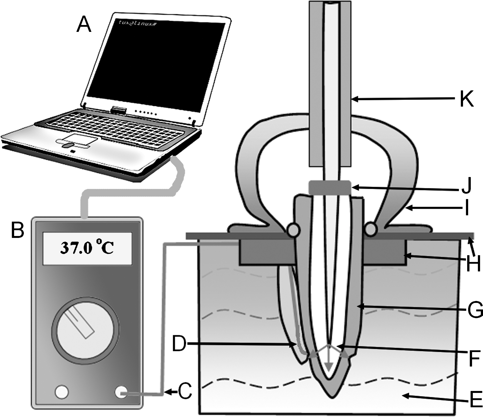
Experimental schematic. (A) Computer interface. (B) Digital multimeter. (C) Thermocouple. (D) Thermocouple positioned 2 mm from the apex and held in place by sticky wax. (E) Thermoregulated water bath. (F) Modified conical tip with lateral emission profile (arrows). (G) Prepared tooth. (H) Rubber dam with grid holder. (I) Rubber dam clamp. (J) Silicone stopper. (K) Polyamide coating of fiber optic.
Lasers and optical fibers
Two lasers were used; the first was the Waterlase MD Er,Cr:YSGG laser system (2780-nm wavelength, Biolase Technology, Irvine, CA) at a panel setting of 1.25 W (62.5 mJ/pulse) at 20 pulses/s, with no air or water. The laser tips used with the Waterlase MD was a plain (unmodified) Z4 tip (400-μm diameter and 28-mm length) or the same tip modified using hydrofluoric acid etching to obtain a conical shape. The modification was undertaken using the tube etching technique at room temperature with 50% hydrofluoric acid 5 for 2.5 h to give a terminal diameter of 33 μm, with the process controlled by continuous examination using an Olympus dissecting microscope (Olympus Optical Co., Tokyo, Japan) at a magnification of 30X. To obviate concerns regarding residual hydrofluoric acid etch solution affecting the delivery of the laser beam, immediately after the completion of the etching process, all fibers were placed for 60 s in saturated sodium bicarbonate solution to neutralize residues of etchant and then rinsed with distilled water.
The second laser used was the KEY3 laser system (2940-nm wavelength, KaVo, Biberach, Germany) at a panel setting of 200 mJ/pulse (4 W) and 20 pulses/s, with no air or water. The tips used with this laser were a plain (unmodified) three-ring tip (470-μm diameter and 28-mm length) or the same tip modified using hydrofluoric acid to obtain a conical shape. Tube etching for the KEY3 germanium (Ge)-doped glass fibers was undertaken for 90 min to give a terminal diameter of 33 μm. A representative example of a conical fiber tip for the KEY3 laser is shown in Figure 2.

Conical fiber tip used with the KEY3 laser. Long cone fiber tip created by etching. Scale markings along the image are in millimeters. The inset shows the emission profile of the diode laser aiming beam.
The average of three forward and lateral emissions from plain and conical fibers were assessed using a power meter (Ophir Nova II display and a Ophir 30A-V1-SH smart head sensor, Ophir Optics, Wilmington, MA), with the fiber tips placed at 10 mm forward of and 2 mm laterally from the sensor head.
The root canal was filled with water as an irrigating solution, and the fiber tip was then placed 1 mm short of the working length (and thus 2 mm short of the apex) and the laser activated. During activation, the fiber tips were kept in constant circular motion and withdrawn coronally at a rate of 1 mm per second. Only the apical 4 mm of the root canal was subjected to laser treatment. The lasing cycle was repeated 10 times (total irradiation time 60 s). Between each cycle, the canals were irrigated with water at room temperature (21°C) for 5 s. Data were captured every 0.1 s. The maximum and minimum temperatures of each experimental run were then recorded and analyzed.
To measure dentine thickness, the teeth were dried on the bench and then grooved vertically on their mesial and distal surfaces using a diamond disc without water. They were then split into two and the root canal walls observed for charring. The section of the root where the thermocouple had been placed was sectioned horizontally at the location of the thermocouple, and the thickness of the dentine at that spot recorded using an ocular grid in an Olympus dissecting microscope at a magnification of 30X.
All data were analyzed for normality using the Kolmogorov and Smirnov test and variations in the data sets assessed using Bartlett's test. Finally, group data were analyzed using the Kruskal-Wallis test [non-parametric analysis of variance (ANOVA)].
Results
When assessed using a laser power meter, the panel settings used for both systems gave average forward emission output powers of 1.03 and 1.10 W from the terminal end of bare (unmodified) optical fibers for the KEY-3 and Waterlase MD system, respectively. The conical fibers showed 48 + 5% and 49 + 5% lower forward emission and 452 + 69% and 443 + 64% greater lateral emissions than conventional plain fiber tips, for the KEY3 and Waterlase MD systems, respectively. The average thickness of dentine at the recorded site for all groups was approximately 1 mm (Table 1). The Kruskal-Wallis (nonparametric ANOVA) analysis of recorded temperatures at the external root surfaces of both laser types and fiber designs showed no statistically significant differences. Recorded temperature plots for 10 cycles revealed that temperature reductions after irrigation occurred and persisted until the start of the next cycle of laser treatment (Fig. 3). The maximum increase in temperature seen on the root surface was approximately 2°C in all groups in the study. The greatest reduction in temperature caused by irrigation was also approximately 2°C. Light microscopic images of the apical third of the specimens in all groups showed no carbonization within the root canal.
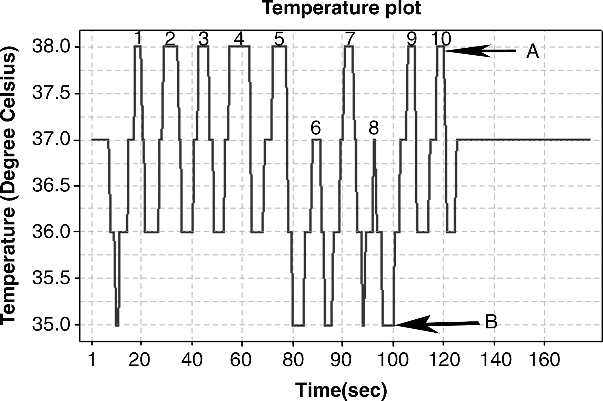
Time course of thermal changes during lasing. This plot shows the rise and fall in temperature over 10 numbered cycles of ablation, in this example using a Biolase modified conical tip. (A) Maximum temperature increase and (B) greatest temperature reduction seen.
Discussion
The objectives of this study were to investigate the thermal changes on the root surfaces of the teeth during laser irradiation with Er:YAG or Er,Cr:YSGG lasers using two different designs of optical fiber. The conical tips produce a fan-shaped emission profile that could disperse more energy onto the walls of the root canal than conventional bare-ended tips. This lateral emission capability has previously been linked to efficient removal of smear layer and may also be of value for preparing root canal walls. 4 Of interest in this particular study were the temperature effects of these modified tips on the apical external root surface compared with those of conventional tips.
Past studies of the thermal effects of lasers have indicated that, during root canal treatment, use of appropriate laser parameters is important for addressing the major concern of thermal damage to periodontal and alveolar bone cells. Alveolar bone is sensitive to temperatures above 47°C (10°C above body temperature), 6 whereas the temperature threshold for the periodontal ligament is 42°C (5°C above body temperature). 7 Accordingly, Machida et al. 8 have stated that the temperature threshold for laser root canal treatments to be done safely without damage to the periodontal tissues is 7°C above body temperature. On this basis, using the laser parameters employed in the present study, with both of the fiber tips, the treatments used with this study should be safe for root canal applications. The caveat for such a conclusion is that the fibers be used with an irrigation protocol, which appears essential for attenuating the thermal effects of lasing.
Several studies have shown that the dispersion pattern of conical optical fibers 9 –12 is at a large angle, and hence such fiber designs would transfer far more laser energy onto the walls of the root canal. This raises a concern that the temperatures achieved at the root apex would be greater with the conical fibers than with bare fibers. It appears likely that the attenuating effect of delivering room-temperature irrigant fluid between lasing cycles completely counters the temperature increase that otherwise would be expected. In preliminary experiments, we noted that temperature increases with these lasers of 2°C were commonplace if lasing was undertaken but not followed by irrigation (data not shown). The observation of a temperature reduction in the present study is consistent with the cooling phenomena seen in previous studies 13 that have documented temperature reductions from water irrigant used during coronal cavity preparation with the Er:YAG laser.
In the present study, irrigation quickly offset conduction of heat from laser energy through the root to the periodontal ligament. This reduction in temperature could prevent heating of the periodontal apparatus, provided that the irrigants used were cooler than body temperature; in this case, the irrigant was at room temperature (21°C). A similar drop in temperature would not occur if warmer irrigants were used. It is also emphasized that water spray was not used with either laser. Thus, the use of irrigation should help to attenuate the known risks of thermal injury when lasers are used in proximity to the periodontal ligament or bone. 14 –17 The supporting periodontal apparatus has been shown to be sensitive to temperatures greater than 47°C, whereas temperatures of 60°C or more stop blood flow permanently and cause bone necrosis. 6 A threshold temperature increase of 7°C is commonly regarded as the highest biologically acceptable value in order to avoid periodontal ligament damage. 8,18
The present results regarding thermal stress from lasing compare favorably with earlier work. For the Er,Cr:YSGG laser, reported temperature elevations on the root surface range from as high as 37°C when lasing was undertaken without cooling to 8°C when cooling was employed. 19 Lower temperature elevations in the absence of cooling have been reported when low average powers are used. 15,16,20
Most studies that have assessed root surface temperatures have used a thermocouple attached to the root surface with heat-conductive heat sink compound, with changes measured from a baseline of room temperature 15 or body temperature. 21 Thermographic cameras have also been used to record temperature changes. 20,22 A technical problem of interference by surrounding environment exists when one side of the thermocouple is exposed to the surrounding environment. This can be overcome by covering the noncontacting surface of the thermocouple with wax, 23 as was undertaken in the present study.
Conclusion
The present study recorded temperature patterns for two different lasers and two different fiber tip designs under simulated oral conditions, taking into consideration the environmental influence on the recording thermocouple. The study indicates that conical fiber tips can be used safely with either laser type under the present experimental conditions for various root canal procedures. There is recent evidence that conical tips provide more-effective smear layer removal in vitro than plain tips, 4 and the present study revealed that greater risks of thermal injury did not accompany this improvement.
Footnotes
Author Disclosure Statement
No competing financial interests exist.