
Abstract
Introduction
Whilst a number of anecdotal reports support the use of laser phototherapy for lymphoedema management, strictly controlled and well-designed trials are few in number. The most rigorously designed study was by Carati et al. 3 who investigated 64 patients with BCRL. Using a 904 nm laser (5 mW power output) at 1.5 J/cm2 to 17 points in the axilla, 31% of those who received two cycles of laser phototherapy (three times/wk for three weeks) had marked tissue softening and significant reductions in affected arm volume at two to three months post-treatment. A diversity of theories exists to explain the mechanisms by which laser phototherapy produces its effects, including photo-acceptor stimulation within cellular mitochondria and production of reactive oxygen species 11,12 with subsequent cellular or physiological responses. From the literature, it is evident that when a variety of different cell types (both normal and malignant species) are exposed to laser irradiation of different wavelength and dose combinations, changes in cell proliferation are observable (Table 1).
These findings have not deterred some from the application of laser phototherapy in malignant clinical arenas such as chemotherapy- or radiotherapy-induced oral mucositis 19 and BCRL. Few long-term safety studies have been undertaken, although it has been suggested that laser phototherapy has no effect on survival or disease recurrence in such patients. 20,21 However, we concur with Werneck et al. 16 who suggested that “clinically, irradiating a cancer lesion may result in increased tumor progression”. Given the fact that research (as summarized in Table 1) suggests that tumor cell growth in culture can be stimulated with laser phototherapy, and that clinical application of laser phototherapy in malignant (clinical) states is increasing, the effect of laser phototherapy on malignant cells in vivo requires more critical review from the perspective of safety and efficacy of application.
In seeking to identify effective and safe dosing parameters for the treatment of BCRL, the aims of this research were to examine the response of two common human breast cancer cell lines and two immortalized human mammary epithelial cell lines in vitro to a range of commonly used doses of laser phototherapy at wavelengths of 780 nm, 830 nm, and 904 nm. This approach would permit determination of whether a dose-response or a wavelength-response relationship exists when different cell lines (breast cancer, melanoma, and immortalized mammary epithelial cells) are irradiated with a range of laser light parameters.
We also aimed to determine whether laser irradiation has the capacity to malignantly transform normal NIH3T3 cells 13 when compared to NIH3T3 cells malignantly transformed by transfection to express the H-Ras-GFP oncogene.
Materials and Methods
Cell culture
A human breast cancer cell line (MCF-7 – adenocarcinoma), a human melanoma cell line (MDA-MB-435S/M14), and immortalized human mammary epithelial cells (SVCT; and Bre80hTERT) were utilized in this study. MCF-7 and MDA-MB-435S cells were grown in Dulbecco's modified Eagle's medium (DMEM) containing 4500 mg/L D-glucose, 584 mg/L L-glutamine, and 110 mg/L sodium pyruvate supplemented with 10% heat-inactivated fetal calf serum (FCS), 50 U/mL penicillin, 50 μg/mL streptomycin, 25 mM HEPES buffer (4-(2-hydroxyethyl)-1-piperazineethanesulfonic acid), pH 7.4, and 0.05 mM 2-mercaptoethanol. SVCT and Bre80hTERT cells were grown in Roswell Park Memorial Institute (RPMI) 1640 medium supplemented with 10% FCS, 50 U/mL penicillin, 50 μg/mL streptomycin, 25 mM HEPES, pH 7.4, 0.05 mM 2-mercaptoethanol, 2 mM Glutamax, and 2 mM Na-pyruvate, and passaged every 3–4 days or as required. For cell proliferation experiments, to avoid scatter of laser to adjoining wells on the culture plate, cell lines were plated into sterile, black-sided, clear-based 96-well tissue culture plates (Costar Cat. No. 3603) at 104 cells per well for single laser exposure experiments, and 5×10³ cells per well for two or three consecutive laser exposures. When cells were loaded into the 96-well plates, colorless (phenol red-free) DMEM or RPMI medium (200 μL per well, Invitrogen Pty. Ltd., Victoria, Australia) was employed rather than regular media to ensure that no interference with the laser light occurred.
Laser irradiation procedure
After 16–17 hours, cultures of cells were exposed to one, two, or three applications of laser irradiation using wavelengths of 780 nm continuous (50 mW; Spectra-Medics Pty. Ltd. Gallium-Aluminum-Arsenide, Sydney, Australia), 830 nm continuous (30 mW; Medeleq Pty. Ltd. Gallium-Aluminum-Arsenide, Nerang, Australia) or 904 nm pulsed (90 mW, Spectra-Medics Pty. Ltd., Sydney, Australia). Irradiation exposures were performed at energy densities of 0.5, 1, 2, 3, 4, 10, and 12 J/cm2 for the 780 nm laser and 0.5, 1, 2, 3, 4, 10, and 15 J/cm2 for the 830 nm and 904 nm lasers. Each experiment included six replicates for each application of laser and 12 replicates of negative/untreated controls. The irradiating probe was secured perpendicularly over each well at a total distance of 15 mm above the cells. For single exposure experiments, cells were irradiated, incubated for 24 hours in normal culture conditions, and then assessed for cell growth. For experiments utilizing two and three repeated exposures, the irradiation applications were separated by 24 hours, before a further incubation for 24 hours before cell proliferation was assessed.
Cell proliferation assays
Cell proliferation was assessed using the XTT (sodium 3,3′-{1-[(phenylamino)carbonyl]-3,4-tetrazolium}-bis(4-methoxy-6-nitro)benzene sulfonic acid hydrate) assay (Bio Vectra, Cat. No. 2525, Charlottetown, Prince Edward Island, Canada). 22 Phenazine methosulfate (PMS) (N-Methyl phenazonium methyl sulphate, Sigma-Aldrich Pty. Ltd., Sydney, Australia, Cat. No. 68600) was used as electron acceptor to increase the efficiency of the reaction. 23 Cells receiving XTT were incubated at 37°C in 5% CO2 for up to 2 hours with absorbance readings measured at t = 45 min onwards in a FLUOstar OPTIMA (BMG LABTECH, Melbourne, Australia) plate reader at 490 nm.
Transfection and transformation assay
NIH3T3 cells were transfected with an H-Ras-GFP plasmid encoding the Ras oncogene using GenJet DNA In Vitro Transfection Reagent (SignaGen Laboratories, catalogue number SL100488, Gaithersburg, MD) following the manufacturer's instructions. After transfection for ∼18 hours, cells were examined by fluorescent microscopy to determine whether the transfection was successful. H-Ras-GFP transfected cells were selected with the antibiotic Geneticin (G418) added to the culture at 1,000 μg/mL for approximately 3 weeks. Untreated NIH3T3 cells (negative control) were loaded into a 96-well microtiter plate alongside cells given a single exposure of irradiation (15 J/cm2) from the 904 nm laser at a density of 5 × 10³ cells per well. Irradiated NIH3T3 cells were cultured for 3 weeks and compared with the positive transformed and negative control cells to determine whether laser irradiated cells demonstrated any change in morphology.
Statistical analyses
The Statistical Package for the Social Sciences (SPSS) Program Version 14.0 (Chicago, IL) was used to determine levels of significance in cell proliferative outcomes to laser irradiation. All data sets were normalized by calculating the mean of the controls for each plate, then calculating an overall mean for all controls on all plates. The multiplier used for normalizing was obtained by dividing the mean of the controls from each plate by the overall mean from all the controls. One-way ANOVA with LSD post-hoc tests and Mann-Whitney U tests were performed to determine the level of significance when comparing each energy density of irradiated cells versus the negative (untreated) controls, determined as either P < 0.05, P < 0.01 or P < 0.005. Results of the Mann-Whitney U tests are not shown, but reflected similar overall outcomes to those from the one-way ANOVA analysis.
Results
Minimal changes occurred in the growth rates of either Bre80hTERT (mammary epithelial) or MDA-MB-435S (melanoma) cells treated with a single exposure of laser irradiation, irrespective of the laser wavelength or the range of energy densities utilized. No significant changes in the relationship between proliferative rate and the energy densities of laser irradiation for either the Bre80hTERT or MDA-MB-435S cell lines were detected when linear regression analysis was performed (Table 2).
The statistical significance of each laser energy density applied to the four cell lines is shown. Overall significance (F) values from the regression analysis are also shown with significance assigned at (P < 0.05).
Increase in cell proliferation compared to untreated control.
Decrease in cell proliferation compared to untreated control.
Significantly different from untreated control, P < 0.05.
Significantly different from untreated control, P < 0.01.
Significantly different from untreated control, P < 0.005.
(one-way ANOVA, LSD post-hoc test).
Figs. 1, 2, and 3 are included as typical examples illustrating the significance and accuracy of the data and to show how the data presented in Table 2 were derived. SVCT cells (mammary epithelial cells) treated with a single exposure of laser irradiation at 780 nm demonstrated statistically significant increases in cell proliferation at energy densities of 0.5 J/cm2 (P < 0.05), 1 J/cm2 (P < 0.01), 2 J/cm2 (P < 0.05), 3 J/cm2 (P < 0.005), 4 J/cm2 (P < 0.05), and 12 J/cm2 (P < 0.005) (one-way ANOVA) (Fig. 1). Regression analysis demonstrated an increasing linear relationship between the absorbance values and energy densities of laser irradiation (P < 0.05); with 12 J/cm2 displaying a 13.2% increase in SVCT proliferation compared with untreated control cells. SVCT cells given a single exposure of laser irradiation at 830 nm and 904 nm also produced statistically significantly increased proliferation at specific energy densities (Table 2).

Proliferation of SVCT cells after one exposure to 780 nm laser. Cells were treated with doses of laser between 0.5 and 12 J/cm2 or left untreated (0 J/cm2). Mean absorbance values and standard error are shown for each dose of laser. The line of best fit from the linear regression is displayed as a dashed line. The dotted line is the baseline control line. One-Way ANOVA, LSD Post-Hoc test: ♦ Significantly different from untreated control, P < 0.05; ♦♦ Significantly different from untreated control, P < 0.01; ♦♦♦ Significantly different from untreated control, P < 0.005. *Linear regression is significant, P < 0.05.
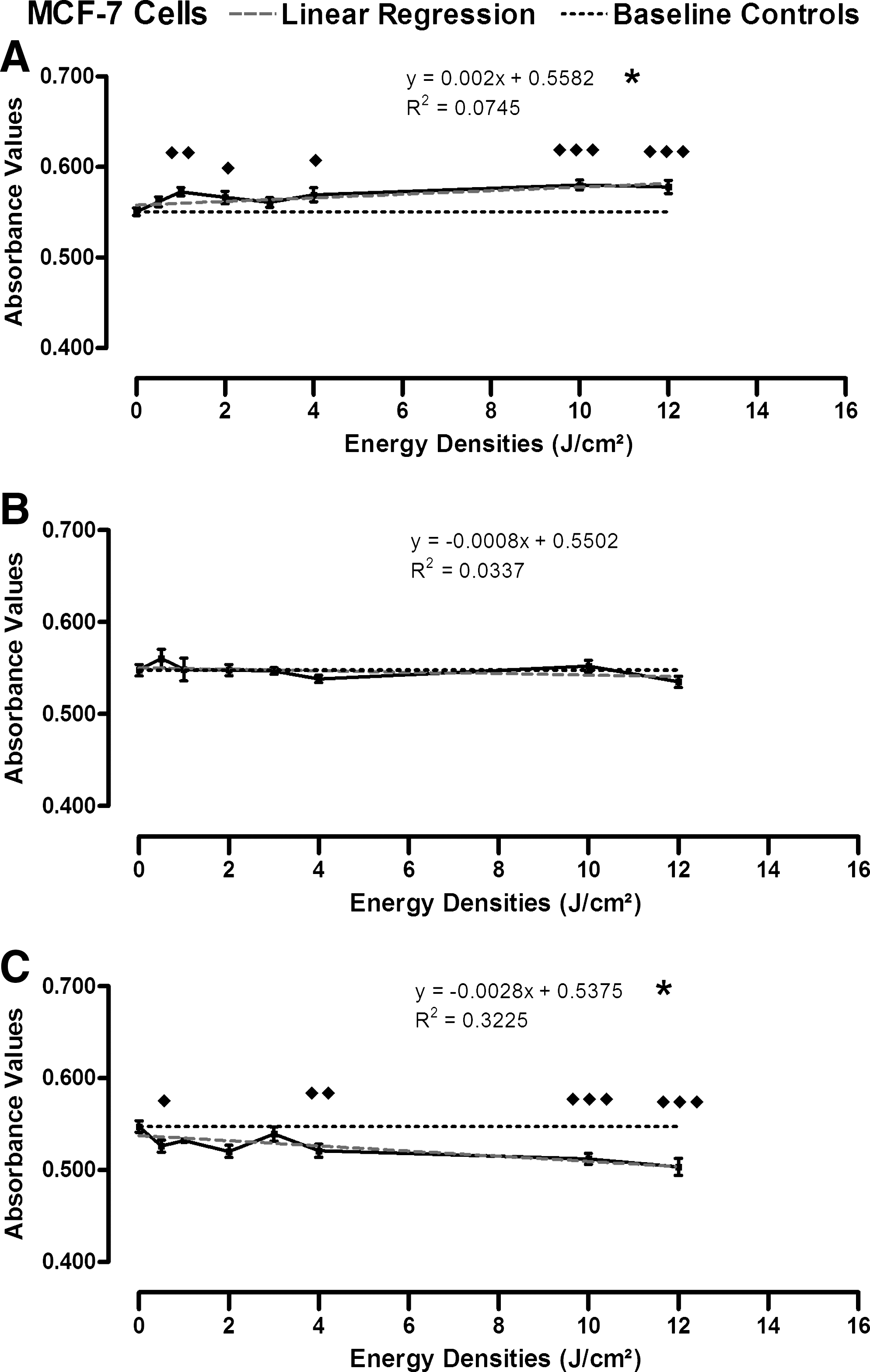
Proliferation of MCF-7 cells after exposure to 780 nm laser.
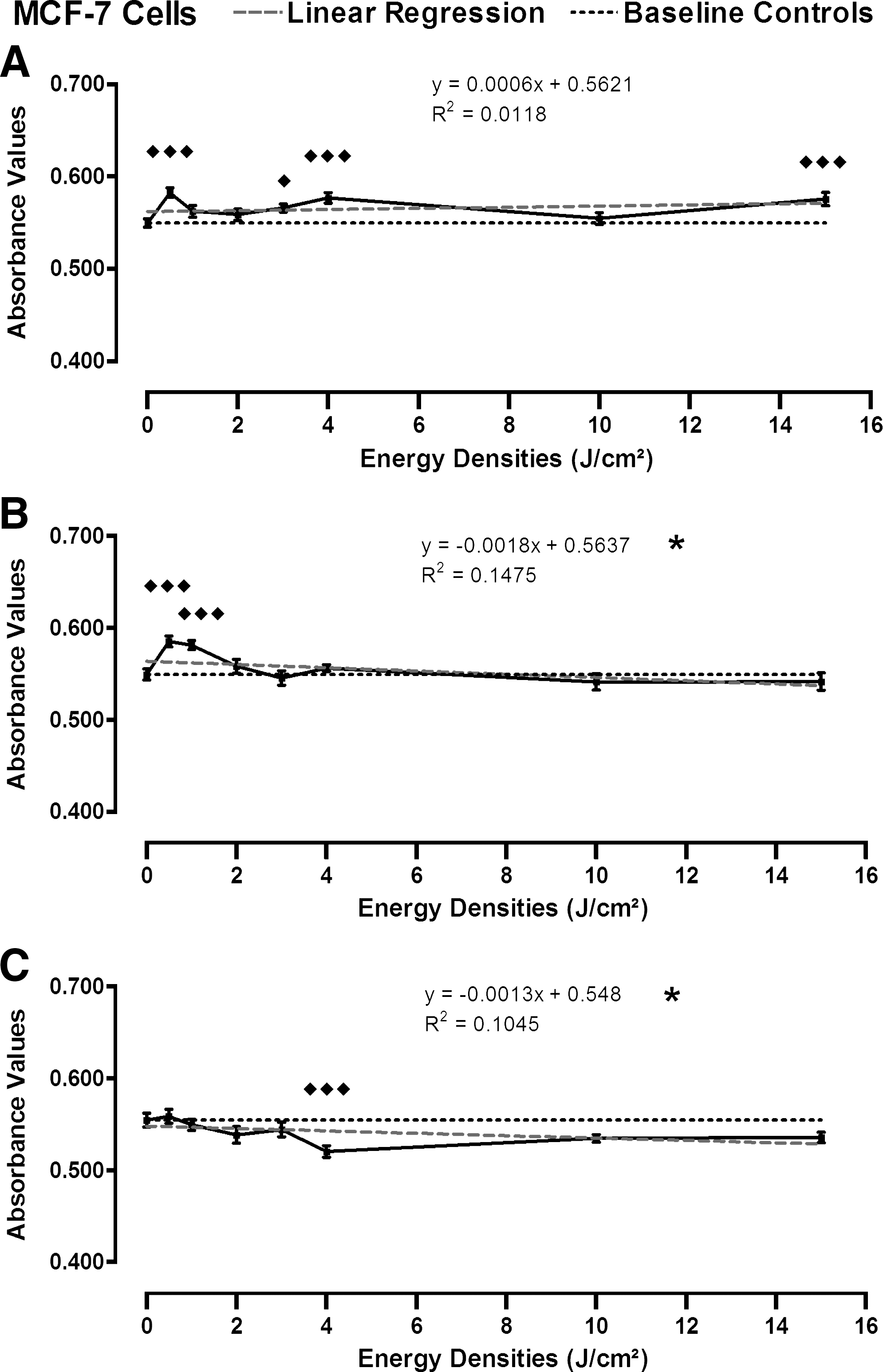
Proliferation of MCF-7 cells after exposure to 904 nm laser.
When MCF-7 cells (breast adenocarcinoma cells) were treated with a single exposure of laser irradiation at 780 nm on four separate occasions and the results were combined (Fig. 2A), significant increases in proliferation occurred at energy densities of 1 J/cm2 (P < 0.01), 2 J/cm2 (P < 0.05), 4 J/cm2 (P < 0.05), 10 J/cm2 (P < 0.005), and 12 J/cm2 (P < 0.005) (one-way ANOVA). Regression analysis demonstrated a clearly increasing linear relationship between proliferation and energy densities of laser irradiation for MCF-7 cells treated with a single exposure from the 780 nm laser (P < 0.05) and there was a 4.90% increase in cell proliferation at an energy density of 12 J/cm2 when compared with untreated controls (Fig. 2A).
When MCF-7 cells were treated with two exposures of laser irradiation at 780 nm, no change in proliferation of the cells at any energy density was observed, which resulted in there being no significant change in linear relationship between the absorbance values and the energy densities of laser irradiation (P > 0.05) (Fig. 2B). However, MCF-7 given three exposures from the 780 nm laser produced a significant decrease in proliferation at energy densities of 0.5 J/cm2 (P < 0.05), 4 J/cm2 (P < 0.01), 10 J/cm2 (P < 0.005), and 12 J/cm2 (P < 0.005) (Fig. 2C). Regression analysis of the data revealed a linear relationship between decreasing proliferation and increasing energy densities of laser, with an 8.04% decrease in cell proliferation at 12 J/cm2 compared with the untreated controls (P < 0.05) (Fig. 2C).
MCF-7 cells treated with one, two, or three exposures of laser at 830 nm produced significantly increased proliferation at some of the individual energy densities tested, but regression analysis revealed no significant overall change in the linear relationship between proliferation and energy densities of laser irradiation (Table 2).
MCF-7 cells were treated on three separate occasions with a single exposure of laser irradiation at 904 nm and the combined results (Fig. 3) demonstrated a statistically significant increase in proliferation at energy densities of 0.5 J/cm2 (P < 0.005), 3 J/cm2 (P < 0.05), 4 J/cm2 (P < 0.005), and 15 J/cm2 (P < 0.005). Despite the increases in proliferation, no significant change in linear relationship between proliferation and the energy densities of laser irradiation was detected (Fig. 3A).
When the MCF-7 cells were treated with two and three exposures from the 904 nm laser, statistically significant changes in proliferation were apparent, with two exposures producing a significant increase in proliferation at energy densities of 0.5 J/cm2 (P < 0.005) and 1 J/cm2 (P < 0.005) (Fig. 3B). In comparison, decreased proliferation resulted from three exposures from the 904 nm laser at an energy density of 4 J/cm2 (P < 0.005) (Fig. 3C). Linear regression analysis of data for both the double and triple exposures demonstrated a linear relationship, with a decrease in proliferation associated with increasing energy densities (P < 0.05; Fig. 3B and 3C).
Transfection and transformation assay
The NIH3T3 cells were successfully transfected, with a large percentage of GFP-positive cells, indicating high efficiency transfection with H-Ras-GFP. The construct containing the H-Ras-GFP fusion protein has been previously characterized by Dong et al., 24 who successfully grew transformed foci of fibroblast cells transfected with H-Ras-GFP as tumors in mice. Fig. 4 illustrates the changed morphology of the NIH3T3 cells once transformed from normal into malignant cells displaying one of the most common characteristics of malignant cells, which is loss of contact inhibition and formation of transformed foci.
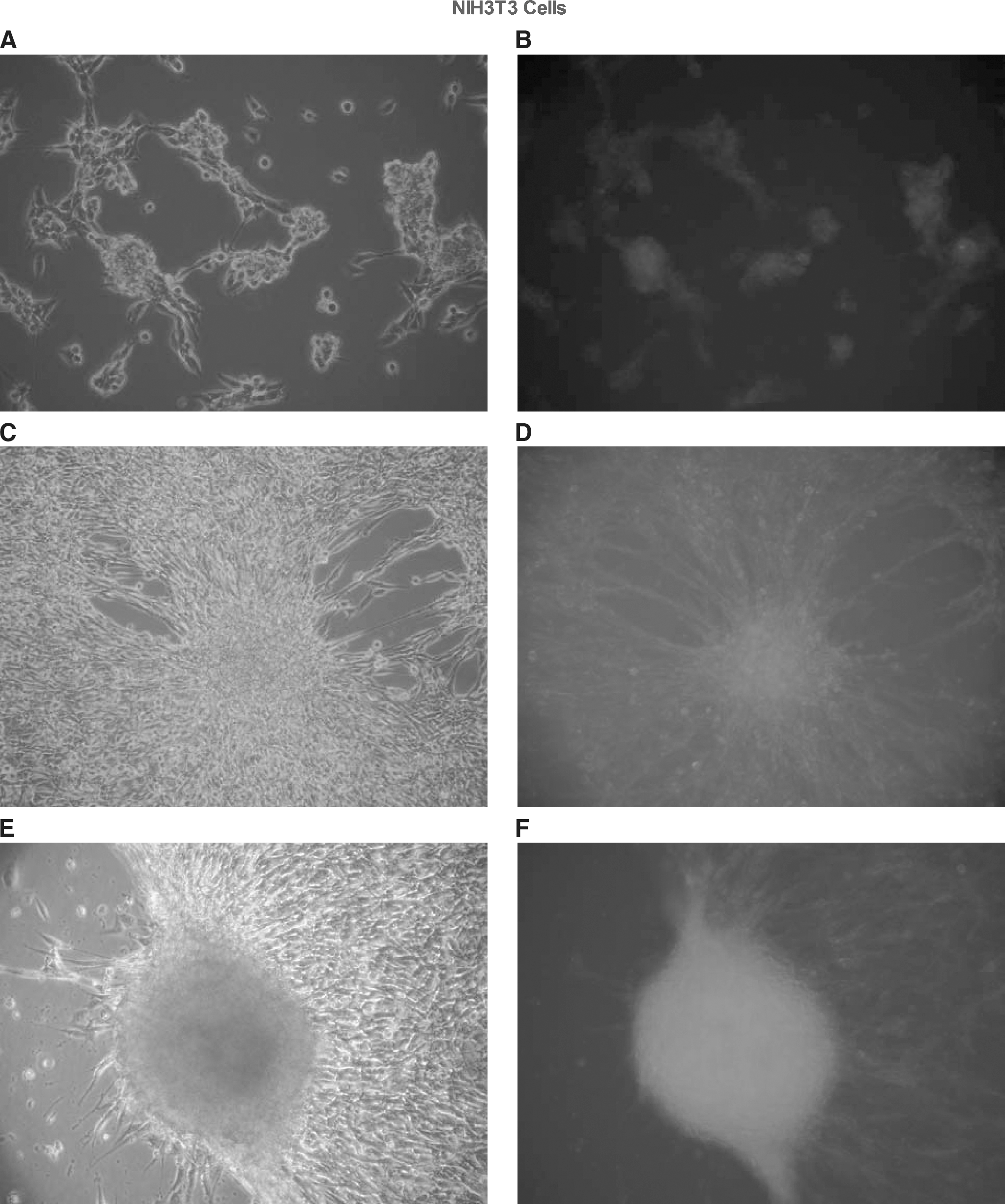
NIH3T3 cells transfected with H-Ras-GFP, three weeks post-transfection.
Fig. 4 (C & E) demonstrates elongation of the cells around the foci, which appear to be drawn into a mound of transformed cells. When the cells are cultured at a lower density, the transformed cells show a more circular morphology (Fig. 4C) when compared with the control NIH3T3 cells (Fig. 5A). In contrast, no morphological changes were observed in the irradiated NIH3T3 cells when compared with untreated cells (Fig. 5).

Control (untreated) and laser treated NIH3T3 cells. The laser treated NIH3T3 cells were exposed to one dose of 15 J/cm2 from the 904 nm laser.
Discussion
This is the first report to methodically investigate the effects of repeated exposures of a range of doses and wavelengths of laser phototherapy commonly used in clinical practice, in an attempt to identify the effects on human breast cells and cancer cell lines. In the clinical setting, patients usually receive multiple treatments with the same laser device. Therefore, the present study is novel in that we also investigated the effects of repeated exposures on the MCF-7 breast cancer cell line. Use of specific wavelength and dose combinations of laser phototherapy resulted in effects ranging from either stimulation or inhibition to no change in cell growth.
Importantly, 904 nm laser phototherapy at 15 J/cm2 did not transform the mouse fibroblast cell line (NIH3T3). Thus, no change in cell morphology was detected by microscopy in comparison to the highly transformed phenotype of the GFP-H-Ras transfected cells. This result indicates that laser phototherapy at the parameters used is most likely not mutagenic in regard to cellular transformation and induction of phenotypes associated with malignancy.
In particular, it was instructive to test the two immortalized mammary epithelial cell lines (SVCT and Bre80hTERT), and compare the results to studies conducted on other “normal” cell lines and cancer cells. Intriguingly, the two immortalized and non-malignant Bre80hTERT cells and SVCT mammary epithelial cell lines showed contrasting effects when laser irradiated. The Bre80hTERT cell proliferation was not significantly affected by one exposure from the 780 nm and 830 nm lasers whereas 904 nm laser at a dose of 10 J/cm2 produced a highly significant inhibition of cell proliferation (Table 2). By contrast, SVCT cell proliferation was significantly increased when treated with most of the tested doses using the 780 nm and 904 nm lasers (Table 2), as well as doses of 3 J/cm2 and 15 J/cm2 from the 830 nm laser.
The above findings are consistent with those of others 25 in which it was reported that C2 cells (normal mouse skeletal myotubes) showed no change in mitotic rate using 630, 640, or 805 nm lasers, increased rates of mitosis after exposure to 4 J/cm2 from a 635 nm laser, but inhibited mitosis at 20 J/cm2. Sroka et al. 25 also found increased rates of mitosis in normal bladder epithelial cells (HCV29) treated with doses between 2–12 J/cm2 from 410 nm, 635 nm, or 805 nm lasers. Hence, it would appear that the growth of several normal cell types can be affected by laser phototherapy.
In a comparison of normal human osteoblasts to malignant osteosarcoma cells with a range of different doses and wavelengths, only 10 J/cm2 from an 830 nm laser was able to significantly enhance osteoblast proliferation, whereas doses of 1 J/cm2, 5 J/cm2, and 10 J/cm2 from a 780 nm laser significantly decreased proliferation. The malignant osteosarcoma cells were unaffected by 830 nm laser irradiation. 18 This finding is similar to those of the present study with the MDA-MB-435S melanoma cells, where proliferation was relatively unaffected by 780, 830, or 904 nm laser irradiation (Table 2). Sroka et al. 25 investigated MCF-7 breast cancer cells as we did, providing a means for directly comparing our results with theirs. They found that 630, 635, or 805 nm laser irradiation decreased rates of mitosis with increasing doses from 0 to 20 J/cm2. This observation markedly differs from our results with MCF-7, where a single exposure to 780 nm showed an increasing dose response relationship that was highly significant (P = 0.0001, Table 2). The markedly contrasting effects between the 805 nm results of Sroka et al. 25 and our 780 nm outcome may highlight the considerable importance of differences resulting from subtle changes in laser wavelengths.
The 830 and 904 nm lasers also led to increased proliferation after one exposure whereas three exposures from the 780 or 904 nm lasers significantly inhibited MCF-7 proliferation (Figs. 2C, 3C). This was similar to the inhibition reported in Sroka et al. 25 with their single exposure. Only a few other studies exist that have investigated the effects of repeat exposures of laser phototherapy on malignant cells. 13,17 Pinheiro et al. 13 applied 635 or 670 nm laser irradiation to H.EP.2 cells (squamous cell carcinoma [SCC] Type 2 laryngeal carcinoma cells), and found that multiple exposures from the 670 nm laser over 7 days with doses ranging between 0.04 J/cm2 and 4.8 J/cm2 greatly increased cell number when compared with the 635 nm laser and untreated controls. The importance of optimizing the cell culture medium to prevent experimental error was also demonstrated in Pinheiro et al.'s 13 study and hence, specific attention was paid to this aspect of our study to ensure reliability of the results. Although results may be affected by other factors such as growth limitation by cell contact inhibition in the culture environment, we believe our experimental design negated such potential concerns, since they were not found in the negative control cultures. De Castro et al. 17 used repeat exposures (at 48 h) of laser at 685 or 830 nm on malignant oral KB carcinoma cells and found reduced cell proliferation after an initial increase (attributed to laser phototherapy), with a further minor stimulatory effect after a second irradiation of 685 nm laser.
The results of our study are consistent with general findings of other investigators 18,25 and suggest that the breast cancer, melanoma, and immortalized mammary epithelial cell lines we employed behave similarly as reported for other normal or malignant cell types. Thus, we conclude that the response to low-level laser irradiation, in terms of initiating growth stimulatory effects, depends on the individual cells targeted and the range of doses, which can be subtle and quite specific. Overall, our results indicate that some dose and wavelength combinations result in significantly increased growth of breast cancer cells. However, further in vivo research is required before making definitive statements regarding the effects and/or safety of specific wavelengths used for laser phototherapy in BCRL patients. Until such studies have been concluded, it may be prudent for clinicians to remain circumspect regarding use of either potentially stimulatory or inhibitory doses of laser phototherapy in unresolved malignant clinical states.
Footnotes
Author Disclosure Statement
No competing financial interests exist.