
Abstract
Introduction
According to White et al., 2 the smear layer is a negative factor in root canal sealing because this layer of organic/inorganic debris forms an interface between the sealer and the root canal walls, reducing the material's adhesion to dentin. Several chemical substances have been used as endodontic irrigants for cleaning and disinfection of root canals, as well as removal of the smear layer formed during biomechanical preparation, among which is ethylenediaminetetraacetic acid disodium salt (EDTA). 3
In addition to the routinely used chemical substances, other surface-altering means have been investigated for removal of smear layer from root dentin walls, such as laser irradiation. Studies have demonstrated the applicability of Er:YAG in endodontics for cleaning of root canals, removal of smear layer from canal walls, and exposure of dentinal tubules. 2,4 –6
Another important step in endodontic treatment is the choice of root canal filling material. Root canal sealers can be classified as calcium hydroxide–containing sealers, resin-based sealers, zinc oxide eugenol-based sealers either containing medication or not, and glass ionomer sealers.
The addition of calcium hydroxide to root canal sealers had the goal of increasing their biologic properties. Apexit (Ivoclar Vivadent, Schaan, Liechtenstein) was developed based on this premise. Apexit is a calcium hydroxide–based sealer containing calcium hydroxide, calcium phosphate, zinc oxide, silicone dioxide, plasticizer, disalicylate esters, and bismuth. It also was reported that this sealer has good flowing, which allows the material to adapt well to root canal morphology. Moreover, the slight setting expansion, in combination with the low solubility, allows good sealing of the root canal. 7
Epoxy resin–based cements have also given a good performance as root canal sealers. 8,9 AH Plus sealer penetrates deeper into these microirregularities because of its high flow rate and longer setting time. The entanglement of the sealer with the dentinal tubules allied to the cohesion between the sealer molecules increases the resistance to removal and/or displacement from dentine surface, which means in higher adhesion. 1,5 AH Plus sealer has good physicochemical properties, low solubility and disintegration, 8 good adhesion, 9 antimicrobial action, 10 and good biologic properties. 11 Advances in adhesive technology have reinforced the search for means to minimize apical and coronal marginal leakage by increasing the sealing between the filling material and the root canal walls. 12 A dual-curable methacrylate resin sealer (Epiphany; Pentron Clinical Technologies, Wallingford, CT) was developed for use with a self-etching primer and in association with a new thermoplastic synthetic polyester polymer-based root canal–filling material (Resilon; Resilon Research LLC, Madison, CT) that replaces gutta-percha. Obturation with the Epiphany/Resilon system is claimed to create a tight seal with the dentinal tubules within the root canal system. In essence, it produces a “monoblock” effect, in which the core material (Resilon), sealer (Epiphany), and dentinal tubules become a single solid structure. 13 –15 In vitro 13 and in vivo 16 studies have demonstrated a good resistance of the Epiphany/Resilon monoblock system to bacterial leakage.
Epiphany is supplied in a catalyst/base paste presentation in which the components are individually packaged in an automix dual-chamber syringe with a disposable tip for easily dispensing the ready-to-use material directly into the root canal. Despite the manufacturer's claims of a simplified operative technique, the automix syringe seems not to promote an optimal homogenization of the catalyst and base pastes, as evidenced by the presence of visible traces of both components in the mixed material dispensed from the syringe tip.
The purpose of this in vitro study was to evaluate comparatively the adhesion of the endodontic sealers Epiphany (dispensed from the automix syringe supplied by the manufacturer or prepared by hand mixing), Apexit Plus, and AH Plus to human root canal dentin treated with distilled water, 17% EDTAC, 1% NaOCl, and Er:YAG laser, by using the push-out test.
Materials and Methods
One hundred twenty-eight extracted sound human maxillary human canines with a single canal, no internal calcifications or accentuated flattening, and fully developed roots with minimal length of 15 mm were used in this study.
By using a water-cooled double-faced diamond disk (KG Sorensen, Barueri, SP, Brazil), the teeth were sectioned transversally at the cementoenamel junction and apically at the root end to leave an ∼8-mm-thick cylinder that was then centered inside an aluminum ring (16 mm diameter and 8 mm high) and embedded in acrylic resin. The aluminum rings containing the dentin cylinders were placed in a parallelometer, and their coronal and apical surfaces were flattened and made parallel, until a final length of 8 mm was obtained. The root canals of each specimen were prepared by using a tapered diamond bur (893-047; Brasseler, Savannah, GA) at a low-speed hand piece, which was attached to the arm of the parallelometer. This arm was lowered to a depth previously determined by a silicone stop, and space for sealer placement was created with the following standardized dimensions: larger diameter, 3.3 mm; smaller diameter, 2.6 mm; and length, 8 mm. During preparation, canals were irrigated with distilled water.
The specimens were randomly assigned to four groups of 32 teeth each, and root canal dentin was submitted to the following treatments: Group I, irrigation with 20 ml distilled water and drying with sterile absorbent paper points (Dentsply-Herpo, Petrópolis, RJ, Brazil); GII, irrigation with 5 ml 17% EDTAC (EDTA + Cetavlon) for 5 min followed by a final flush with 20 ml distilled water and drying with sterile absorbent paper points; GIII, irrigation with 5 ml 1% sodium hypochlorite (NaOCl) for 30 min (changing the solution every 5 min), followed by a final flush with 20 ml distilled water and drying with sterile absorbent paper points; GIV, irradiation with Er:YAG laser (Opus 20; Opus Dent, Tel Aviv, Israel) at 16 Hz, 400-mJ input (240-mJ output), and 0.32-J/cm2 energy density. Er:YAG irradiation was performed with a sapphire tip of 17 mm length and 1.3 mm diameter attached to an angled handpiece according to a helicoidal kinematics and using apical–cervical movements at a constant speed of ∼1 mm/s during 1 min (four cycles of 20 s each). These laser parameters were selected because they represent the best result of a previous investigation into this subject. 1 During laser irradiation, the specimens received a constant irrigation with 20 ml distilled water, and thereafter, they were dried with sterile absorbent paper points. 17
The 32 specimens of each group were divided into four subgroups (n = 8), according to the root canal sealer used: Apexit Plus (Ivoclar/Vivadent), AH Plus (Dentsply-DeTrey, Konstanz, Germany), and Epiphany (Pentron). Epiphany was either dispensed directly from the tip of its automix dual-chamber syringe, as supplied by the manufacturer (denominated the Epiphany/automix syringe) or prepared by hand mixing the catalyst and base pastes on a glass plate for 15 s until an homogeneous paste was obtained (Epiphany/hand mixed).
The specimens were filled with the sealers by using a lentulo drill (Maillefer, Ballaigues, Switzerland) mounted in a low-speed handpiece. For the subgroups Epiphany/automix syringe and Epiphany/hand mixed, before sealer placement, Epiphany self-etching primer was applied to root dentin with the assorted accessory points supplied by the manufacturer, and excesses were removed with microbrush disposable tips (KG Sorensen) and absorbent paper points (Dentsply-Herpo). The sealer was light cured for 40 s to provide an immediate root canal seal, according to the manufacturer's instructions.
The specimens were placed immediately at 37°C and 95% humidity for a period 3 times greater than the regular setting time of the sealer, according to ANSI/ADA Specification No. 57 (Apexit Plus, 90 min; AH Plus, 520 min; and Epiphany, 50 min). Subsequently, the specimens were submitted to the bond strength push-out test in an Instron 4444 universal testing machine (Instron Corporation, Canton, MA) that was equipped with load cell, an oscillating system, and a spring adapter. The machine was calibrated at a constant speed of 1 mm/min. The load required to cause failure of the bond was recorded in kN and transformed in MPa. Figure 1 shows a schematic illustration of the sample during the push-out test.
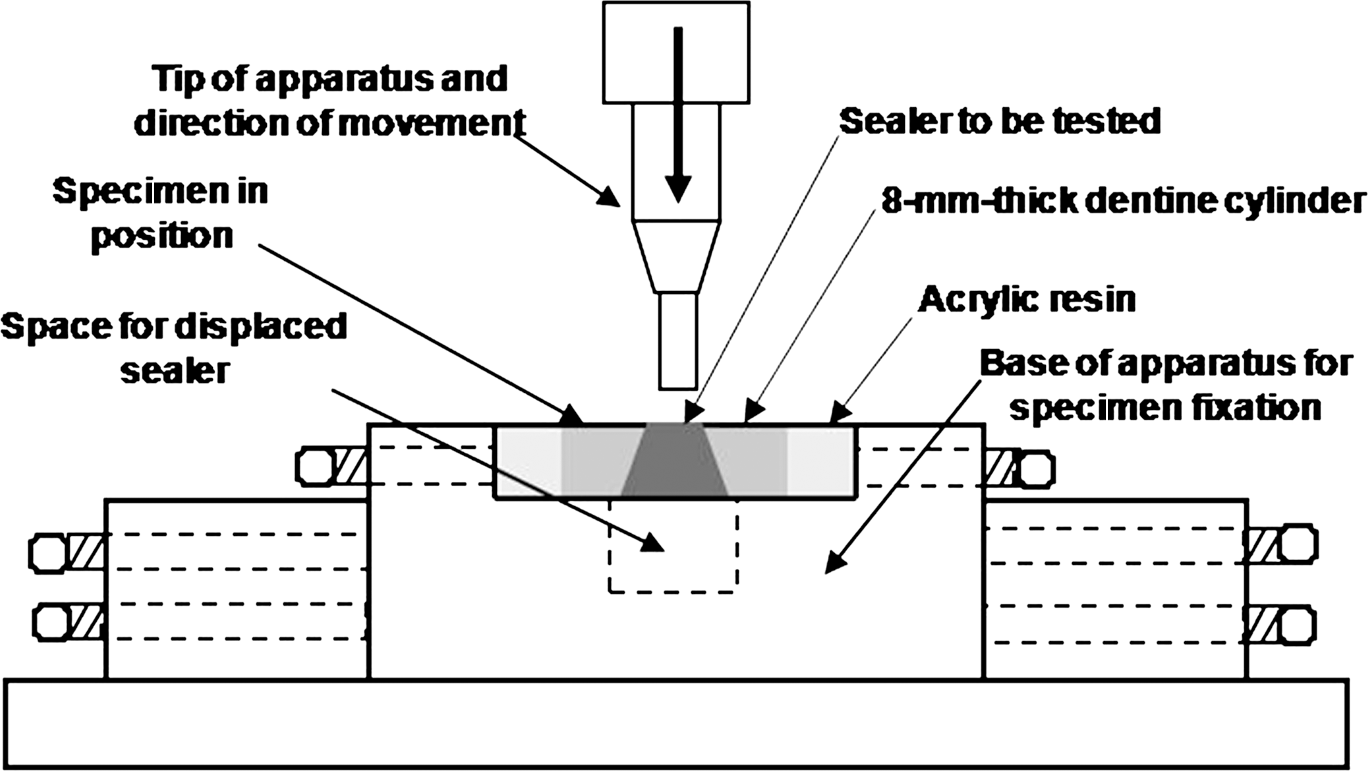
Schematic illustration of the sample during the push-out test.
Data were analyzed statistically with two-way analysis of variance. Tukey's post hoc test was then applied to determine which sealers and root surface treatments were different from each other.
Results
Push-out bond strength means (in MPa) and standard deviations are given in Table 1.
ANOVA showed statistically significant difference (p < 0.01) among the root canal sealers, except for Epiphany/hand mixed and Epiphany/automix syringe, which had statistically similar results to each other (p > 0.01). The highest adhesion values (load required for sealer displacement from dentin) were obtained with AH Plus (p < 0.01).
A statistically significant difference (p < 0.01) was found among the root dentin treatments. When distilled water was used as an irrigant, Apexit Plus showed significantly lower adhesion than the other sealers (p < 0.01). AH Plus, Epiphany/automix syringe, and Epiphany/hand mixed showed no statistically significant differences from each other (p > 0.01). When root dentin was treated with 17% EDTAC, Apexit Plus had significantly lower adhesion than the other sealers (p < 0.01). AH Plus was significantly different from Epiphany/automix syringe and Epiphany/hand mixed (p < 0.05) and had the highest adhesion values of all materials. When 1% NaOCl was used as an irrigant, Apexit Plus had significantly lower adhesion values than the other sealers (p < 0.01). AH Plus presented the highest adhesion values and was significantly different from Epiphany/hand mixed (p < 0.05). No statistically significant difference was found between AH Plus and Epiphany/automix syringe (p > 0.05). When root dentin was irradiated with Er:YAG laser, Apexit Plus and Epiphany/automix syringe showed statistically similar results to each other (p > 0.01) and the lowest adhesion values of all sealers. Both materials differed significantly from AH Plus, which presented the highest adhesion, and from Epiphany/hand mixed, which presented intermediate adhesion values. AH Plus and Epiphany/hand mixed had significantly different adhesion values from each other (p < 0.01).
Discussion
Adhesion has been studied since the development of the experimental model proposed by Grossman. 18 This model was improved by Ørstavik, 19 who used a universal testing machine to standardize the test, making it reproducible and more reliable. From the method proposed by Ørstavik et al., 19 a variety of surfaces have been used for testing. Previous studies analyzing the surface submitted to the test included dentin discs obtained from crowns of third molars, 20,21 coronal dentin from the cervical region of molars, 5 and gutta-percha discs. 20 –22
Sousa-Neto et al. 1 developed a method that permits evaluating the adhesive capacity of endodontic sealers by using as a test surface the root canal dentin of root cylinders. This experimental model allows understanding how adhesion to root dentin occurs under conditions closer to those of clinical use. The sealer is placed in direct contact with the root canal dentin in its original anatomic shape, instead of a flat surface obtained from tooth crowns, which presents a different tubule arrangement. Therefore, when the specimen is filled with sealer, the material adapts to the canal shape and penetrates into the dentinal tubules, promoting a mechanical retention similar to that obtained in a root-filled tooth. The force obtained with this model is thus derived from shear strength rather then pure tensile strength, in the same way as that in the present study.
Some factors directly related to the endodontic treatment may interfere with sealer adhesion, including root canal preparation and cleaning, the filling technique, and the type of sealer. 1 Regarding the root canal cleaning, great concern has been expressed with respect to the presence of smear layer on the dentinal walls. Removal of smear layer before root canal filling has been advocated to allow a better sealer penetration into the dentinal tubules, increasing the mechanical interlocking and providing an intimate contact of the sealing material with the dentin surface. 1,5 Smear layer removal has been performed with chemical substances, such as EDTA, 3 and dental lasers, such as Er:YAG laser. 4,5,23
Dentin irradiation with Er:YAG laser not only removes the smear layer, leaving clean walls and open dentinal tubules, but also promotes morphologic changes, increasing the dentin area and forming irregularities, as demonstrated in SEM analyses. 5,23 Another effect caused by the laser-induced surface heating is liquefaction of hydroxyapatite crystals and alteration of the carbonate amount in dentin, 24 which may improve sealer adhesion to root dentin.
The root dentin treatments proposed in the present study produced significantly different adhesion values of the sealers to root canal dentin. AH Plus had the highest adhesion, regardless of the surface treatment. As an epoxy resin–based sealer, AH Plus has better penetration into the microirregularities because of its creep capacity and long setting time, which increases the mechanical interlocking between the sealer and the root dentin. This, allied to the cohesion among sealer molecules, increases the resistance to removal and/or displacement from dentin, 5 which can be translated as higher adhesion. In addition, unlike other sealers, epoxy resin–based sealers are able to penetrate into the dentinal tubules exposed by smear layer removal, partially filling them, and forming tags similar to those that occur with adhesive systems. 5
Apexit Plus presented the lowest adhesion values regardless of the treatment performed on the root canals walls. It has been reported that Apexit presents low adhesion to dentin, 19,20,25 which is justified by the low cohesion of its molecules, 21,25 although sealer penetration into the dentinal tubules may occur in the absence of smear layer. Although Apexit Plus is a new proposal of the manufacturer, the trend to a low adhesion to dentin was observed in the present study.
Regardless of the mode of preparation (Epiphany/automix syringe or Epiphany/hand mixed), Epiphany sealer presented intermediate adhesion values in relation to the other sealers. No statistically significant difference was noted between the adhesion of the sealer used by following manufacturer's instructions and the sealer prepared by hand mixing, except for the Er:YAG laser-irradiated subgroups, in which Epiphany/hand mixed presented better results. The new generation of methacrylate-based root canal sealers, developed for use with a self-etching primer, increased the expectations regarding a better performance on adhesion to dentin and coronal and apical marginal sealing. However, in the present study, the adhesion of Epiphany to root dentin was not significantly higher than that of AH Plus, which is also a resin-based sealer but is not used with an system adhesive. The results obtained with Epiphany sealer may be attributed to physicochemical interferences during sealer polymerization and to the interaction of the self-etching primer with the root dentin walls submitted to different treatments. 26
According to Franco et al., 27 the oxygen inhibits vinyl polymerization in composite resins, and 40–60% of the carbon bonds remained unsaturated. 28 This rationale had been described by Rueggeberg and Margeson, 29 who stated that the oxygen produces a fine polymeric film with a low polymerization degree. It is likely that the presence of this layer inhibited Epiphany setting at sealer/dentin interface and within the dentinal tubules.
Failures at the sealer–dentin interface may also occur because of the polymerization of the methacrylate-based resin sealer immediately after its placement into the root canal. 12 The coronal photoactivation of the sealer, after following the manufacturer's instructions, may reduce its creep capacity. A higher sealer flow would allow a greater contact with the primer and hence a greater mechanical interlocking with dentin. 26 Tay et al., 12 in an SEM analysis, observed that primer was found in all root canal thirds (cervical, middle, and apical), which reinforces the statement that failure recorded at the sealer/dentin interface is more likely to be related to the sealer and not to primer application.
Another aspect that could interfere in the polymerization reaction of a root canal sealer is its incomplete photoactivation in the whole extension of the specimen, which results in the presence of unreacted residual monomers in the deepest portion of the specimen.
Epiphany showed the closest behavior to that of AH Plus when 1% NaOCl was used as a root canal irrigant. In this case, the smear layer was not completely eliminated from root canal dentin because this amorphous layer is composed of organic and inorganic debris, 30 and NaOCl acts selectively on the removal of organic particles. 31 The excellent organic tissue-dissolving property of NaOCl is due to the presence of sodium hydroxide and hypochlorous acid in its composition, but this substance cannot dissolve inorganic particles, and therefore, does not effectively remove the smear layer formed on canal walls after biomechanical preparation. In addition, NaOCl removes the demineralized collagen matrices that allow the formation of a hybrid layer and impede the creation of a weakened zone of demineralized dentin not infiltrated by resin monomers. 32
Epiphany sealer had its lowest adhesion values when root dentin was treated with 17% EDTAC. According to Hülsmann et al., 3 EDTA is able to act on tooth mineral matrix and promote removal of the smear layer formed during biomechanical preparation, which allows a better penetration of sealers into the dentinal tubules, increasing the contact surface of the filling material with dentin. This, however, was not observed in the present study, which leads us to assume that the chelating agent interfered with the polymerization reaction of the Epiphany self-etching adhesive system.
The use of distilled water as root-canal irrigant seemed not to have interfered with Epiphany adhesion to dentin, probably because this sealer is a hydrophilic resin–based material, and its self-etching adhesive system has an acidic primer that penetrates the smear layer, demineralizes the superficial dentin, and forms a hybrid layer with the smear layer incorporated to it. 26,33
The best result of Epiphany was obtained when the sealer was hand mixed and applied to Er:YAG laser-treated dentin surface, and this subgroup also had lower adhesion values than those obtained with AH Plus in laser-irradiated specimens.
Another aspect that should be considered in the analysis of the obtained results is that the goal of this study was to evaluate the adhesion of Epiphany to root dentin submitted to different treatments. Therefore, the Epiphany/Resilon system was not used in this experimental model because its monoblock effect could have interfered with the adhesion values as reported previously. 33
Regarding laser treatment, it is important to highlight that some probable melting areas that could be present in the laser-irradiated dentine can change surface permeability, microleakage, and decrease the adhesion values of filling material. Conversely, the removal of the smear layer and fissures on dentine surface can facilitate the mechanical interlocking of endodontic materials and improve bond strength. 26
Laser application in endodontics can increase with the development of a thinner, more flexible, and durable laser fibers. Further in vitro and in vivo research is certainly required to search for the optimal and safe laser parameters for each specific purpose before this promising alternative technology becomes routine in clinical practice.
Conclusions
Under the tested conditions and according to the obtained results, it may be concluded that AH Plus presented the highest adhesion to root dentin; Apexit Plus presented the lowest adhesion to root dentin; and Epiphany/automix syringe and Epiphany/hand mixed presented intermediate adhesion values. Overall, the mode of preparation of Epiphany (manufacturer's automix syringe or hand mixing) did not to influence sealer adhesion to root dentin.
Footnotes
Author Disclosure Statement
No competing financial interests exist.