
Abstract
Introduction
Do Nascimento et al. showed in their histological study that low-level laser therapy (LLLT) at 670 and 685 nm using a dose of 10 J/cm2 was more effective when higher intensity was combined with a shorter wavelength and/or lower intensity with a higher wavelength. 7 Hence, it may be suggested that an inverse relationship exists between wavelength and intensity. This study was evaluated histologically. Since tensile strength (TS) measurement has been shown in numerous experimental studies 8 to be an effective method of wound-healing evaluation, we used it to evaluate the effect of LLLT.
The relationships between basic LLLT parameters and their effects on wound healing are not yet fully clarified. Therefore, in the present experiment we compared two different power densities (4 vs. 15 mW/cm2) and wavelengths (635 vs. 670 nm) achieving an equal daily dose of 5 J/cm2 to extend our knowledge about LLLT and its impact on skin wound healing.
Materials and Methods
Animal model
Ten-month-old male Sprague-Dawley rats (n = 40) weighing 450–500 g were used in the experiment and randomly divided into five groups (eight rats per group): (1) sham irradiated control group (SIC); (2) 635 nm laser-treated group at 4 mW/cm2 (L-635/4); (3) 635 nm laser-treated group at 15 mW/cm2 (L-635/15); (4) 670 nm laser-treated group at 5 mW/cm2 (L-670/4); and (5) 670 nm laser-treated group at 15 mW/cm2 (L-670/15). Under general anesthesia (ketamine 40 mg/kg, xylazine 15 mg/kg, tramadol 5 mg/kg) and aseptic conditions, a full-thickness skin incision, 4 cm long, was performed on the back of each rat and immediately closed using an intradermal running suture (5/0 Chiraflon, Chirmax, Prague, Czech Republic).
Low-level laser therapy
The wound of the laser-treated rats (L-635/4, L-635/15, L-670/4, L-670/15) was irradiated daily for 7 d using GaAlAs and AlGaInP diode lasers with 635 nm and 670 nm wavelengths, respectively (Maestro/CCM, Medicom Praha, Prague, Czech Republic). The shape of the beam was oval, approximately S = 1 cm2 (r1 = 10 mm, r2 = 3 mm), with one point/area per wound irradiated in continuous mode at a distance of 15 cm. One treatment session took place per day, lasting 5 min 33 s for groups treated at 15 mW/cm2 and 20 min 50 s for groups treated at 4 mW/cm2, to achieve the total daily dose of 5 J/cm2. Meanwhile the SIC group was sham irradiated and served as the control group. During the treatment, all rats were restrained in a Plexiglas® cage with a circular opening over the wound.
Wound TS measurement
The device for measuring wound-breaking strength was constructed in our laboratory. Briefly, it is based on a specially shaped horizontal arm pulling one side of a sample with the opposite side fixed to a measuring tip of a force meter unit (OMEGA Engineering, Inc., Stamford, CT). The moving arm is driven by a high-precision stepper motor MDI-17 (Intelligent Motion Systems, Inc., Marlborough, CT) through a linear slider.
The method is described in detail in our previous study. 9 Briefly, all animals were killed 7 d after surgery by ether inhalation. Two 1-cm-wide skin strips were then removed from each wound and placed lengthwise between the clamps of the TS testing device. Pulling was performed perpendicularly to the original direction of the incision.
The maximal breaking strength was measured for each sample. The TS was calculated by using the following formula: TS = MBS/A, where TS = tensile strength (g/mm2), MBS =maximal breaking strength (g), A = wound area (mm2).
Statistical analysis
Data are presented as mean ± SD. ANOVA followed by the Tukey–Kramer test were used to compare the differences between groups. Significance was accepted at p < 0.05.
Results
Figure 1 shows the results of wound TS measurements. The lowest wound TS results were measured in SIC rats (10.5 ± 2.8 g/mm2). Slightly higher wound TS results were measured in group L-670/15 (11.5 ± 2.5 g/mm2) and group L-635/4 (11.7 ± 4.3 g/mm2), while significantly higher results were found in group L-670/4 (15.8 ± 4.4 g/mm2) and group L-635/15 (15.9 ± 4.8 g/mm2). The differences were significant between certain groups (p < 0.01: SIC vs. L-635/15, SIC vs. L-670/4; p < 0.05: L-635/4 vs. L-635/15, L-635/4 vs. L-670/4, L-635/15 vs. L-670/15, L-670/4 vs. L-670/15).
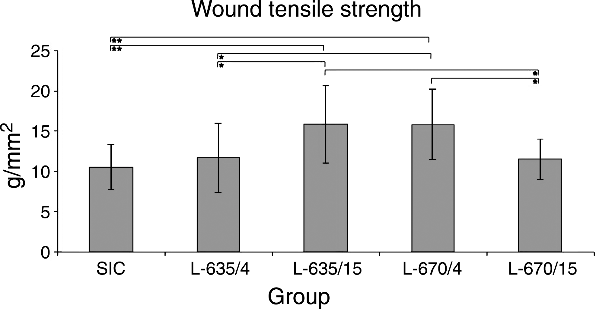
The lowest wound TS results were measured in SIC rats, with slightly higher wound TS results found in groups L-670/15 and L-635/4, and significantly higher wound TS results found in groups L-670/4 rats and L-635/15 (*p < 0.05; **p < 0.01).
Discussion
Interestingly, an inverse relationship between wavelength and intensity in LLLT at 670 and 685 nm was suggested by do Nascimento et al. in their preliminary wound-healing investigation. 7 The results from our current investigation support this suggestion. However, in our study this effect was observed at a lower dose and a different wavelength. In contrast to do Nascimento's investigation at 670 nm, we found a similar effect at 635 nm, while their observation at 685 nm was recorded in our study at 670 nm. Therefore, further detailed investigations need to be performed to clarify this discrepancy.
Previously, by evaluating the effect of LLLT at 635 nm during the proliferative phase, we observed that epithelization and collagen synthesis were significantly accelerated in a power-density-dependent manner, 4 which explains, from histological point of view, our present results at 635 nm. On the other hand, in contrast to do Nascimento's results, Erdle et al. recorded in their investigation that LED light at 670 nm stimulates wound healing in the same manner as was observed in our current study. 7,10 It was suggested that the possible biostimulatory mechanism may be based on the fact that a lower density of light delivers the energy for a longer time period and more continuously. However, the question of why LLLT at 635 nm improves wound healing at a higher density remains open.
Conclusion
Our study demonstrates that both tested red lasers are able to increase wound TS and thereby accelerate wound healing. Whereas radiation at 635 nm improves wound healing by using higher tested power density, radiation at 670 nm improves healing by using lower power density. Since a shorter time is needed to achieve the daily dose of 5 J/cm2 when higher power density is used, it may be suggested that LLLT at 635 nm and 15 mW/cm2 might have a better clinical outcome.
Footnotes
Acknowledgments
We thank F. Filický for his technical assistance. This study was supported by the Slovak Grant Agency (VEGA 1/4228/07), Slovak Research and Development (APVV 2-0682-07), and Šafárik Univerzity (VVGS 53/09-10 & 54/09-10).
Author Disclosure Statement
No competing financial interests exist.