




Abstract
Introduction
Alterations in salivary-gland composition and salivary-gland hypofunction have been associated with elevated fasting blood-glucose concentrations (hyperglycemia) and neuropathy. 4 –8 According to reports of the Expert Committee on Diagnosis and Classification of Diabetes Mellitus (DM), DM consists of a group of metabolic diseases characterized by hyperglycemia, resulting from disorders in insulin secretion, insulin action, or both. 9 Type 1 and 2 diabetic patients may report xerostomia and hyposalivation, along with other clinical signs and symptoms of salivary-gland dysfunction. 5 –7 Patients with hyposalivation show a higher risk of infection, carious lesions, and taste alterations, in addition to inadequate preparation of food for digestion. 3 Moreover, when associated with symptoms of xerostomia, hyposalivation leads to a decrease in the patient's quality of life.
Diabetic state has also been associated with an increase in free radicals. 10 Reactive oxygen species (ROS) are generated during the metabolism. However, an excess of ROS is toxic. Thus, the enzymatic and non-enzymatic antioxidant systems are responsible for eliminating an excess of ROS production. The enzymatic system consists of superoxide dismutase (SOD), catalase (CAT), and glutathione peroxidase (GPx) enzymes. SOD converts the superoxide anion in hydrogen peroxide, which is converted into water by CAT or used by GPx. 11,12 In addition, the peroxidase, an enzyme with antimicrobial properties, also uses hydrogen peroxide in the chemical reaction of thiocyanate (SCN−) oxidation, leading to the production of hypothiocyanate (OSCN−), which inhibits bacterial growth. 13
Several salivary gland alterations in diabetic rats have been reported previously by our research group. 4,8,14 Some of these data showed that hyperglycemia increases the activity of CAT 4,14 and peroxidase in the PG. 14 In addition, we confirmed 14 some previous works 15 –17 that showed the accumulation of lipid droplets in the PG of diabetic animals, which can also be related to salivary-gland hypofunction.
We used laser phototherapy (LPT) in the aim of improving some biochemical alterations observed in the salivary glands of STZ-induced diabetic rats. Our results showed that LPT was able to reduce CAT activity in diabetic rats to the same value found in non-diabetic animals not subjected to irradiation. 14 In addition, LPT was also able to decrease the accumulation of lipid droplets found in the PG. 14
Thus, the purpose of the present study was to analyze the effect of laser irradiation on the amylase and the antioxidant enzyme (CAT and peroxidase) activities, as well as on the total protein concentration of the SMG of diabetic rats.
Material and Methods
Experimental design and diabetes induction
The experimental protocol used in this investigation was approved by the Bioethics Committee of Animals, at the School of Dentistry at the University of São Paulo. Ninety-six adult female Wistar rats weighing approximately 200 g were used. The animals were caged individually and had free access to water and solid food. The care and handling of the animals throughout the study were conducted in accordance with the principles for animal experimentation established by the Brazilian College of Animal Care.
The animals were randomly divided into eight groups: four diabetic groups (D0, D5, D10 and D20) and four non-diabetics groups (C0, C5, C10 and C20). DM was induced by a single intraperitoneal injection of 60 mg/Kg of STZ (Sigma Chemical Co, St. Louis, MO) dissolved in 0.1 M sodium citrate buffer (pH 4.5). In non-diabetic animals, only the citrate buffer was used. The development of DM was confirmed by blood glucose analysis, using the blood-glucose meter Accu Chek Advantage (Roche-Diagnostics®, Mannheim, Germany), 72 h after the STZ injection, in animals fasted overnight. Rats that had a blood-glucose level higher than 14 mM (250 mg/100 ml) were considered diabetic.
Twenty-nine days after diabetes induction, the animals were anesthetized by an intraperitoneal injection of chloral hydrate (400 mg/kg b.w.) and sodium diethylbarbiturate (50 mg/kg b.w.). The areas corresponding to the SMG were shaved and demarked before irradiation or simulation. The animals in groups D5 and C5, D10 and C10, and D20 and C20 were subjected to laser irradiation, according to Simões et al. 14 The rats in the non-laser groups (D0 and C0), on the other hand, were subjected to a placebo procedure.
On the following day, both the diabetic and non-diabetic rats were euthanized. The euthanasia procedures were always carried out in the morning (9–11 a.m.) to minimize the circadian rhythms. The SMGs were then removed, clamped between aluminum tongs, precooled in dry ice, and stored at −80°C until being used.
Laser irradiation
Laser irradiation was conducted using an InGaAlP diode laser (Quantum, Ecco Fibras®, São Paulo, Brazil) working at a wavelength of 660 nm, according to Simões et al. 14 The irradiation mode was punctual, transcutaneous, and in contact and perpendicular to the skin surface. The power was 90 mW. The laser was applied on an area of 1.1 cm2, corresponding to the position of both SMG.
For irradiated groups, the laser irradiation was performed in continuous-wave mode; the laser-beam spot was 0.017 cm2, the doses were 5 J/cm2, 10 J/cm2, and 20 J/cm2, and the time of laser irradiation was 1, 2, and 4 s for groups D5 and C5, D10 and C10, and D20 and C20, respectively. 14 Groups D0 and C0 consisted of placebo groups, where the irradiation was simulated by positioning the laser tip on the tissue. 14 Sixty-four points were necessary to evenly cover the entire submandibular area of 1.13 cm2. 14 The output power was measured using a power meter (Coherent Molectron®, Santa Clara, CA).
Biochemical analysis
For all biochemical parameters (total protein concentration and amylase, peroxidase, and catalase enzyme activities) the SMG was homogenized at 10% w/v in 10 mM sodium phosphate buffer (pH 6.0) and then centrifuged for 10 min at 1540 g. The supernatant was used.
The protein concentration was measured using Folin's phenol reagent, as described elsewhere; 18 bovine serum albumin was used as standard. The Beckman DU-68 (Beckman, Fullerton, CA) spectrophotometer readings were taken at 660 nm. The amylase activity was measured after the incubation of the samples in 1% starch solution in 20 mM phosphate buffer, pH 7.0, for 5 min at 30°C. 19 The reaction was stopped by adding an alkaline solution of dinitrosalicylic acid, and the mixture was then maintained in boiling water for 5 min. After the mixture was diluted in distilled water, the intensity of the developed color was measured at 530 nm in a Beckman DU-68 spectrophotometer. Maltose was used as standard. One unit of enzymatic activity corresponds to the amount of enzyme that produces 1 μmol of the product in 1 min.
The peroxidase activity was assayed in a medium containing 10 mM phosphate buffer, pH 6.0, supernatant, 10 mM o-dianisidine, and 2.1 mM hydrogen peroxide, as described elsewhere. 20 The absorbance was measured in a Beckman DU-68 spectrophotometer at 460 nm. Lactoperoxidase was used as standard. The catalase activity was investigated in a medium containing 50 mM phosphate buffer, pH 7.0, supernatant and hydrogen peroxide at 100 mM. The catalysis of the H2O2, observed spectrophotometrically was revealed by the decrease in its absorbance at 240 nm. The difference in absorbance per unit time was the corresponding measurement of the catalase activity. 21
In order to perform statistical analysis, data were presented as mean ± standard deviation (SD). Based on the normal distribution of all parameters studied under different conditions, the analysis of variance (ANOVA) and Tukey tests were carried out, setting the level of significance at 5%.
Results
After a single injection of STZ, the rats became diabetic, with a blood-glucose concentration >14 mM (data not shown).
An increase in the catalase activity (Fig. 1) was observed in the diabetic animals not subjected to laser irradiation, when their results were compared with the non-diabetic animals (p < 0.05) that had not received any laser application. However, results obtained from the diabetic rats subjected to irradiation with 5, 10, and 20 J/cm2 (0.10 ± 0.03, 0.09 ± 0.01, and 0.08 ± 0.02 U/mg protein, respectively) were not significantly different with regards to catalase activity when compared to the non-diabetic groups not irradiated (0.07 ±0.01 U/mg protein). Regarding the non-diabetic animals, laser irradiation had no effect upon catalase activity.
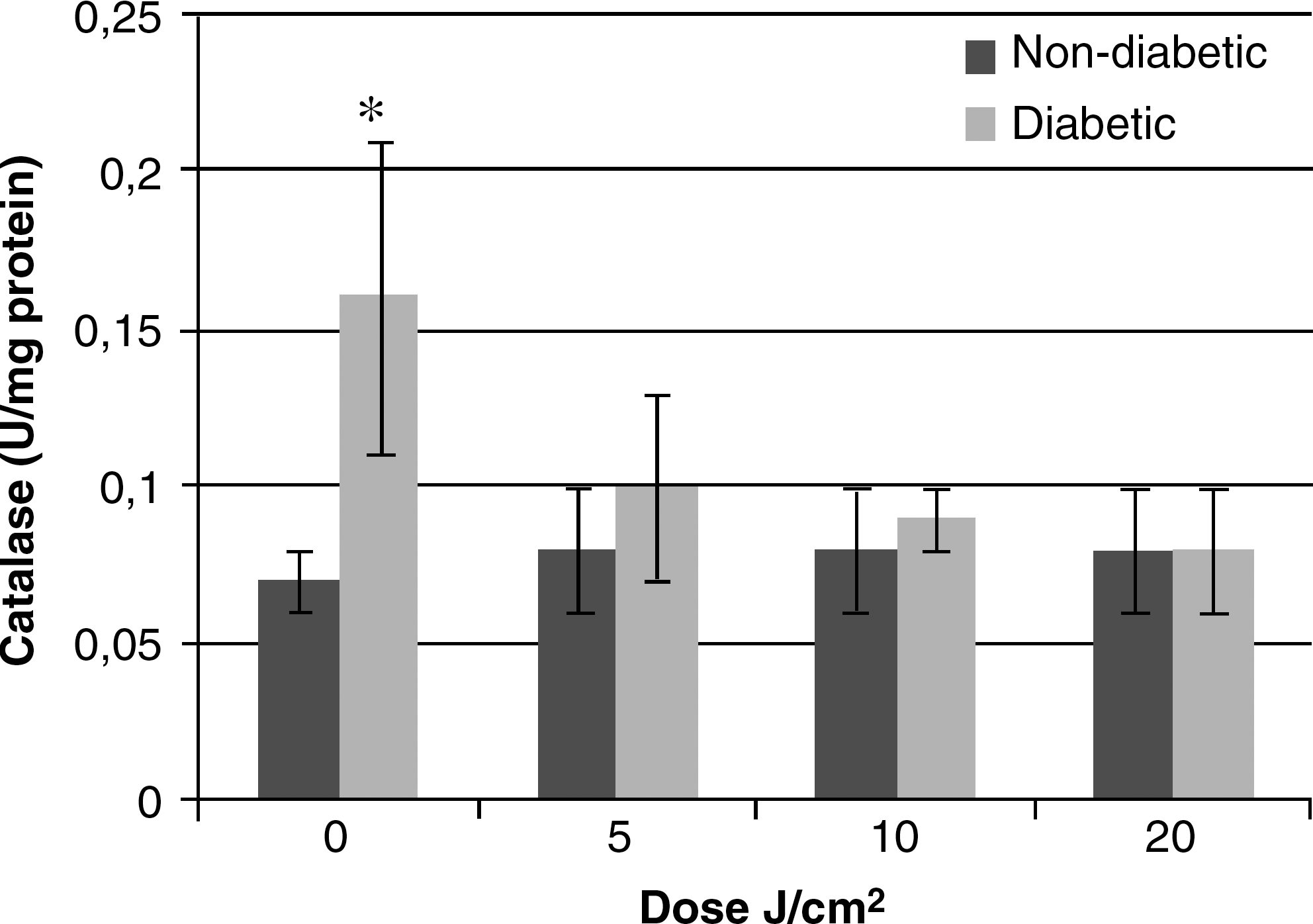
Catalase activity of the SMG from diabetic and non-diabetic rats, subjected to different radiation doses (0, 5, 10, and 20 J/cm2). * indicates that catalase activity from diabetic groups was different to non-diabetic animals that received the same dose of irradiation (p > 0.05) (11 ≤ n ≤ 12).
Concerning the salivary peroxidase activities, similar to the catalase activities, there was an increase (p < 0.05) in value for the diabetic animals (group D0) when compared to the non-diabetic group (C0) (0.16 ± 0.05 and 0.08 ± 0.04 μg lactopereroxidase/mg protein, respectively) (Fig. 2). However, in contrast to what was observed for the catalase activity, the peroxidase activity in diabetic rats had decreased to values found in the non-diabetic animals (p > 0.05) using the irradiation of 5 J/cm2 (0.11 ± 0.04 μg lactopereroxidase/mg protein) (Fig. 2). In addition, the dose of 10 J/cm2 increased the peroxidase activity of the non-diabetic animals (0.15 ± 0.08 μg lactoperoxidase/mg protein).
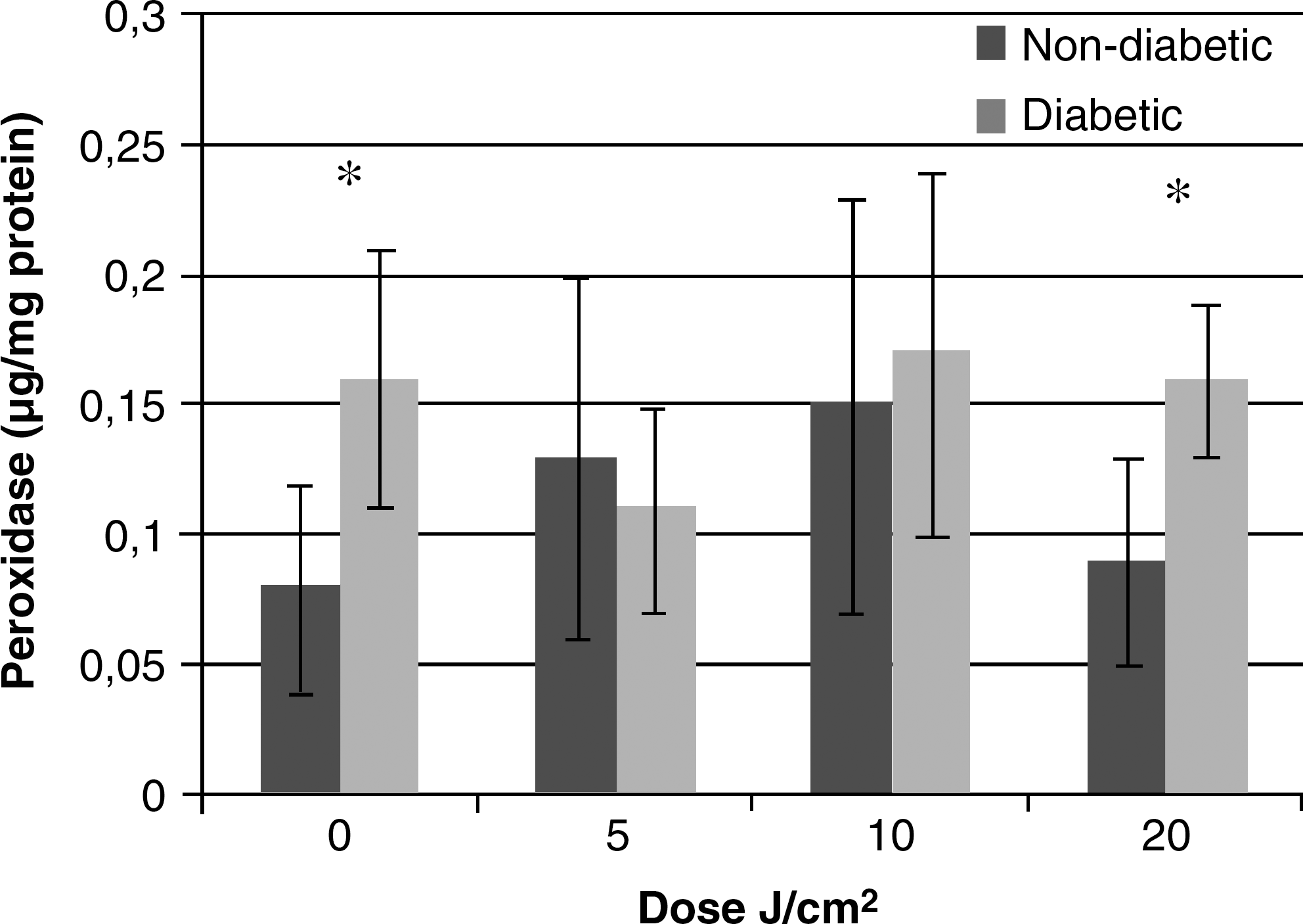
Peroxidase activity of the SMG from diabetic and non-diabetic rats, subjected to different radiation doses (0, 5, 10, and 20 J/cm2). * indicates that peroxidase activity from the diabetic groups was different to non-diabetic animals that received the same dose of irradiation (p > 0.05) (10 ≤ n ≤ 12).
The amylase activity in the SMG of the diabetic animals had not been influenced by laser irradiation (data not shown). However, for the non-diabetic animals, the dose of 10 J/cm2 was responsible for its increase (p < 0.05).
The diabetic animals not irradiatiated (D0) showed a decrease in the total protein concentration in the SMG when compared to the non-diabetic animals not subjected to laser irradiation (C0) (3.82 ± 0.44 and 4.94 ± 0.76 mg/mg tissue, respectively) (Fig. 3). However, 5 J/cm2 was able to increase the total protein concentration in the diabetic animals (4.38 ± 0.63 mg/mg tissue), reaching values similar to the non-diabetic rats (4.42 ± 1.17 mg/mg tissue) irradiated with the same energy density (p > 0.05).

Total protein concentration in the PG from diabetic and non-diabetic rats, subjected to different radiation doses (0, 5, 10, and 20 J/cm2). * indicates that protein concentration from the diabetic groups was different to non-diabetic animals that received the same dose of irradiation (p > 0.05) (n = 12).
Discussion
This investigation clearly shows that DM alters the antioxidant enzymatic defense of rat SMG, as observed previously for the PG. 14 Moreover, the data showed that low-power laser irradiation had some effects upon these altered parameters.
DM is a metabolic disease that affects many body systems and organs, including the oral cavity. A higher incidence of oral diseases related to poorly controlled DM has been reported, and their severity is also a cause of concern, since saliva is essential for the maintenance of oral health, and a decrease in its output may result in deleterious consequences to the carrier of the disease. In our previous study, we observed an increase in the salivary flow rate of irradiated rats and suggested that LPT should be studied as an auxiliary therapy for the hypofunction or inflammatory process of salivary glands. 18 In addition, it was also observed that LPT was able to decrease the catalase activity and accumulation of lipid droplets in the PG of diabetic rats. 14,22
Considering that many diabetic patients complain of a dry mouth or xerostomia and that several studies on experimental diabetes have shown that hyperglycemic rats had their salivary-gland morphology and function modified, the aim of this study was to investigate whether red diode irradiation is able to improve some biochemical parameter changes observed in the SMG of diabetic rats.
Numerous studies have reported the effects of experimental diabetes, induced by either STZ or aloxan, on the structure and functions of the salivary glands of animals, 4,23,24 including the antioxidant parameters in various tissues. 4,24,25 Studies based on aloxan-induced DM in rats showed a significant decrease in DNA, RNA, and amylase content in rat SMG and an increase in the activity of peroxidase. 26 On the other hand, STZ-induced DM in rats showed an increase in catalase activity in the PG, 4 kidney, 25 muscles, 27 heart, and brain. 28 A decrease in amylase and glycogen phosphorylase activities and an increase in the glycogen synthase, peroxidase, and catalase activities of the salivary glands of diabetic rats have been already reported in the literature. 4,8,26,29
Like our previously study evaluating the PG, 10 the present work has shown that specific activities of peroxidase and catalase were higher in diabetic SMG than in non-diabetic when laser irradiation was not applied. A diabetic state has been associated with an increase in free radicals (oxidative stress). Thus, the increase of catalase and peroxidase activity is acceptable, once their functions are related to the reduction of H2O2 concentration. However, in the presence of laser irradiation, the catalase activity for groups D5, D10, and D20 showed the same value for non-diabetic groups that had not been subjected to laser irradiation (C0). These results suggest the effect of laser irradiation on oxidative damage. In further studies, the analysis of H2O2 concentration should be carried out in order to understand better the oxidative stress and antioxidant defense of the tissue subjected to laser irradiation.
The peroxidase activity did not change after the laser treatment given to the diabetic groups. These results are also in accordance with our previously published data. 14 However, it is important to highlight that peroxidase is a biomarker of the SMG and that, in the present study, we can observe a tendency to decrease its activity with 5 J/cm2 of energy density, although these data were not statistically significant.
The effect produced by the LPT is based on the capacity of modulation of diverse metabolic processes, through the conversion of the luminous energy on biochemical and photophysical processes, which transforms the laser light into useful energy for the cell. 30 The visible laser is absorbed by chromophores in the respiratory chain of the mitochondria, which causes an increment in the ATP production, resulting in an increase in cellular proliferation and protein synthesis. 30
Considering that different data on catalase and peroxidase activities after laser irradiation were observed, it is important to highlight the differences between the structures of these enzymes. Peroxidase is a monomeric heme-enzyme. 31 On the other hand, catalase is a homotetramer, which contains a heme and ferriprotoporphyrin IX at each active site, and NADPH binding, 32 which can absorb the light. 33 Artykhov et al., using a catalase from bovine liver that was dissolved in phosphate buffer and irradiated with He–Ne laser, showed that laser irradiation changed functional and structural properties of catalase. However, further studies are necessary to confirm if laser irradiation can act directly on the protein structure.
In addition to enzyme structure differences, the PG and SMG metabolisms are also different. The PG is an aerobic tissue, while the SMG is predominantly an anaerobic tissue. Such results suggest that laser irradiation can act differently on the PG and SMG, as well as acting differently in dependence of the enzyme.
The effect of laser irradiation upon catalase and peroxidase activity (antioxidant enzymes), as well as upon radical species of oxygen (ROS) should be further studied, since antioxidant system alterations is one of the main causes of diabetic complications.
Conclusion
Based on the results of the present study, red laser irradiation decreased the catalase activity of the SMG of diabetic rats.
Footnotes
Acknowledgments
The authors wish to express their gratitude to the Special Laboratory of Lasers in Dentistry (LELO) for providing the laser equipment, and to CNPq (the Brazilian National Council for Scientific and Technological Development) and FAPESP (the State of São Paulo Research Foundation) for their financial support.
Author Disclosure Statement
No competing financial interests exist.