
Abstract
Introduction
When a flap undergoes ischemia, hypoxia causes depletion of the adenosine triphosphate (ATP) levels, failure of the Na+ and K+ pump, cellular edema, vascular permeability increase, and build-up of proinflammatory substances. During reperfusion, when oxygen is reintroduced, the production of reactive oxygen species (ROS) occurs via xanthine oxidase on the endothelial surface and via the neutrophyles oxydase adenosine dinucleotide phosphate (NADPH) system. The subsequent neutrophyl infiltration, as well as its adhesion to endothelium and activation with liberation of inflammatory mediators, is the major cause of the tissue lesion. The chain reaction formed amplifies the cell membrane lesion, which provokes microcirculation thrombosis, vasoconstriction, and necrosis of both endothelial and parenchymal cells, resulting in flap failure. 4
The mechanism of cellular damage is tied with lipoperoxidation of the cellular membrane. This process may be indirectly measured using the malondialdehyde (MDA) concentration, its stable final product. 5 MDA concentration gives circumstantial evidence of the ROS presence and activity, the reason why it is frequently used to test inhibitor drugs, in the ischemic lesion and reperfusion. 6
In an attempt to avoid flap loss caused by ischemia, researchers have investigated techniques aimed at increasing blood flow in the flaps. Although a range of pharmacological agents, such as vasodilators, calcium channel blockers, prostaglandin antagonists, anticoagulants, antioxidants, and anti-adrenergic drugs, have been demonstrated to reach such a goal, some of them can trigger adverse systemic effects, thus making their use unviable in clinical practice. 7,8 Acupuncture, electroacupuncture, and polarized and unpolarized low-frequency electrical currents are alternative treatments similarly used to avoid complications from the reconstructive procedure. 9 However, since these nonpharmacological resources are not exclusively aimed at increasing blood flow, their effectiveness in increasing flap viability may be significantly reduced.
The biostimulant effects of laser, which influence cell functions such as stimulation or inhibition of biochemical, physiological, and proliferative activities, have been reported for 40 years. The extent of the effects depends on both the wavelength and doses used.
Low-level laser therapy (LLLT) is a therapeutic treatment able to increase blood flow and promote angiogenesis, and thus is widely used for accelerating cicatrisation of wounds. 10 As a result of these effects, the efficiency of LLLT in improving skin-flap survival began to be investigated. Many authors have demonstrated that the diode laser may improve flap survival by both eliciting proliferation of new blood vessels around the irradiated sites and increasing reperfusion. 11 –14
According to Stadler et al. (2000) 15 LLLT also activates superoxide-dismutase (SOD) delivery, thus helping the inhibition of free-radical action. Some authors state that free radicals are important ischemia mediators, favoring tissue destruction. The radical may take part in the chain reactions and cause cell membranes and intracellular peroxidation, leading to irreversible cellular damage. 4,5
In addition, in the reviewed literature, we did not find any study evaluating MDA concentration in cutaneous flaps after LLLT application, but there are studies evaluating the oxidative stress (MDA) behavior and the antioxidant defense (values of the total antioxidant ability) in fragments of ischemic random cutaneous flaps in rats, and those studies concluded that the presence of necrosis in the distal portion of flaps is a consequence not only of the oxidative aggression, but also of the reduction of the local antioxidant defense ability. 6
Based on the principle that LLLT promotes the supply of both oxygen and nutrients to match the needs of the proliferative phase of the tissue-repair process, activating the SOD delivery and more, the literature points to several discrepancies in terms of laser parameters, especially those regarding the ideal wavelength. Thus, we aimed to find out which wavelength is the most efficient in treating ischemic flaps, as well as investigating the effect of 830 nm and 670 nm diode lasers on the MDA concentration in random skin-flap survival.
Materials and Methods
The study was approved by the Commission of Ethics in Research of the University of São Paulo (FMRP-USP), in accordance with current legislation.
Thirty (Rattus norvegicus: var. albinus, Rodentia, Mammalia), lineage Wistar, adult male rats were used, weighing between 260 and 320 g. After the laser-therapy sessions, the animals were housed in their respective cages separately, received commercial rations and water ad libitum, and were kept at a temperature of 22–27°C in a dark–light cycle of 12/12 h.
Equipment
The diode laser equipment used in this study was Ibramed® Equipamentos Médicos (Amparo, Brazil) with 830 and 670 nm wavelength, 30 mW power, and 0.06 cm2 beam diameter with continuous wave.
The equipment was calibrated in the São Paulo University Physics Department, USP. Before each application, the calibrator was adjusted. Radiant power determination was performed by attaching the end of the laser diode, at a 90° angle, to the digital potency analyzer Lasercheck® (Coherent®, Staunton, VA) sensor, calibrated and used according to the manufacturer's recommendations. Three consecutive measurements were collected, and the mean value was used.
Groups and experimental protocol
Laser irradiation was performed immediately after surgery and for 4 consecutive days, totaling 5 consecutive days of irradiation, always at the same time of day. Irradiation was applied by means of punctual contact for 72 s per session, with the probe being positioned perpendicularly to the skin flap. The site of laser application was located 2.5 cm from the cranial base of the skin flap 16 (Fig. 1).

Irradiation site.
All 30 rats were weighed and randomly divided into three groups of 10 animals. The number of animals in each group was determined by statistical planning. In group 1 (control), animals underwent sham irradiation. In group 2, animals underwent diode laser irradiation at 830 nm. In group 3, animals underwent diode laser irradiation at 670 nm. For the experimental groups, the energy density (dosimetry) was set at 36 J/cm2 (total energy = 2.52 J and potency density =0.5 mW/cm2). After the irradiation sessions, the animals were again housed in the cages, receiving water and standard pelleted chow ad libitum.
Surgical technique
The animals were anesthetized by intraperitoneal injections of an even mixture of tiletamine and zolazepam (50 mg/kg), remaining anesthetized during surgery and laser sessions. Following anesthesia induction, the rats were positioned on a flat surface with members extended, and a digital trichotomy was performed on their back. The skin flap was anatomically planned on each animal's back by using a plastic template, 10 cm long and 4 cm wide. The inferior angles of the scapulas and the ilio of the pelvis served as reference. The flap was elevated in a caudocranial direction and included the deep and superficial fasciae, the panniculus carnosus, and the skin. 17 A plastic sheet (10 × 4 cm) was placed between the flap and the donor site. 18,19 Afterwards, the flap was sutured in the original position by using 4-0 monofilament nylon thread with simple stitches 1 cm apart. 17
Immediately after the surgical procedure, all groups were kept anesthetized for a further 30 min while they underwent the diode laser irradiation, with group 1 (control) at the same time undergoing a simulation of the procedure.
On day 7 postoperative, animals were anesthetized and submitted for evaluation of their necrosis area percentage using the paper-template method. 20 A sample of skin was then collected to perform biochemical assessments of MDA concentration, expressed as μM × 10−2/mg of protein. MDA concentration was performed using the tiobarbiturate method. 21 The investigator did not know which group each skin sample was from.
In order to standardize the sample collection, a pachymeter was used and 1.0 cm width was established. The starting line of transition between the viable and necrotic tissue and the left edge of the flap was adopted as the reference. The length was 1.5 cm starting from the transition line longitudinal to the cranial base of the skin flap (Fig. 2). Immediately after, euthanasia was performed by excessive inhalation of ethylic ether.
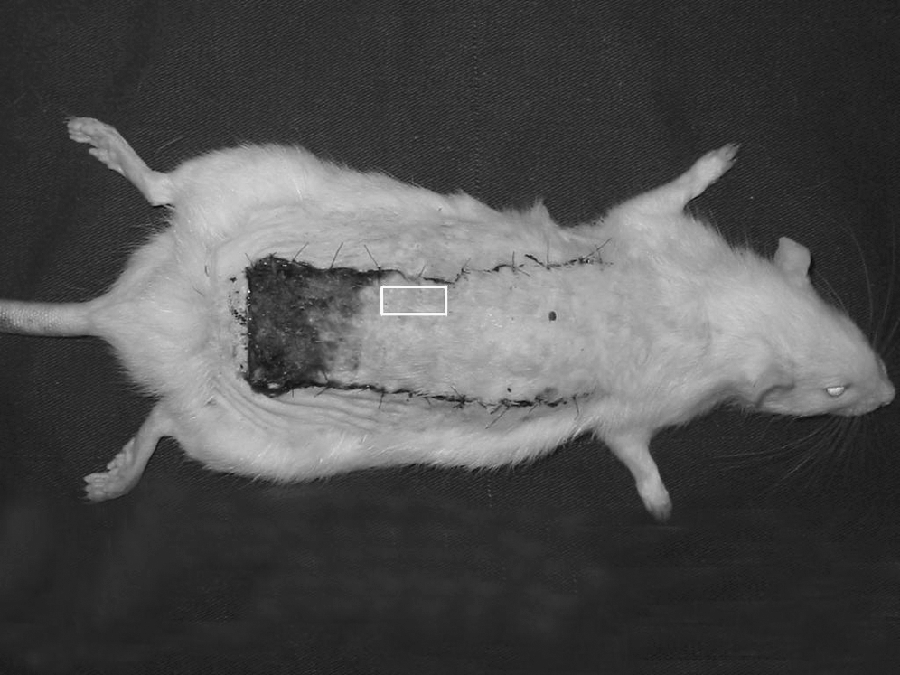
Representation of skin-flap sample collected for the MDA analysis.
Method for estimating percentage of necrotic area in distal portion of flaps
The limit between viable tissue (characterized by soft skin, reddish, warm, and haired) and necrotic tissue (stiff, dark, cool, and hairless skin) was demarcated on the animals.
20
A mold of the entire flap and the necrotic area was drawn and cut on transparent paper, and was checked in a precision balance (± 0.001 g error). Then, the following equation was used:
Statistical analysis
ANOVA (variance analysis) was performed to determine whether statistically significant differences existed among the three groups, in terms of necrosis percentage and MDA concentration. Pearson's correlation coefficient was then applied to determine whether a statistically significant relationship existed among necrosis percentages and the value of MDA concentration. The significance limit for all of these values was set at 0.05, in a bilateral test.
Results
Results indicate that the experimental groups (groups 2 and 3) showed statistically significant differences compared to group 1 (p < 0.01) in terms of the necrosis percentage. Values seen in group 1 were the highest of all the groups. Differences between groups 2 and 3 were not statistically different (p > 0.05) using the paper-template method (Table 1).
Group 1–control; group 2–830 nm laser; group 3–670 nm laser.
The statistical analysis of the MDA results showed that groups 1 and 3 presented statistically significant differences (p < 0.05). Results for group 3 were not as high. Comparing groups 1 and 2, differences were not statistically significant (p > 0.05), and likewise between groups 2 and 3 results were not statistically significant in terms of the MDA concentration (p > 0.05) (Table 2).
Group 1–control; group 2–830 nm laser; group 3–670 nm laser.
As detailed in Table 3, a positive correlation was found for evaluating MDA and necrosis, showing that as the values of one variable increase, the other also increases, and difference was statistically significant (p < 0.01).
p < 0.05.
Discussion
LLLT is being studied more and more in both laboratories and therapeutic practice, and nowadays it is one of the most-used biostimulating ways for rehabilitation, which is contributing to a better understanding of its principles and applications. Scientific and clinical evidence is, however, scarce, and in most cases it is contradictory, which justifies the need for and relevance of investigations involving this therapeutic tool. 22
LLLT was first used with the aim of speeding up the scarring process of ulcers of several etiologies. Several studies were performed and evidenced many of the laser's action mechanisms, such as angiogenesis induction, blood-flow increase, collagen synthesis, fibroblast proliferation, and SOD activation delivery. 23,24
Based on this knowledge, we decided to investigate the effects of the blood-flow increase yielded by laser irradiation on the viability of ischemic random skin flaps.
In the literature, some papers show the efficacy of LLLT in cutaneous flap viability, which corroborate the results achieved in the present study. 11,13,25 –27 However, a wide discrepancy can also be found in the standardization of parameters for the application of LLLT, making it difficult to compare results, as well as interpret some of the action mechanisms of this therapeutic tool.
It is well known that LLLT efficacy is dependent on a number of variables, such as wavelength, power, flow, transversal section of the transmitter beam area, total issued energy, application technique, and treatment frequency. 28
Only two trials have evaluated the action of 670 nm laser on the viability of random cutaneous flaps in rats. Assis et al. (2005) 29 compared lasers with 670 nm (30 mW power) and 904 nm (15 mW power) wavelengths in the random cutaneous flap viability in rats. However, the same flow (16 J/cm2) was used for both types of laser. A significant decrease occurred in the necrosis area of the irradiated groups compared to the control group. However, results showed that the 670 nm laser, operating with 30 mW power, was more effective than the 904 nm laser, with 15 mW power. Bossini et al. (2008) 30 studied the 670 nm laser using 3, 6, 12, and 24 J/cm2 flows. They concluded that all experimental groups had statistically significant values when compared to the control group, and the one with the 24 J/cm2 flow had the smallest area of necrosis.
These studies are contradictory for variables such as the number of irradiated points, the amount of applications of laser radiation, and the flows used. Assis et al. (2005) 29 applied radiation on just one point located 2.5 cm away from the flap base, for 5 consecutive days. In the Bossini et al. (2008) 30 study, the application of laser radiation was spread over 24 points, distributed on and around the flap, based on the study by Pinfildi et al. (2005) 25 who found the smaller necrosis area when the laser application was applied on and around the random cutaneous flap in rats.
In terms of the 830 nm laser, only four papers were found evaluating the action of laser on random flap viability in rats, 11,13,26,27 but these also showed contradictions for some variables. Kami et al. (1985) 11 used equipment with 15 mW power, 0.02 cm2 beam area, and 16 J/cm2 dosimetry. Kubota and Ohshiro (1996) 13 used a laser with 60 mW power, 830 nm wavelength, and 36 J/cm2 dosimetry. The beam area was not mentioned in their report.
Kubota (2002) 27 used a laser with 100 mW power, 0.0054 cm2 beam area, and 185 J/cm2 dosimetry. In spite this very high dose, a statistically significant outcome was reached. Prado et al. (2005) 26 used equipment with 30 mW power, 36 J/cm2 flow, and 0.06 cm2 irradiation beam area, with a continuous beam. Despite the differences between the equipment, all authors found significant results.
On this basis, the purpose of this study was to compare lasers with 670 and 830 nm wavelengths using the same number of irradiated points, as well as the same power, flow, area of the transversal section of the beam, total energy issued, application technique, and treatment frequency.
We found that there was no statistically significant difference in the percentage decrease of the necrosis area between either wavelength. However, when compared with the control group, both lasers showed a statistically significant reduction.
In order to study necrosis and its prevention, we used a dorsal random cutaneous flap from the cranial base, as proposed by McFarlane, DeYoung, and Henry (1965), 17 as the experimental model. In this model, a flap 10 cm long ×4 cm wide generally presents a necrosis percentage between 25% and 50% from its distal portion.
To assure and standardize the flap ischemia degree, we adopted the approach of placing a plastic film between the flap and the donor bed to avoid the flap revascularization, as proposed by Ugland (1966) 18 and reviewed by Kaufman et al. (1985). 19
In this study, a percentage of the flap necrosis area was assessed 7 days postoperative through the paper-template method, as first described by Sasaki and Pang (1980). 20 This method was used because it is quick and easy to perform, with an error of less than 5%, and all that is required is a piece of translucent paper and a precision scale. 3
The experimental groups of the study were irradiated at the same time for 5 consecutive days, since we based our work on a study by Prado et al. (2005), 26 who also irradiated experimental groups for 5 consecutive days.
In order to choose the dosimetry, studies investigating LLLT effects in cutaneous flaps were examined. Kami et al. (1985) 11 and Assis et al. (2005) 29 used 16 J/cm2 and reached satisfactory results. Kubota and Ohshiro (1996) 13 and Prado et al. (2005) 26 used 36 J/cm2, and they also found efficacy in the reduction of the tissue necrosis area. Some authors compared different fluencies. For example, Bossini et al. (2008) 30 studied the 670 nm laser with 3, 6, 12, and 24 J/cm2 fluencies, and concluded that the 24 J/cm2 group had the lowest necrosis area with a dose-dependent result, i.e., the higher the dose, the lower the tissue necrosis area. Therefore, the dosimetry used in this study was 36 J/cm2, according to the investigations by Kubota and Ohshiro (1996) 13 and Prado et al. (2005) 26 who also used this higher dose, which was enough to yield a flap necrosis area reduction.
The application technique used was the punctual contact one, according to studies by Kami et al. (1985), 11 Amir et al. (2000), 14 and Pinfildi et al. (2005). 25 According to some authors, the fact that the application technique allows contact between the laser source and the skin surface during irradiation increases the penetration depth due to the reduction of both reflection and energy dispersion. 11
The studies in the literature evaluating LLLT in random cutaneous flaps used one laser radiation point. 26 There are other studies that used 18 radiation points, 11 and others, such as Pinfildi et al. (2005), 25 which applied 27 and 54 laser points around, inside, and outside of the flap. All of these authors reached a satisfactory result. Another study by Prado et al. (2008) 16 aimed to evaluate the ideal site for the laser application. The authors concluded that the group receiving only one point on the cranial basis (2.5 cm) presented the smallest necrosis area.
Thus, in the present study, the application of laser radiation occurred on a point located at 2.5 cm from the flap base, according to the experimental model for LLLT on random ischemic cutaneous flap in rats proposed by Prado et al. (2008), 16 and to stimulate cranial basis vases, instead of stimulating angiogenesis in regions cut to lift the flap.
There are reports in the literature stating that LLLT is able to increase the blood stream in microcirculation, because it increases the release of nitric oxide–a powerful vasodilator–at the irradiated site. 22 Moreover, it may promote both the delivery and secretion of angiogenesis-related growth factors, 10 thereby allowing the supply of oxygen and suitable nutrients to meet the needs of the proliferation stage of the tissue repair process. 31
Some experimental studies indicate that the exogenous administration of Vascular Endothelial Growth Factor (VEGF), which is both an angiogenesis-related and vascular permeability increase-related growth factor, 30 may promote local angiogenesis induction, as well as favoring cutaneous flap viability. 32
LLLT also activates SOD delivery, so helping the inhibition of free-radical liberation. Some authors state that free radicals are important ischemia mediators, by favoring tissue disruption. These radicals may play a role in chain reactions and may cause peroxidation of cell membranes and intracellular proteins, and they may also cause irreversible cell damage. 15,33,34
The structural damage to the cell membrane is the key point of the ischemic lesion. MDA is the final product of lipoperoxidation and has been considered as a tissue lesion marker. 5,6
In the searched literature, there are no studies evaluating the MDA concentration found in ischemic cutaneous flaps after the application of LLLT. However, we found studies evaluating the oxidative stress behavior (MDA) and the antioxidant defense (values of total antioxidant capacity) in fragments of ischemic random skin flaps in rats, such as the study by Cymrot et al. (2004), 35 who concluded that there is a decrease in the local antioxidant defense capacity.
In this study, we found that group 3, laser irradiated with 670 nm wavelength, had a lower concentration of MDA than the control group. Group 3 also showed a lower percentage in the necrosis area.
The diode laser, as well as LLLT, is able to increase blood flow without a perceptible temperature increase. This fact allows the application of LLLT at any stage of disease progression, but mainly during the immediate postsurgical period. 27 However, its exact mechanism remains unknown.
Within the literature, some hypotheses are described that try to explain this finding. For example, the increased synthesis in fibroblasts, which is made possible after the increase in cell mitotic activity, and the proliferation and delivery of basic fibroblast growth factor, thus contributing to neovascularization, which increases the vascular perfusion, 10,23 and a possible mechanism of the autonomic nervous system modulation promoted by LLLT. 27
As we can see, there are many studies that comprise investigations with LLLT, but at the same time, there is also a lack of standardization and parameters, which makes comparing results and understanding some of the mechanisms involved across the studies quite difficult.
It is hoped that the present study may contribute toward the scientific literature, showing a more effective wavelength (670 nm) to apply laser radiation for the random cutaneous flap viability and increase of the local antioxidant defense capacity, in an attempt to minimize tissue necrosis.
Further studies are, however, needed to verify both the importance and dependency of each of the laser parameters, and the possible influences exerted on biologic responses, in order to improve the laser therapy specificity, as well as the elaboration of protocols for safer and more effective treatments.
Conclusions
LLLT is an efficient way of increasing the random cutaneous flap viability in rats. No significant difference was found between the 830 nm and 670 nm lasers, but the 670 nm laser was effective in reducing the MDA tissue concentration.
Footnotes
Acknowledgments
The authors gratefully acknowledge funding from the CNPq (Conselho Nacional de Desenvolvimento Científico e Tecnológico) and FAEPA, University of São Paulo (Fundação de Apoio ao Ensino, Pesquisa e Assistência).
Author Disclosure Statement
No competing financial interests exist.