
Abstract
Objective:
The objective of this work was to explore a new modality of cell tracking that uses multifunctional nanoparticles. The tracking of transfused cells in vivo is an important step to study the therapeutic course and mechanism of cell therapeutics.
Conclusions:
PNIPAM-coated multifunctional nanoparticles showed potential for labeling cells and for tracking cells both in vitro and in vivo with the use of fluorescence and magnetic resonance. This new modality of cell tracking has the merits of simplicity and reliability.
Introduction
Materials and Methods
Nanoparticles
PNIPAM-coated Fe3O4–SiO2–CdTe nanoparticles were synthesized and characterized as described in our previous report. 11 In brief, the photoluminescent QDs (λem = 600 nm) and magnetic particles were synthesized and then combined to form Fe3O4–SiO2–CdTe particles with silicon shells. The nanoparticles were finally coated by the polymer PNIPAM. PNIPAM is a well-known thermosensitive polymer with a volume phase transition temperature; it is swollen and hydrophilic in water at temperatures below lower critical solution temperatures (32–34°C) and shrunken and hydrophobic at higher temperatures. The diameters of these prepared nanoparticles are about 150 nm, measured by scanning electron microscopy (SEM). 11
Biological samples
Chinese hamster ovary (CHO) cells, obtained from the Cell Bank of the Shanghai Science Academy, were used in experiments. Cells were incubated in Dulbecco's modified Eagle's medium, containing calf serum (10%), penicillin (100 U/mL), streptomycin (100 μg/mL), and neomycin (100 μg/mL), in a fully humidified incubator at 37°C with 5% CO2. The cell labeling was performed by adding nanoparticles to cell dishes at a final concentration of 0.1 mg/mL and incubating for 30 min at 37°C in an incubator. Then cells were washed with phosphate-buffed saline (PBS) to remove unbound nanoparticles and were then ready for further experiments.
Kun-Ming mice were used as the animal model to track the labeled cells. By injecting labeled cells into mice, they could be monitored in vivo.
Imaging measurements
The images of labeled cells were measured in a confocal laser scanning microscope (Olympus, FV-300, IX71, Tokyo, Japan) by a photomultiplier tube and a 580–640 nm band-pass filter, excited with 488 nm Ar+ laser. Differential interference contrast (DIC) images were recorded simultaneously in a transmission channel to exhibit the cell morphology. A water immersion objective (×60) and a matched pinhole were used in experiments.
The in vivo MRIs of labeled cells in mice were scanned and recorded under T2 weighted (TR = 4000 msec, TE = 45 msec) gradient echo pulse sequences in a 4.7 Tesla MRI machine (Oxford System).
Results
Cellular binding of nanoparticles
The thermosensitive property of the nanoparticles can be used to control cellular binding. When cells were incubated with nanoparticles (0.1 mg/mL) at 25°C for 30 min, very few nanoparticles adhered on the cell surface (Fig. 1), because at this temperature the surface PNIPAM of nanoparticles is hydrophilic and does not have affinity for cell membranes. In contrast, many nanoparticles were bound on the external surface of cells when the incubation temperature increased to 37°C for 30 min, as shown in Fig. 2, demonstrating that the hydrophobic PNIPAM favored cellular binding at this higher temperature. The result reflects that cellular binding of these PNIPAM-coated nanoparticles is easily controlled by changing only the temperature.
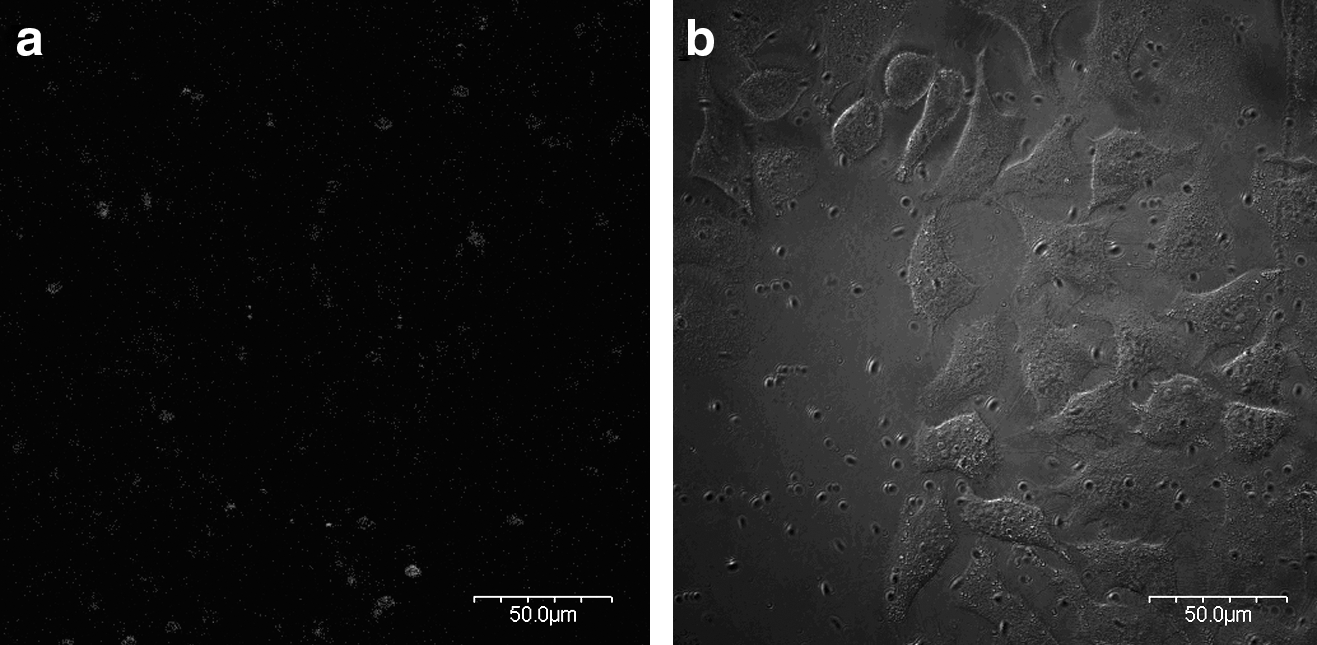
The cellular binding of nanoparticles at 25°C: (
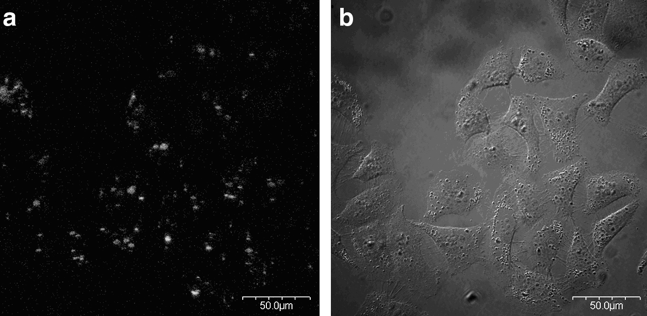
The cellular binding of nanoparticles at 37°C: (
The stability of the cellular binding of nanoparticles
Figure 3 is a confocal image of the typical cellular binding of nanoparticles. As predicted, the nanoparticles only bound on the outer membrane but did not enter cells, which may cause fewer side effects to cells. The labeled cells were continuously cultured in the incubator for different time periods until 48 h post-labeling, and their images were recorded with the microscope in the different periods accordingly (Fig. 4). As seen in Fig. 4, the labeled cells continued to divide, based on a dividing period of about 8 h for these CHO cells, demonstrating that the nanoparticles did not affect the cell viability and confirming the low toxicity of this labeling modality. Figure 4 also shows that the cell labeling of nanoparticles is very stable because the photoluminescence can still be seen in divided cells though the PL intensities gradually decreased. Therefore, these nanoparticles could provide a safe and reliable way to label living cells.
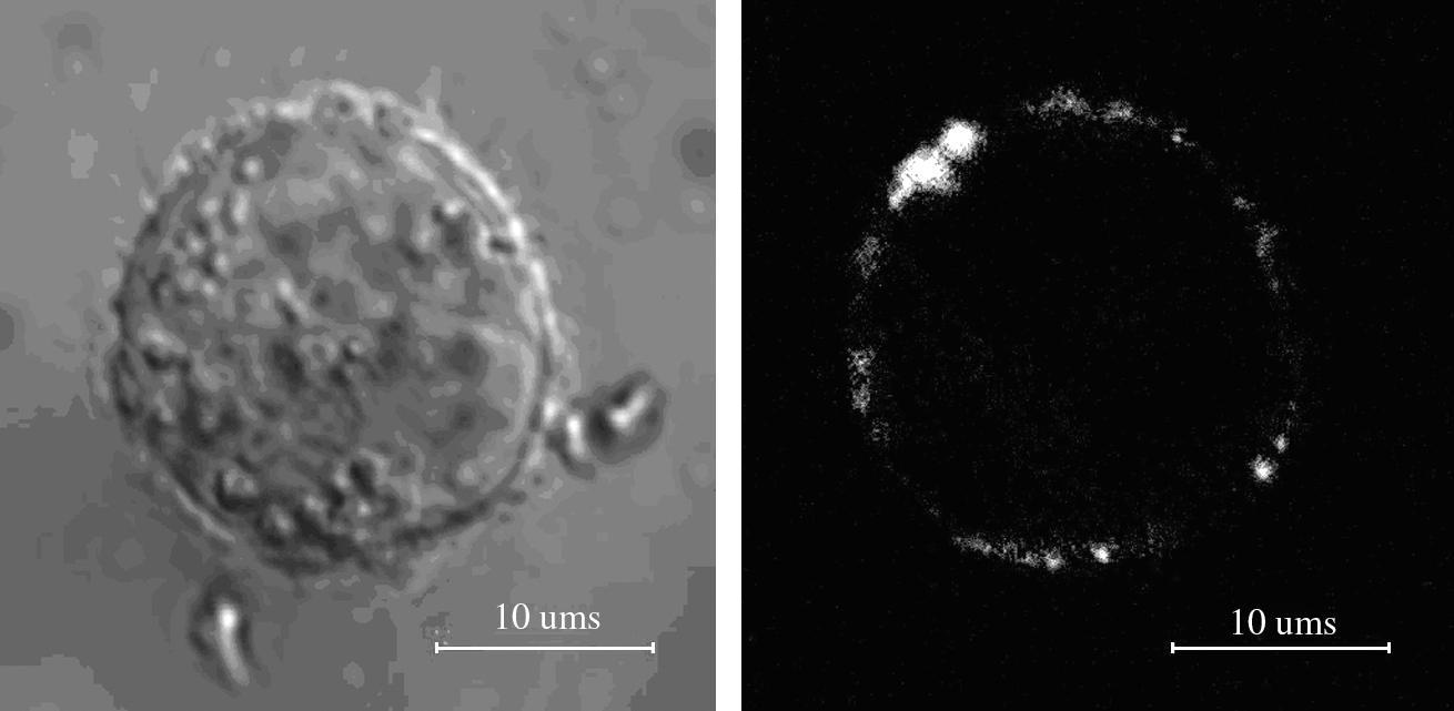
The nanoparticle-labeled Chinese hamster ovary (CHO) cell: (
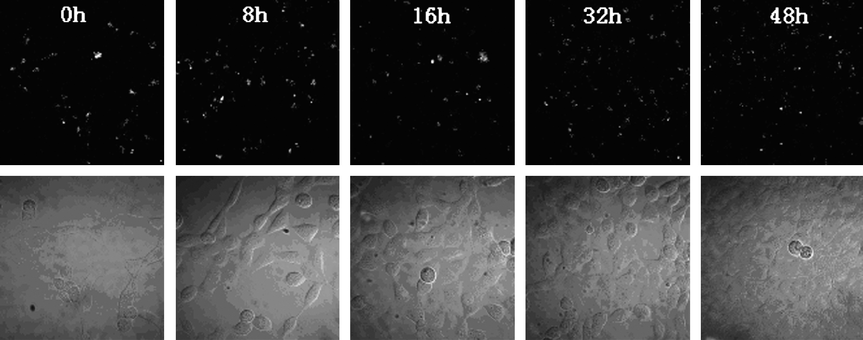
The images of nanoparticle-labeled CHO cells at different times post-labeling: (
The manipulation of labeled cells with the magnetic field
The nanoparticles contained not only the photoluminescent QDs but also Fe3O4 magnetic particles. The magnetic particles inside nanoparticles can be used to manipulate the nanoparticles as well as the labeled cell. The labeled cells and unlabeled cells (control) were placed together in PBS. The labeled cells and control cells suspended in the cell dish could be distinguished from the photoluminescence and DIC images as shown in Fig. 5, because the nanoparticle-labeled cells emit luminescence while control cells have no emission. When a magnet was added with the direction of the magnetic field paralleling the plane of the cell dish producing a gradient of 300 Gs/cm in cell suspension, the labeled cells were forced to move along the direction of the magnetic field while the control cells had no response (Fig. 5). This result indicates that these nanoparticles could be used not only for cell tracking but also for manipulating labeled cell to the targeting area.

The movement of the labeled cell in a magnetic field with a gradient of 300 Gs/cm. The control cell has no response to magnetic field. (
In vivo imaging labeled cells with MRI
The other merit of these multifunctional nanoparticles is tracking the nanoparticle-labeled cells with MRI. The labeled cells and control cells were injected into opposite parts of a mouse, respectively. Two hours later this mouse was scanned by MRI (Fig. 6). The dark area near the left surface, indicated by the left arrow, indicates the labeled cells. On the right side of the image, where the control cells were injected as indicated by the right arrow, no signal changes can be found. The results show that the nanoparticle-labeled cells can be specifically imaged with MRI.
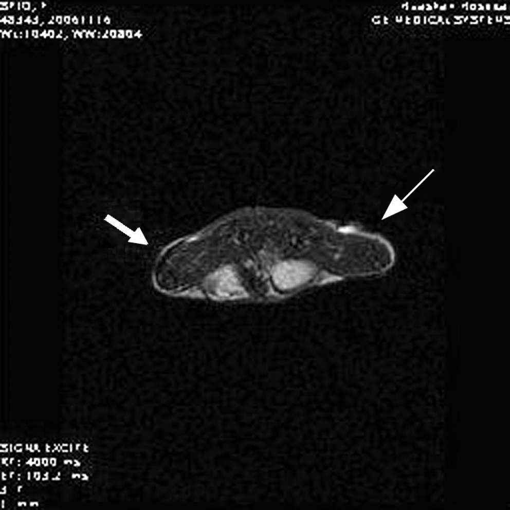
The T2 weighted magnetic resonance imaging (MRI) result in a mouse. The field is 6 × 6 cm. The labelled cells have been injected into the left side of the mouse as the left arrow indicates and the control cells have been injected in the right side of the mouse as the right arrow indicates.
Discussion
Unlike common labeling agents, our nanoparticles have functional PNIPAM on the surfaces. The thermoresponsive characteristic makes PNIPAM undergo a volume phase transition from a hydrophilic state to a hydrophobic state when the temperature becomes higher than the critical temperature (32–34°C). Above this temperature, the PNIPAM easily binds to the external membrane of cells because the lipid components of the plasma membrane are hydrophobic too. Because the binding sites are the external surfaces of cells, these nanoparticles cause the lowest side effect on cells. Due to the temperature-controlled hydrophilic/hydrophobic properties, labeling with these nanoparticles could generally be suitable for any kind of living cell. The QDs and magnetic materials contained in nanoparticles also facilitate the cell tracking both in vitro and in vivo via photoluminescence and MRI. The ability to use two detection techniques enhances the tracking ability for labeled cells.
Conclusion
We designed PNIPAM-coated Fe3O4–SiO2–CdTe multifunctional nanoparticles to track labeled cells both in vitro and in vivo. Our results show that the merits of these nanoparticles are outstanding, and suggest that the PNIPAM-coated multifunctional nanoparticles could be a new modality for cell labeling and cell tracking.
Footnotes
Acknowledgments
Financial support from the Shanghai Municipal Science and Technology Commission (06ZR14005) and the National Natural Science Foundation of China (10774027) is gratefully acknowledged.
Author Disclosure Statement
The authors declare that no competing financial interests exist.