
Abstract
Introduction
In experimental models present in the literature, it is possible to observe the production of various forms of lesions and to note that the contraction speed depends not only on the lesion, but also on its form and the age of the animal. Circular lesions heal slowly, and therefore, are an excellent study model for the contraction of granulation tissue.
The explanation for the contraction phenomenon was initially attributed to the shrinking of preexisting collagenous fibers, which became thick after healing. Various ideas about the contraction of the wound were brought to light.
In 1971, Gabianni et al. 1 observed in granulation tissue that fibroblasts underwent intrastructural morphologic modulation peculiar to smooth muscle cells, and that this was responsible for the contraction of this tissue. Thereafter, fibroblasts with contraction characteristics were called myofibroblasts.
These cells have been widely described in various physiologic and pathologic conditions. In 2003, Medrado et al. 2 initially demonstrated that GaAlAs laser increased cutaneous wound contraction, due cellular myofibroblasts. Consistent results were described by using different wave lengths and other light modes. 3 Few scientific studies have characterized possible interferences of photobiomodulation on the formation of myofibroblasts, associated with antiinflammatory responses during cutaneous repair.
In the dental clinic, dexamethasone is used to reduce pain, edema, and postsurgical trismus. 4 This antiinflammatory action is due to the decrease of various inflammatory mediators. The drug inhibits cytokine production, such as IL-1, IL-2, and TNF-α and its receptors, interferes with lymphocyte membrane adhesion molecules, which regulate the activation and movement of these cells, 5 besides reducing the proliferation of T cells. 6 Dexamethasone reduced inflammation through inhibition of edema, polymorphonuclear and mononuclear cells, and deposition of collagenous fibers.
The present study aimed to verify how low-level laser therapy (LLLT)m, associated with dexamethasone, can act as a photobiostimulator resource on tissue repair, especially on myofibroblasts and on cells that synthesize fibrous elements.
Methods and Materials
This research was approved by the Committee for Ethics in Animal Experimentation, Bahia Foundation for Science Development (FBDC), Salvador, Bahia, Brazil (no. 03/2006).
Eighty male Wistar rats weighing 200 to 250 g were randomly divided into four groups of 20 rats each. Each group was subdivided into five subgroups according to the animal death schedule (1, 3, 5, 7, and 14 days). The animals were kept at a conventional animal station during the course of the experiment, under standard temperature conditions (22°C to 25°C), relative humidity (40% to 60%), and exposure to artificial light for 8 to 10 h/day day. The rats were fed with a standard pelleted laboratory diet and water ad libitum and were free of ectoparasites and endoparasites.
The animals were anesthetized with tiletamine chloride and zolazepam chloride (Zoletil 50; Virbac, São Paulo, Brazil; 50 mg/kg bw). Under aseptic conditions, a trichotomy was performed, and, subsequently, a 6-mm diameter punch was used to inflict a round wound on the dorsal skin of each rat. After procedure, the animals entered one of the experimental groups as follows:
Histology
The animals were killed by an overdose of anesthetic. A section of the cutaneous tissue around the lesion and muscular tissue was removed. Half of the cutaneous tissue was fixed in a 10% buffered formalin solution for a minimum of 18 h. Then the tissues were processed for hematoxylin and eosin, immunohistochemistry, and Sirius red specific for collagen.
Immunohistochemistry
Paraffin-embedded 3 μm-thick sections were obtained and assembled on slides previously treated with aminopropyl-triethoxy-silano and incubated with monoclonal anti-α-actin antibodies of smooth muscle (1:800) and anti-desmin (1:100) (DAKO, Carpinteria, CA). Antigenic recovery was obtained by microwave treatment. All procedures were in accordance with the antibody manufacturer's instructions.
All incubations were performed at room temperature. Primary antibodies were incubated from 30 min to 24 h, according to the manufacturer's instructions; the secondary antibodies were incubated for 20 min; the enzyme conjugated with streptavidin was incubated for 20 min; and the DA peroxidase substrate was incubated for 5 min. After development, slides were counterstained with Gil's hematoxylin and covered with Canadian balm. Sections from control animals were treated identically. Sections containing smooth muscle tissue were used for both positive and negative controls; however, the primary antibody was omitted from the reaction for negative controls.
Transmission electron microscopy
For the ultrastructural analysis, the other half of the wound tissue was fixed in 2.5% glutaraldehyde and 0.1 M cacodylate buffer (1:1) for 1 h at 40C. Postfixation was performed with a 2% osmium tetroxide and 0.15 M cacodylate (1:1) for 1 h at 40C. The fragments were dehydrated in successive dilutions of acetone and embedded in PolyBed 812 resin (Polysciences, Warrington, PA). These blocks were then sectioned by using a Reichert-Supernova ultramicrotome (Leica, Austria). Selected sections were submitted to ultrafine cuts and stained with uranyl acetate and then lead citrate for analysis with transmission electron microscopy (EM-109, Zeiss, Germany) at 50 kV. Analyses of the results obtained from this assessment were described qualitatively.
Semiquantitative analysis
Changes affecting presence of edema, polymorphonuclear cells, mononuclear cells, collagen, actin-α, and desmin were evaluated by blind evaluation of coded slides with the following criteria: absent (0), slight (+), moderate (++), and intense (+++). To define these scores histologically, three criteria were used: intense, present in ≥50% of the observed region; moderate, present in 25–50% of the observed region; and slight, present in ≤25% of the observed region.
Statistical analysis
Nonparametric Exact Kruskal-Wallis and Exact Fisher tests were used to compare groups. We used the Kruskal-Wallis test, as it is an appropriate test to be used on small samples. Furthermore, ethical constraints prevented its use in a larger sample on this study. Statistical significance was defined as p ≤ 0.05.
Results
The distribution of the degree of edema and polymorphonuclear cell infiltrate is presented in Tables 1 and 2. On the first day, a significant reduction in edema was seen in groups treated with dexamethasone and LLLT when compared with the control group. The presence of polymorphonuclear cells was slight in 100% of the animals from the dexamethasone group, with a significant difference from the sham group in the same period (p = 0.03). On day 3 of the study, all three treated groups had significant reductions of edema and polymorphonuclear cells, compared with the control group (p = 0.04). On the day 5, this group of cells showed significant differences, not only from animals treated with dexamethasone, compared with the control group, but also from the group submitted only to LLLT.
Groups in which significant differences were found.
Groups among which significant differences were found.
Table 3 shows levels of mononuclear cells in healing areas of the different groups. Only the control group had no mononuclear cells on the first day of the study. On day 5, however, 100% of the animals given all types of treatment had a slight infiltrate of mononuclear cells, with a significant difference from the control group. On day 14, only the control group showed a slight presence of histo- and lymphoplasmic infiltrate.
Groups among which significant differences were found.
As in Table 4, the collagenous matrix was more plentiful in the animals submitted to phototherapy throughout the study days, and when associated or isolated from dexamethasone. On day 3, only the laser-treated group had 100% of animals with moderate collagen synthesis, significantly higher than the control (p = 0.048) and dexamethasone-only group (Figs. 1 and 2). On day 7 of the study, ∼25% of the animals submitted to LLLT and LLLT + dexamethasone had intense collagen results (Figs. 3 and 4). On day 14, all animals from these two groups had an intense collagenous filling on the healing area, with significant differences from the other two groups (p = 0.04).

Slight distribution of collagenous fibers. Sham Group, day 3 after surgery. (Red Sirius, scale = 0.1 mm).

Expression of collagenous fibers with most-accentuated organizational pattern. Laser-only group, day 3 after surgery. (Red Sirius, scale = 0.1 mm).
Slight presence of scarce distribution of collagenous fibers. Dexamethasone-only group, day 7 after surgery. (Red Sirius, scale = 0.1 mm).

Moderate presence of collagenous fibers in dilated vessels. Laser + dexamethasone, day 7 after surgery. (Red Sirius, scale = 0.1 mm).
Groups among which significant differences were found.
The analysis of the myofibroblasts was performed with the immunohistochemistry of cells showing actin-α of smooth muscle and desmin (Tables 5 and 6). A predominance of myofilament was observed in animals given to photostimulation, especially on days 5 and 7. The presence of actin-α of smooth muscle was intense in 100% of the animals that underwent laser-only therapy, with a significant difference from sham group, dexamethasone-only, and laser +dexamethasone (p = 0.03).
Groups among which significant differences were found.
Groups among which significant differences were found.
The expression of desmin was less representative. On day 5, this myofilament was strongly identified in 50% of the animals given LLLT, with a significant difference only from the dexamethasone (p = 0.04). On day 7, however, the animals given LLLT exclusively showed intense and moderate immunomarking of desmin, with significant differences when compared with all other groups (p = 0.04).
The ultrastructural study revealed the presence of myofibroblast-like cells supporting immunohistochemical findings. In the control group, we observed, during days 3 and 5 after the lesion, a predominance of fusiform cells similar to fibroblasts, with a slight synthesis activity, distributed in a loose matrix, lightly electrodense. The cells characterized as myofibroblasts were less present and showed dented nuclei and membranes with electrodense regions throughout its borders.
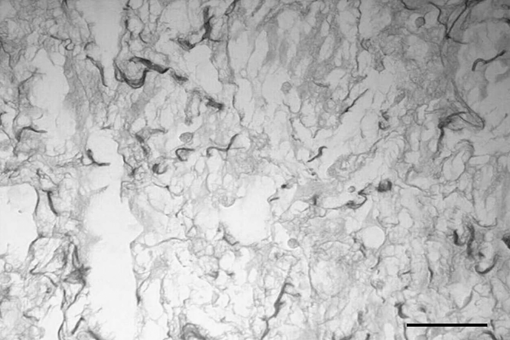
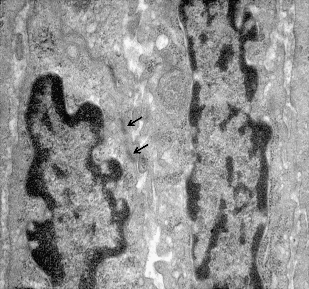
In the laser-only group, the myofibroblasts were bulky, of irregular shape, and showed an intense synthesis process, especially on day 5 (Fig. 5). The presence of fibronexus was observed (Fig. 6). Few of these cells were present through the end of the experiment. In the presence of dexamethasone, the myofibroblasts were scarce and appeared isolated, with rough endoplasmic reticulum and less-developed Golgi complexes. When LLLT and dexamethasone were used, myofibroblasts were more often observed and often were associated with fibroblasts.
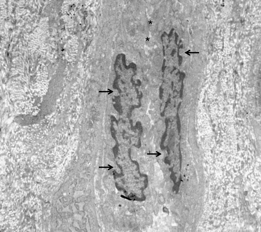
Myofibroblasts exhibiting focal contacts (*), with irregular shaped nucleus and the presence of indentations (arrows). Laser-only group, day 5 after surgery. Electromicrography, 4.400 ×.
Myofibroblasts in detail, seen throughout the membrane, dense bodies (arrows). Laser-only group, day 5 after surgery. Electromicrography, 7.000 ×.
Discussion
The present work evaluated the action of LLLT on the antiinflammatory activity of dexamethasone and scar-tissue genesis, with emphasis on myofibroblast evaluation. This study used circular wounds on the dorsum of rats in an experimental model with the formation of abundant granulation tissue, slow healing, and a uniform distribution of myofibroblasts in the margins of wounds. 7 The laser diode of the AsGaAl was used as the laser of choice, often used in clinical dentistry at a dose previously shown to optimize the phenomenon of healing. 2,8
The results of this evaluation demonstrated that laser irradiation is efficient in accelerating healing during acute and delayed wound responses in rat skin 24 h after surgery. Other studies investigating acute inflammation on paws and pleura of rats, also using red beam length, described the beneficial modulating effect of the laser therapy. 9 –11 The function of the mechanism of the laser on the inflammation is still not completely known, but suppression of IL-1β, IL-2, 12,13 COX-2, 6 RNAm, and TNF-α expression have been hypothesized. 14,15
Groups submitted to LLLT showed greater collagen distribution compared with controls, in accordance with other studies, 16 –20 as well as the dexamethasone-only group. The collagen deposition in irradiated animals and the decrease of mononuclear cells on days 3 through 5 after wounding could be explained by the capacity of the laser locally to increase enzyme activity of mononuclear cells and fibroblasts. 8,21 –23
On days 5 through 7, a predominance of fusiform cells showing smooth muscle actin-α and desmin was observed by electron microscopy in animals exposed to LLLT. We assume that a light-source stimulus not only increased the number of myofibroblasts on the wound, but also activated secretory functions.
Glycocorticoids are capable of acting as immunosuppressors or potent antiinflammatory compounds, depending on the dosage. 24,25 The reduction of collagen synthesis demonstrated a potential negative effect of this drug in tissue repair. These findings were identified after an ultrastructural study performed on days 3 and 5 after use of dexamethasone, when fibroblasts exhibited few organelles and a reduced collagenous matrix. 8 The inhibitory action of dexamethasone treatment was evident in lower levels of positive actin-α and desmin-positive cells.
One of the objectives of this study was to assess the effect of the association of the laser light and dexamethasone in regard to the pattern of the interaction of these therapies in the chronology of cutaneous repair. During the exudative phase, the association of laser light and dexamethasone showed similar results to that seen when laser-only and dexamethasone-only were used. Therefore, no synergic effect was detectable with the use of the association in regard to the inflammatory reaction. It is possible that the use of the laser light alone may replace the use of the dexamethasone.
Laser + dexamethasone treatment resulted in more collagenous fibers and myofibroblasts than found in the dexamethasone-only group. Lasers exert local effects that accelerate metabolism, cellular proliferation, 26 and organization of the extracellular matrix. 3,8,22 As dexamethasone slowed cutaneous repair, phototherapy may have locally stimulated cellular and extracellular components, even in the presence of dexamethasone.
Conclusion
In clinical surgery cases in which dexamethasone therapy is indicated, lasers can be used to improve collagen synthesis and contraction of wounds in soft tissues.
Footnotes
Author Disclosure Statement
No competing financial interests exist.