
Abstract
Introduction
The literature shows that low laser energy densities in phototherapy increase cell proliferation and cell viability, accelerating scar closure in humans 3 ; however, when used in high energy densities, it is able to alter the fibroblasts morphology or may cause damage such as lyses and DNA fragmentation, mitochondrial activity decrease, or plasma membrane damage. 3,4 Moreover, Posten 5 described, in a recent literature review article, a few beneficial effects of laser therapy associated with superficial wound healing in humans, suggesting a better standardization. Further studies must be done by using animal models that have similar characteristics of the human skin. Yamamoto 6 reported that the low-intensity laser therapy stimulates fibroblast proliferation and collagen synthesis through the following mechanisms: changes in biochemical properties of coenzymes dehydrogenase and nicotinamide adenine dinucleotide phosphate (NADH) with a concomitant increase in cellular activity due to the light effects, and increase in the mitochondrial transmembrane potential, which promotes fibroblast activity, stimulating ATP synthesis.
In regard to low-laser intensity effects on lesions, current literature shows that it stimulates proliferation or differentiation or both of muscle cells recruited to participate in the regeneration process, 7,8 blocks the effects of reactive oxygen species (ROS) released, and activates the nuclear factor kappa beta (NF-κB) 9 ; it also increases mitochondrial activity, activates fibroblasts, and stimulates angiogenesis. 10 It has also been reported in the prevention of musculoskeletal degeneration after injury by ischemia–reperfusion, suggesting induction of antioxidant synthesis and that of other cytoprotective proteins. 11
Despite these studies, the laser-therapy action mechanisms are not conclusive, and some controversial aspects remain. It has been observed that not all studies consider parameters such as wavelength, strength, and intensity when evaluating biologic aspects of lasers.
Thus, aiming to inform professionals in regard to the cost–benefit of this therapy, and to comprehend its biologic effects, the objective of this study was to analyze how the number of laser applications can affect the masseter muscle of mice of the HRS/J lineage by a histochemical reaction represented by the succinate dehydrogenase (SDH) enzyme.
Materials and Methods
Animals
Thirty male mice of the HRS/J lineage, weighing ∼35 g, were randomly distributed into groups according to the number of laser applications (three, six, and 10). For each group of laser applications (experimental, n = 5), we also considered the control group (n = 5), which was not irradiated. These animals came from the Animals Experimentation House, School of Dentistry, University of São Paulo (FORP-USP), in five animals per box of polyethylene, at a controlled temperature (24°C to 25°C), and 12 hours of light, daily. Animals received food and water ad libitum. All procedures of this study were approved by the Local Ethics Committee (N. 07.1.547.53.9.) in accordance with international laws.
Laser application
We used the Twin Laser-Mm Optics laser apparatus with 780-nm wavelength, and gallium-aluminum-arseneto semiconductor diode of the Electromyography Laboratory, Department of Morphology, Stomatology and Physiology (FORP-USP) acquired with a FAPESP grant (2006/55567-1) (Table 1). In the experimental group, the irradiation was performed on the middle portion of the posterior region of the left masseter muscle, on alternate days, with an energy density equivalent to 20 J/cm2 (40 mW of power, 20 sec) by following the manufacturer's instructions (Laser Quality Comércio e Serviço LTDA, Meireles, Brazil).
LLLT, low-level laser therapy.
The middle portion of the posterior region of the masseter muscle was chosen because of the great number of fibers in relation to the thickness of this muscle, for easy identification, and mainly by scattering of the laser in muscle tissue. Because this experimental animal has a small muscle, a single point of application was used. This is important because the spot area of application is nearly the size of the surface area of the muscle.
The control group was not irradiated and served as sham-operated controls. All animals, control and experimental, inhaled halothane (2-bromo-2-chloro-1,1,1-trifluoroethane, minimum 99%, Sigma Aldrich, India) with a slight sedation before each laser irradiation (three, six, or 10 laser applications).
Histochemical analysis for SDH
At 72 h after the last predetermined laser application, animals of both groups, experimental and control, were killed by decapitation after an anesthetic overdose with xylazine, 10 mg/kg, and ketamine, 150 mg/kg body weight, by intramuscular injection. The clinical applications, in general, have 48–72 h of effects, according to the literature. Thus, aiming to evaluate the application effects, we waited 72 h for security. If the killing of the animals occurred before this period, the final result could not represent the effects of all the desired applications in groups.
The left masseter muscle was dissected, and the specimen was placed on an aluminum sheet with Tissue-Tek, OCT (optimal cutting temperature), and frozen quickly in isopentane cooled in nitrogen (−150°C), which was kept under −80°C until use. Transverse serial sections, 10 μm thick, were obtained by using a Leica cryostat microtome (−20°C) and stained as described by Nachlas
12
to demonstrate the SDH activity, representing the metabolic muscle pattern. It was captured 15 random microscopic fields of the masseter muscle for each irradiated group and their respective controls, which were used for area determination occupied by the fiber types (light, intermediate, and dark), with the countpoint method, by using a grid composed of 90 dots.
13
The photomicroscope Leica MZ125 (Germany), connected to a digital camera, was used for image captions, and the Image J software (free,
The histochemical analysis was performed by a blinded observer. All quantitative data were expressed as mean and standard deviation. Statistical analysis was performed with ANOVA (SPSS software), with a 95% confidence level.
Results
Histochemical reactions for succinate dehydrogenase (SDH) activity revealed, according to the staining intensity of the superficial bundle of masseter muscle, that this structure in mice is composed of light large fibers, intermediate fibers with a middle diameter with the subsarcolemmal region more stained, and dark fibers with a small diameter. The enzymatic reaction showed an intensity variation in staining between the groups that received three, six, and 10 laser applications (Fig. 1).
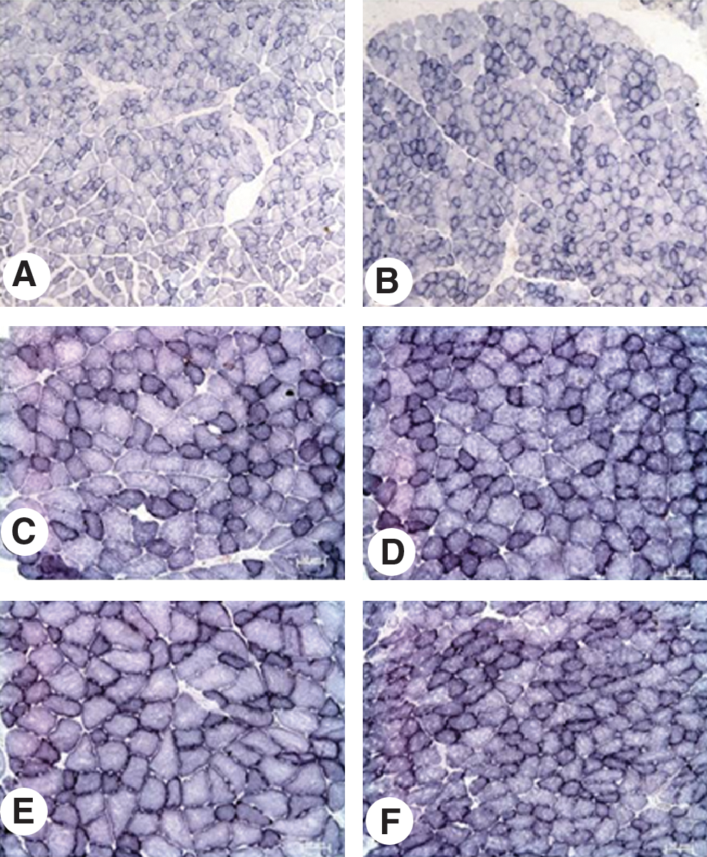
Masseter muscle of the control group (left side of the histologic board) and irradiated group (right side of the histologic board) stained with succinate dehydrogenase (SDH) enzyme, after three (
Areas of different fiber types estimated by the countpoint method showed that the laser promoted the following results: (a) decrease in light-fiber type (low reactivity) (35.91% ± 6.9%; 32.08% ± 6.3%; and 27.88% ± 6.3%), according to the increase of laser applications (three, six, and 10), respectively, with a significant statistical difference (p < 0.05); (b) significant increase (p < 0.05) in the area of the intermediate fibers, with an increasing laser application, three, six, and 10, respectively (11.08% ± 3.9%; 16.52% ± 5.7%; and 15.96% ± 3.9%), although the increase with 10 applications was small; (c) area increase of dark fibers in the group with three laser applications (0.16% ± 0.3%) (p < 0.05), and in groups with six and 10 laser applications, respectively (9.68% ± 6.0% and 9.60% ± 4.0%) (Table 2).
Control (C) and laser irradiated (I) with three, six, and 10 laser applications.
Comparing these data with those of controls, the most significant difference was revealed in the group that received six laser applications, in which the intermediate-fiber area increased in the control group by 14.32% ± 4.25 to 16.52% ± 5.7%, the for irradiated group. Otherwise, the light fibers area decreased from the control (39.40% ± 8.7%) to the irradiated group (32.08% ± 5.8%). Thus, the present data indicate that the number of laser applications increases the metabolic activity of muscle fibers (Table 2).
Discussion
Comparing the data of experimental group with the controls, the most significant difference was revealed in the group that received six laser applications, in which the intermediate-fibers area increased in the control group (14.32% ± 4.2%) from the irradiated group (16.52% ± 5.7%). Otherwise, the light fibers area decreased from the control (39.40% ± 8.7%) to the irradiated group (32.08% ± 5.8%). Thus, the present data indicate that the number of laser applications increases the metabolic activity of muscle fibers.
This study used the Nachlas 12 histochemical technique to demonstrate the activity of the SDH enzyme concentrated in the mitochondria of muscle fibers. With this technique, nitroblue tetrazolium has the specificity to contact the succinate dehydrogenase enzyme system, acting as an important hydrogen acceptor. The product of this reaction results in a pigmented precipitate, strongly linked to the insoluble protein formazan. Thus, it was possible to examine the effects of three, six, and 10 laser applications on the metabolic activity of the muscle, as the intensity of fiber types stained light, intermediate, and dark.
Areas comparison of different fiber types between irradiated groups with three, six, and 10 laser applications, with 20 J/cm2 of energy density, on alternate days in the masseter muscle, showed a significant increase in the metabolic activity of the muscle given six applications. Comparing these results with their respective controls, a significant difference was observed in the group with six applications, in which an increase in the fiber area with intermediate staining was closely associated with the area reduction of light fibers.
In relation to the number of laser applications, no reports in the literature compare the effectiveness of different laser applications. However, Oron 14 reported in a review that multiple and frequent (daily) applications of lasers was less effective than irradiation on alternate days, by using a frog as experimental animal model. The results of the present study revealed no significant difference between six and 10 applications; in this experimental model, it is possible to reduce the number of laser applications to six and obtain the same therapeutic results.
Increased metabolic activity of the muscle induced by three, six, and 10 applications of lasers supports the Lopes-Martins 15 findings in which the laser prevents the development of muscle fatigue in rats during repeated tetanic contractions, in healthy male volleyball players reported by Leal Jr. et al., 16,17 and in patients with myofascial pain. 18,19 These data are confirmed by Nachlas 12 reaction specificity to demonstrate the activity of the SDH enzyme concentrated in the mitochondria of muscle fibers and by reports that the functional ability of muscle is closely associated with activity of mitochondria, the organelle responsible for muscular energy production. 20 In a recent article, Leal Jr. et al. 17 showed that low-level laser therapy (LLLT) might be able to delay the skeletal muscle exercise-induced fatigue, probably by local mechanisms that may include the reduction of oxidative stress or the decreased production of reactive oxygen species. Xu et al. 21 chose a mouse skeletal muscle cell line (C2C12) to study the mechanisms of photobiomodulation and corroborated this fact. They reported that low-intensity laser (LIL) radiation at 0.33–8.22 and 11.22–14.16 J/cm2, respectively, improved and repaired the mitochondrial dysfunction induced by electrical stimulation. Although the exact mechanisms behind the effects of LIL on the metabolism of ROS remain unclear, they showed that the proper dosage of LIL irradiation can induce ROS production, whereas excessive LIL irradiation can improve ROS production. Considering all the described literature until then, the number of laser applications in muscles suggests changes in mitochondrial morphology.
Although the experimental condition, laser type, and energy densities are different in this work, Iyomasa 10 reported a space increase between the inner and outer membrane and dilated mitochondrial crista in irradiated muscle for 7 consecutive days with 5 and 10 J/cm2 of laser-energy density. A great variety of intrinsic and extrinsic factors increase the permeability of the outer and inner mitochondrial membranes to solutes, protons, and metabolites to maintain the ionic homeostasis of cells and organelles, especially in relation to (Ca2+), which may participate in the survival or death of mitochondria. 22
It must be mentioned that different wavelengths and strengths affect photoreceptors at various points of the respiratory chain, suggesting stimulatory or inhibitory effects. 23 Thus, ultrastructural studies are being developed by this research group as an additional tool for a better understanding of the biologic laser effects.
Conclusions
The enzyme SDH activity revealed that the number of laser applications increases the metabolic rate of muscle fibers. The minimal difference in metabolic activity between six and 10 applications of lasers suggests that further analyses must be done to confirm that six applications are enough to produce the same clinical effects, thereby contributing data to professionals from different fields in regard to the cost per benefit of this therapy.
Footnotes
Acknowledgments
This study was supported by FAPESP.
Author Disclosure Statement
No financial conflict of interest exists.