
Abstract
Introduction
The factors that affect healing may be divided into systemic or local; in other words, those factors that influence the inflammatory response. 2 Nutritional deficiencies, including Ca, Mg, K, Zn, Mg, Fe, Cu, I, Cr, active enzymes, antioxidants, vitamins, minerals, and amino acids, have a great effect on wound healing by changing tissue regeneration through interference with inflammatory reactions and immunological function at any point of the healing process. 3 It has been shown that delayed healing may occur in subjects with deficiency in any one of the essential nutrients. 4,5 However, this situation reverts to normality upon the introduction of a diet with appropriate levels of nutrients. 1
Laser phototherapy (LPT) is used in many biomedical applications to promote tissue regeneration, and it has been shown to possess several advantages, including the control of pain, stimulation of the healing process, anti-inflammatory action, increase in the production of collagen, increase in fibroblastic proliferation, and increase in local microvascularization. 6,7
The present work aimed to evaluate histologically the differences in healing of cutaneous wounds of nourished or undernourished rats fed with a Northeastern Brazilian Basic Diet (DBR) following LPT (λ635 nm or λ780 nm).
Materials and Methods
Fifty male and female Wistar rats, on average 21 days old, were divided into two main groups. In Group I, the animals were fed with standard pelleted laboratory diet (Labina®, Purina Nutrimentos, Sao Paolo, Brazil) and the animals of Group II were fed with a Northeastern Brazilian basic diet (DBR, Department of Nutrition of the Federal University of Pernambuco, Recife, PE, Brazil) for 30 days in order to induce undernourishment. The procedures for making the DBR were carried out at the Laboratory of Experimental Nutrition of the Department of Nutrition of the Federal University of Pernambuco (Phaseolus vulgaris = 37.1 g%; salted meat and dried meat = 13.9 g%; Iponaea potatoes = 32.0 g%; Manihot esculenta = 67.4 g%; proteins = 7.88 g%; carbohydrates =69.96 g%; fat = 0.60 g%; ashes = 1.27 g%; and fibers = 7g%).
The surgical procedures were carried out at the Laboratory of Animal Experimentation of the School of Dentistry of the Federal University of Bahia (Salvador, BA, Brazil). The animals, two months old on average, under general intraperitoneal anesthesia (Zoletil50® 0.1 ml/1000g [Virbac do Brazil, São Paulo, Brazil]) had their back shaven and one standardized wound measuring 1 cm × 1 cm was created on the dorsum. No suture was performed.
The animals were divided then into nine subgroups as follows: Group 1 - Control (standard diet); Group 2- Control (DBR); Group 3 - Standard diet X LPT (λ635 nm; 20 J/cm2); Group 4 - Standard diet X LPT (λ635 nm; 40 J/cm2); Group 5 - Standard diet X LPT (λ780 nm; 20 J/cm2); Group 6 - Standard diet X LPT (λ780 nm; 40 J/cm2); Group 7 - DBR X LPT (λ635 nm; 20 J/cm2); Group 8 - DBR X LPT (λ635 nm; 40 J/cm2); Group 9 - DBR X LPT (λ780 nm; 20 J/cm2); Group 10 - DBR X LPT (λ780 nm; 40 J/cm2).
For all the experimental groups, the first application of the treatment was carried out immediately after the surgical procedure, and repeated every 24 hours during the experimental period of seven days. Animal death occurred at the eighth postoperative day. The wounds of groups I and II were irradiated with λ635 nm or λ780 nm laser light (SAEF 20 J/cm2 or 40 J/cm2; 40 mW; φ ∼2 mm; Laser Solutions Tecnologia Ltda, Niterói, Rio de Janeiro, Brazil). Treatment parameters are shown in Table 1. Laser light was applied on four points around the wound (4 × 5 or 10 J/cm2).
The animals were humanely sacrificed and specimens were taken and routinely processed to wax, cut, and routinely stained with H&E and Sirius Red stains and were analyzed under light microscopy by two experienced pathologists. The descriptive and semi-quantitative analyses included: re-epithelization, inflammatory infiltrate, and fibroblastic proliferation. Sirius Red stained slides were used perform descriptive analysis of the collagen matrix.
Results
Control – Nourished
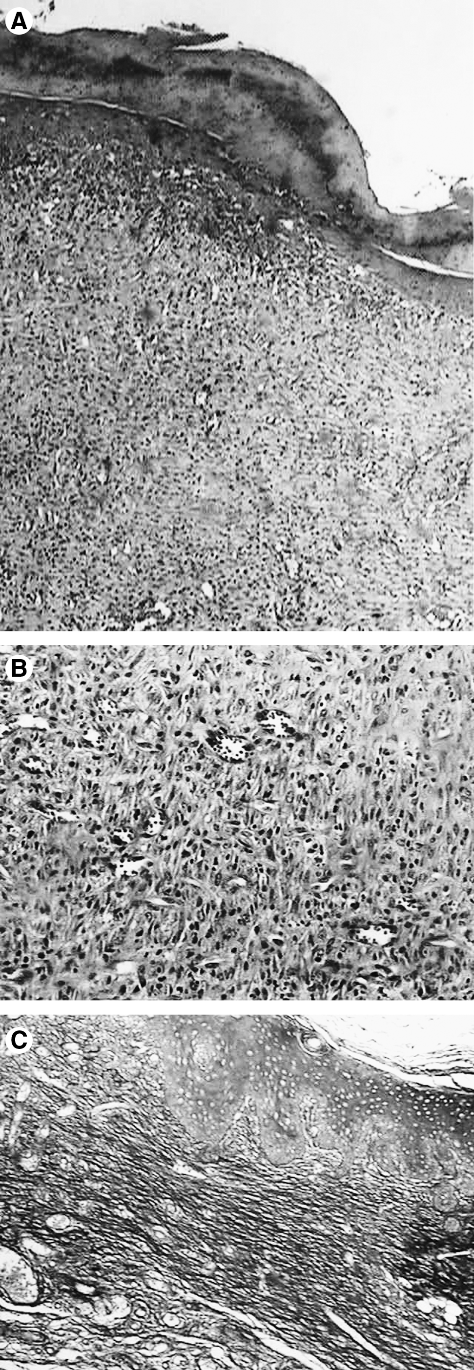
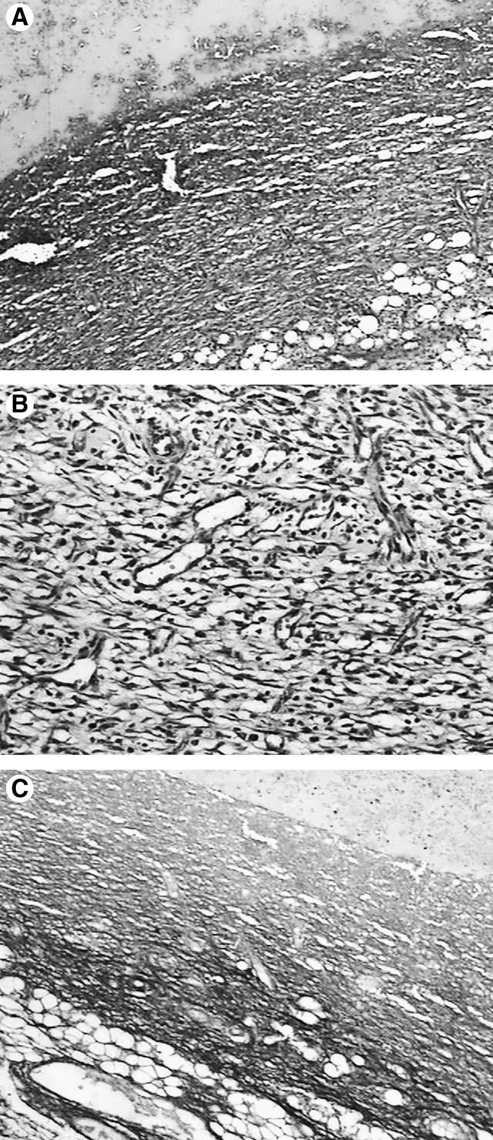
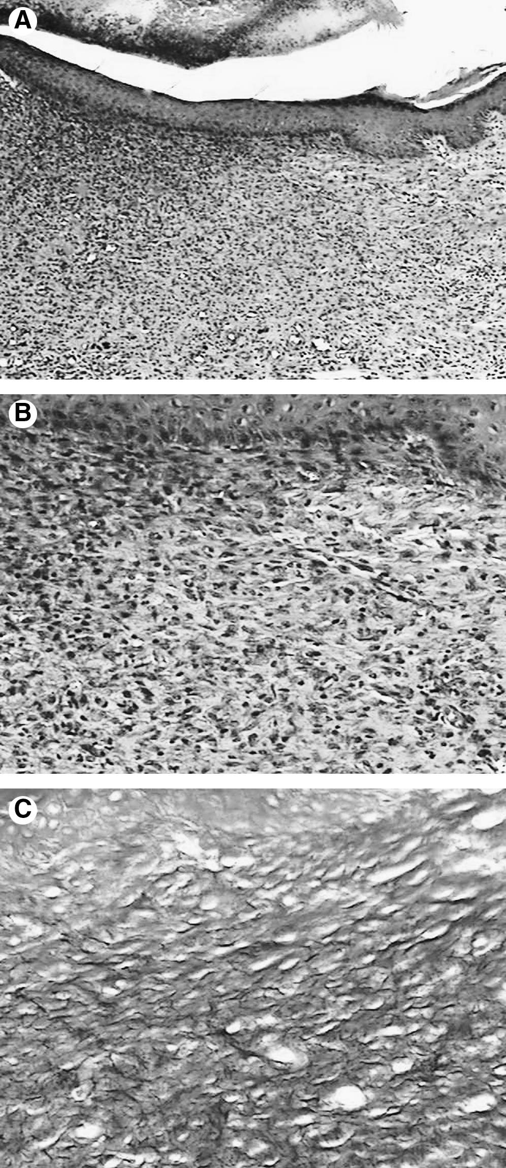
Animals fed with standard diet showed, at the end of the experimental period, wounds partially covered by typical epithelium and a lack of skin appendices (Fig. 1A). All specimens showed, superficially, extensive areas of eosinophilic coagulum, here referred to as crust. Subjacent to the epithelium, granulation tissue was seen that contained young fibroblasts parallel to the surface, producing a collagen matrix. Blood vessel sprouts, usually hyperemic and containing neutrophils, and a moderate mixed and diffuse inflammatory infiltrate were also observed (Fig. 1B). The inflammatory reaction was distributed among bundles of mature collagen fibers regularly disposed as evidenced by Sirius Red stain (Fig. 1C).
λ635 nm – Nourished
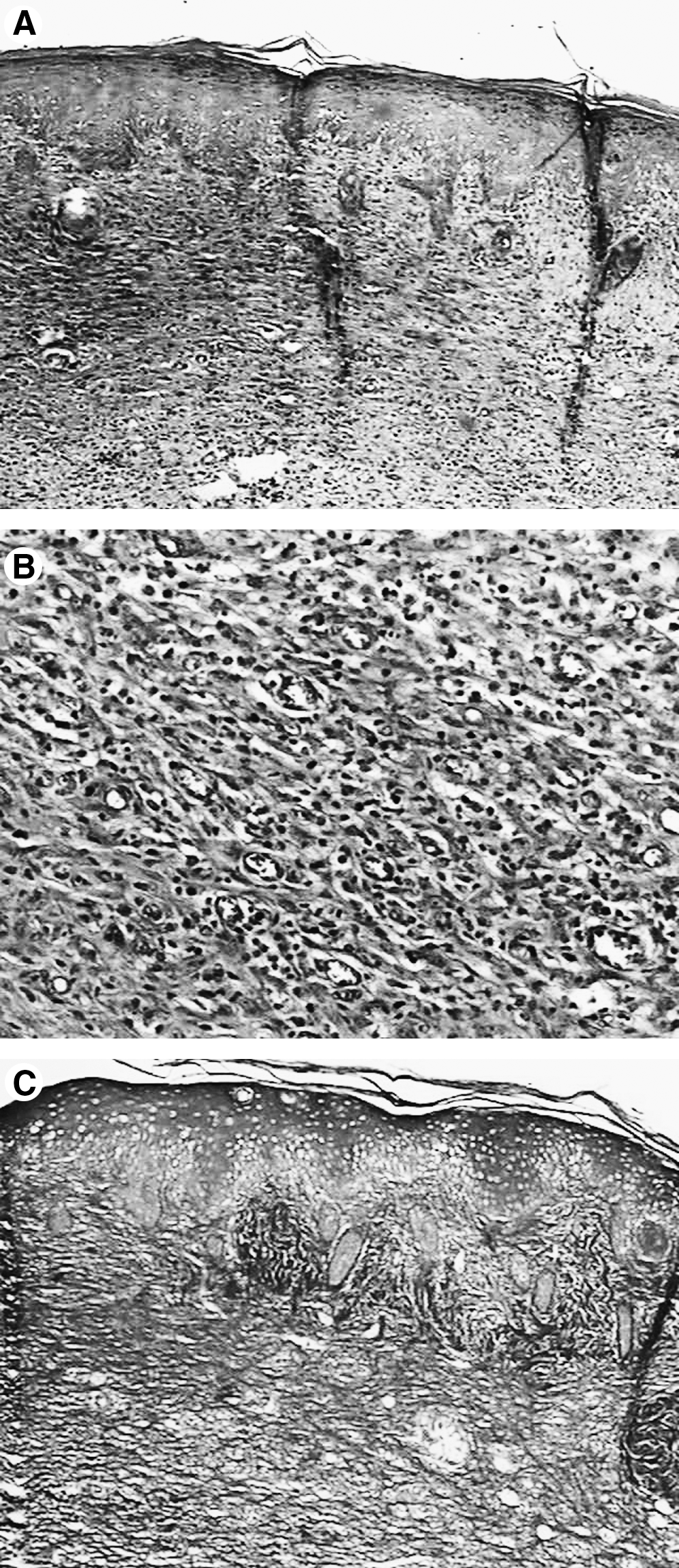
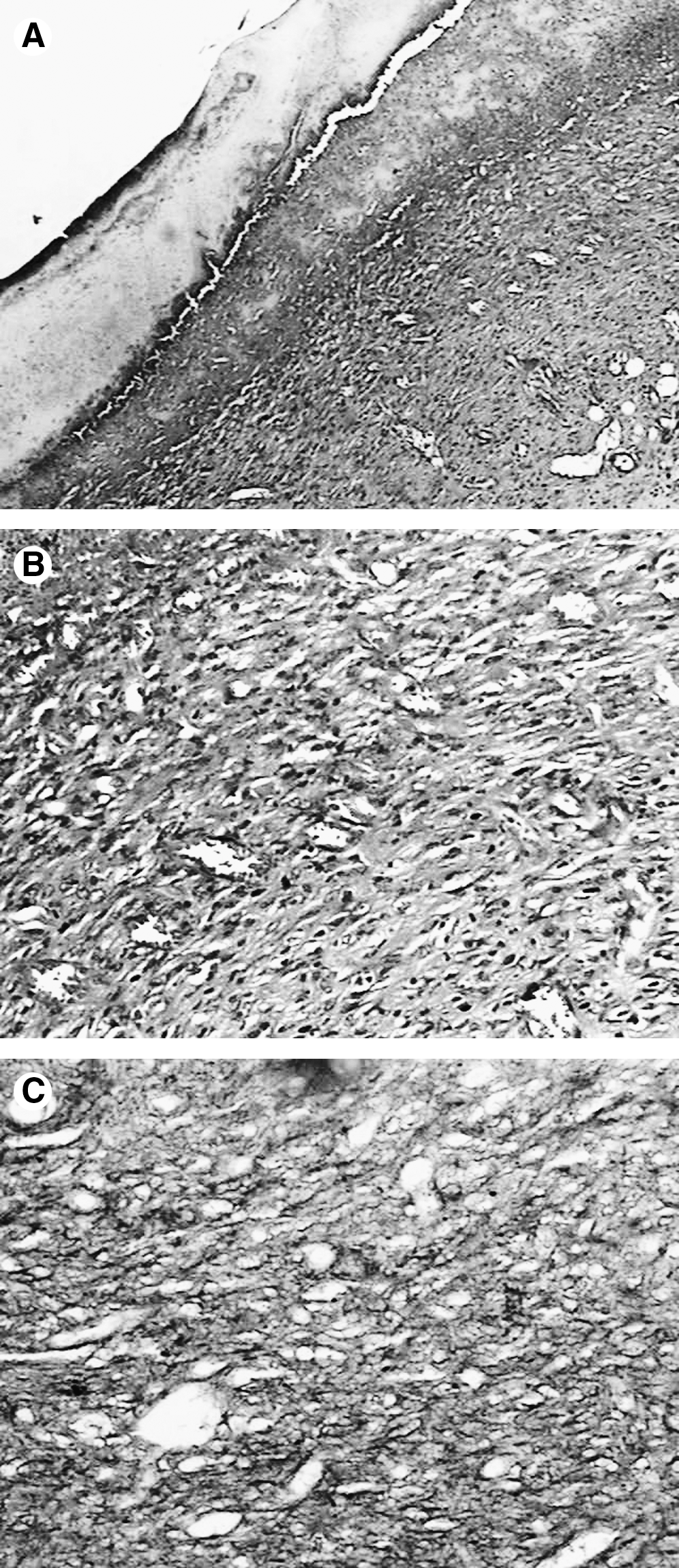
Wounds irradiated with λ635 nm and SAEF of 20 J/cm2 showed extensive ulceration in most cases, and were covered by a thick fibrin coagulum. The central area in two of the specimens showed epithelial pavementing starting from the margins of the wound and partially replacing the crust. In one case, pavementing was complete (Fig. 2A). Underlying the surface were extensive areas of granulation tissue rich in mostly hyperemic sprouting blood vessels as well as young fibroblasts and mixed inflammatory infiltrate (Fig. 2B) within a mature and regularly distributed collagen matrix (Fig. 2C). Discrete interstitial edema was seen in two specimens. In one case, an abscess was present. Increasing the SAEF to 40 J/cm2 resulted in ulceration and crust of variable thickness in four cases. In two specimens, outstanding epithelial pavementing (Fig. 3A) started from the margins of the wound, partially replacing the crust. In another specimen, however, the ulceration was free from crust. Underlying the area of ulceration, a large amount of granulation tissue containing newly formed capillaries and frequently hyperemic young fibroblasts as well as mixed inflammatory infiltrate was detectable (Fig. 3B). In this case, granulation tissue was seen distending the fatty layer of the hypodermis. All of the elements described previously were dispersed in an irregularly distributed collagen matrix in an advanced stage of maturation (Fig. 3C).
λ780 nm – Nourished
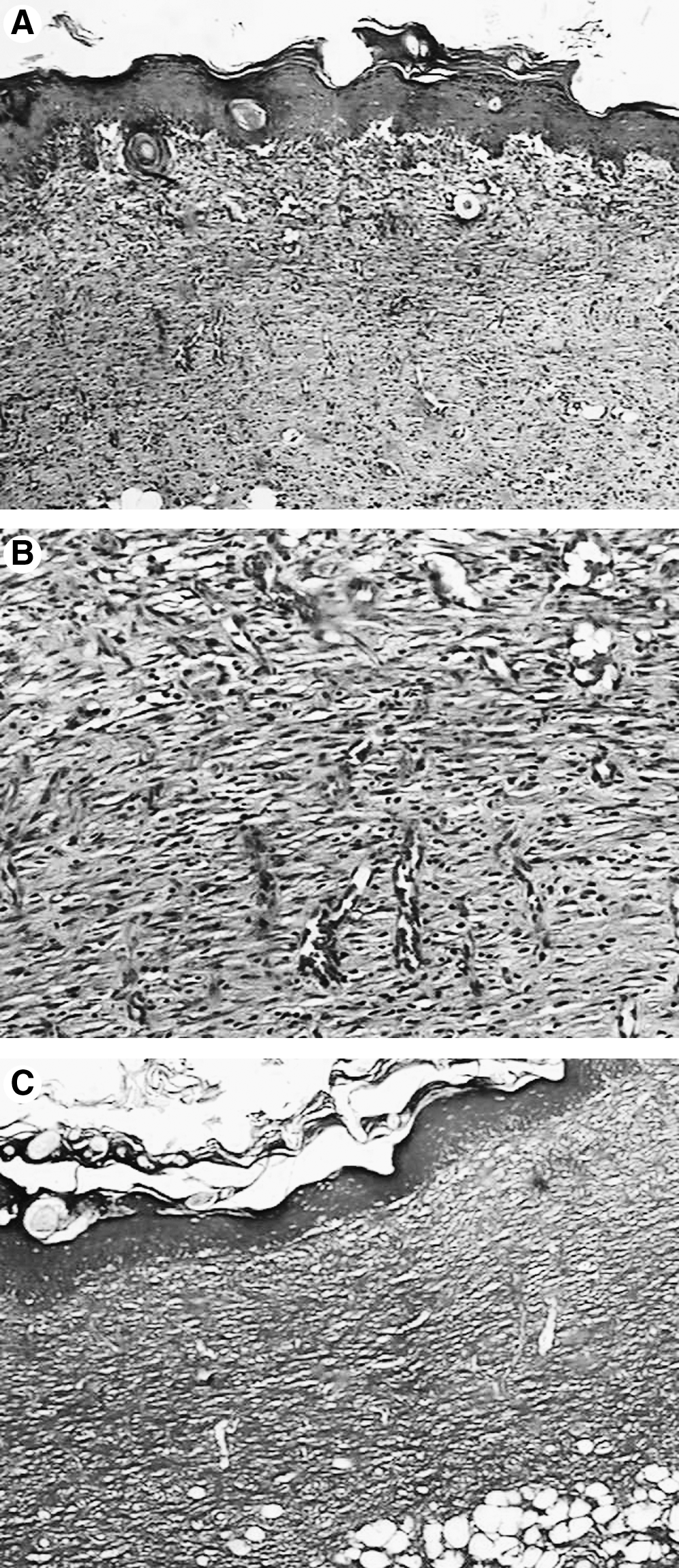
The SAEF of 20 J/cm2 resulted in a wound partially covered by epithelium and without skin annexes (Fig. 4A). The central part of the lesion showed the presence of an ulceration in 40% of the specimens. All specimens showed extensive areas of eosinophilic coagulum, here referred as crust. Underlying the area, a highly vascularized and congested layer of granulation tissue was seen. Within these vessels, neutrophils were present. Edema was seen in most of the specimens (Fig. 4B). A diffuse mixed cellularity inflammatory infiltrate, severe in most cases, was also present. Young fibroblasts secreting a collagen matrix were present on the wounded site (Fig. 4C). One specimen showed the granulation tissue invading the hypodermis, with adipocytes present at the wound surface. The use of SAEF of 40 J/cm2 also resulted in a wound partially covered by epithelium and without skin annexes, with the center of the area still showing the presence of the wound and eosinophilic coagulum, here referred as crust (Fig. 5A). Underlying the area, a highly vascularized and congested granulation tissue layer was seen, and in 20% of the specimens, edema was present. The inflammatory infiltrate of mixed cellularity was diffuse and moderate in 60% of the specimens and discrete and lymphoplasmocitary in the remaining specimens (Fig. 5B). Young fibroblasts secreting a delicate collagen matrix were present in the wound site (Fig. 5C). Thick bundles of collagen fibers were seen at the margins and deeper in the wound site. One specimen showed the granulation tissue invading the hypodermis, with adipocytes present at the wound surface.
Control – Undernourished
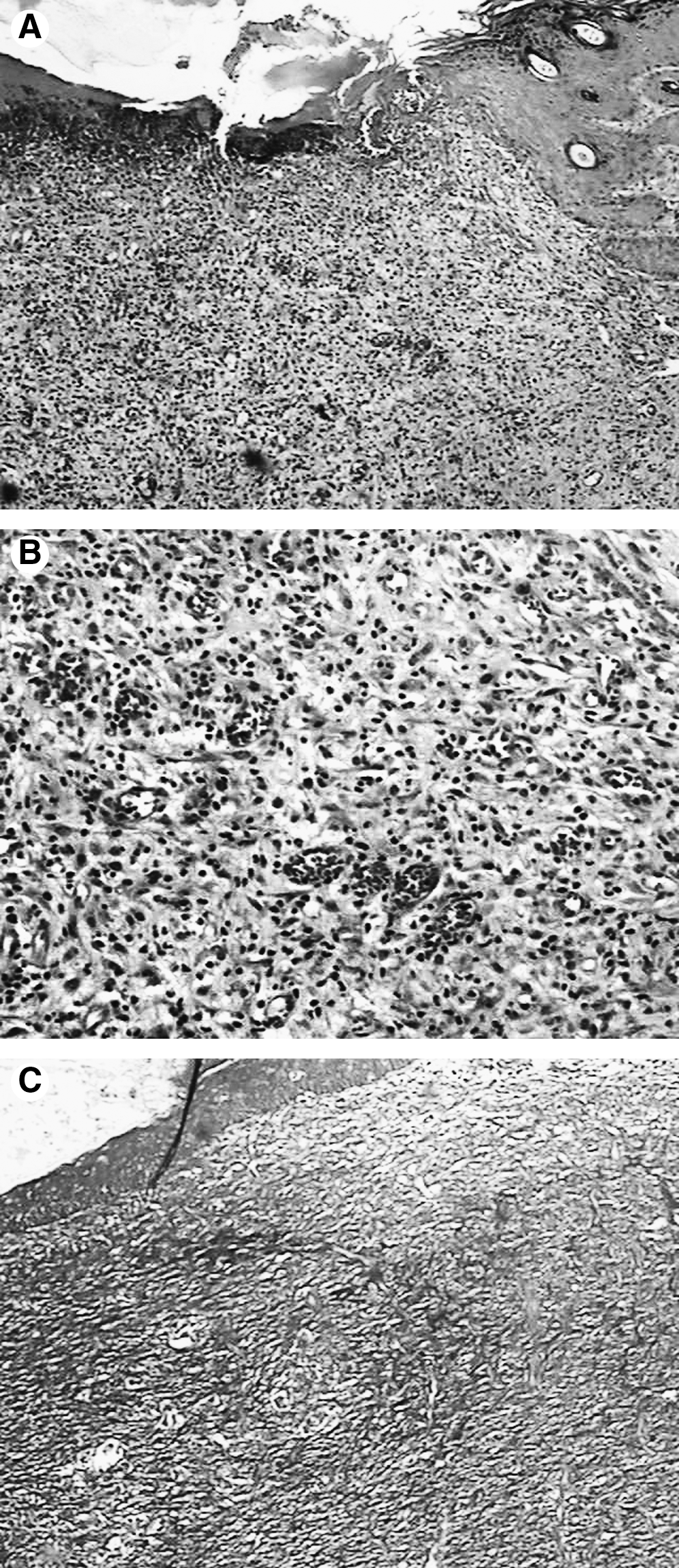
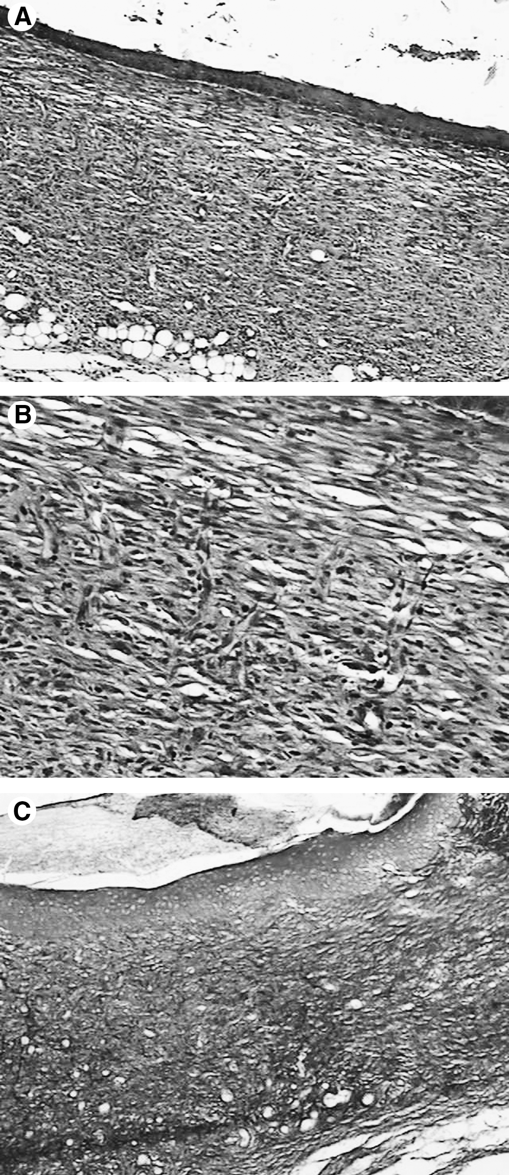
In undernourished subjects (untreated controls), the presence of ulceration covered by crust of variable thickness was observed (Fig. 6A). Underlying this area was an extensive area of granulation tissue containing hyperemic neo-capillaries (Fig. 6B), a moderate number of young fibroblasts, and intense mixed inflammatory infiltrate dispersed in a large amount of an irregularly distributed collagen matrix in maturation, as evidenced by Sirius Red staining (Fig. 6C). In one of the specimens, the collagen fibers were not remarkably positive by Sirius Red staining, demonstrating that the tissue was quite immature. The granulation tissue described reached the hypodermis. In two other specimens, areas of hemorrhagic exudate were observed. Most specimens showed areas of necrosis and, in a few, interstitial edema was present. In most of the specimens, immature multinucleated adipocytes were seen deeper in the wound.
λ635 nm – Undernourished
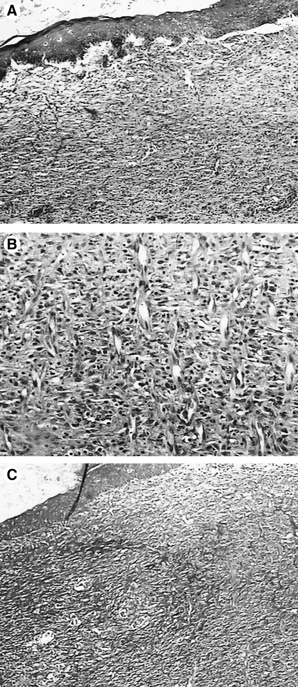
When using SAEF of 20 J/cm2, it was observed that the wound was covered by epithelium of varied levels of keratinization in three cases, exhibiting a plane interface and free from skin appendices (Fig. 7A). Underlying this area, granulation tissue showed the presence of young fibroblasts parallel to the surface, hyperemic neo-capillaries, and a moderate to intense, predominantly chronic, inflammatory infiltrate down to the hypodermis (Fig. 7B). All the cellular elements described in the dermis were distributed in a matrix containing bundles of collagen fibers, which were regularly organized in relation to the cutaneous surface as evidenced by Sirius Red staining (Fig. 7C). Although ulceration covered by crust was observed in two cases, in those initially observed there was also the presence of a thick crust along with the epithelial pavementing. Raising the dose to 40 J/cm2 resulted in complete pavementing free from skin appendices and exhibiting plane interface (Fig. 8A). Underlying this area, granulation tissue was seen that contained young fibroblasts parallel to the surface, as well as hyperemic neo-capillaries and moderate to intense mixed inflammatory infiltrate eventually extending to the hypodermis (Fig. 8B). All cellular elements described in the dermis were distributed in bundles of collagen fibers regularly organized in relation to the cutaneous surface and were in advanced phases of maturation as evidenced by Sirius Red staining (Fig. 8C). Although in two cases the surface presented ulceration covered by crust, in those initially described there was also the presence of crust in spite of epithelial pavementing of the wound.
λ780 nm – Undernourished
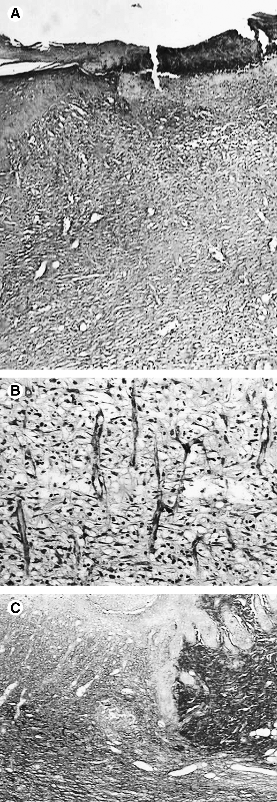
Wounds irradiated with a dose of 20 J/cm2 showed the wounded site filled by granulation tissue. Despite 40% of the specimens presenting epithelial pavementing only, 20% showed complete pavementing of the surface (Fig. 9A). The underlying connective tissue showed a diffuse predominantly lymphoplasmocitary inflammatory infiltrate in many specimens (Fig. 9B). The inflammation was considered discrete and moderate in 20% of the specimens. Areas of residual edema were seen in one case. Discrete hyperemia was observed in several specimens. Young fibroblasts were present and secreted a delicate matrix of oblique distribution within the wounded site (Fig. 9C). Mature adipocytes were seen at the surface in most of the specimens. Some of these cells were multi-vacuolated and were associated with macrophages, showing a feature similar to that seen on fatty change. The increase in the dose showed a wound covered by epithelium and a lack of skin annexes in most cases (Fig. 10A). The central part of the lesion showed the presence of ulceration in 40% of the specimens. All specimens showed extensive area of eosinophilic coagulum, here referred as crust. Some specimens also showed the presence of a crust at the wounded surface. The underlying connective tissue showed the presence of a highly vascularized granulation tissue. In most cases, the blood vessels were congested. A discrete to moderate predominantly lymphoplasmocitary inflammatory infiltrate was present in most specimens (Fig. 10B) and in some of them there were foci of giant cells. The majority of the specimens showed the presence of multivacuolated adipocytes, mostly deeper in the wound site, that were associated with the presence of macrophages and some congested blood vessels. Young fibroblasts and collagen fibers were seen at the site (Fig. 10C).
Discussion
Nutritional deficiencies have significant effects on wound healing in organisms, including alteration of the inflammatory reaction, immunologic function, and the regeneration of tissue; in other words, interfering at any point of the healing process. 3 States of malnutrition will result in deep changes in the process of protein synthesis for the scar, in addition to stimulating greater lysis of collagen. 8 Changes in diet are mostly necessary following extensive surgical procedures in the oral cavity due to the surgical trauma. When the impairment of the oral function is intense, the use of parenteral feeding can become necessary. The use of dietary supplements is not always necessary after small surgeries in a patient who has an appropriate diet. 9
The DBR diet is a model for the induction of severe malnutrition developed in Brazil to induce marasmus, which is still a common disease affecting lower class populations living in deprivation. The disease is one of the three forms of serious protein-energy malnutrition and can be considered to be an adaptation to an insufficient energy intake. Marasmus results from a negative energy balance. This imbalance can result from a decreased energy intake, increased energy expenditure, or both, such as that observed in acute or chronic disease, with victims adapting to the energy deficit with a decrease in physical activity, increase in lethargy, decrease in basal energy metabolism, slowing of growth, and, finally, weight loss.
A considerable reduction in the levels of nutrients, especially proteins, is mostly observed in these patients. Patients suffering from severe trauma or consumptive diseases exhibit deficiencies in collagen synthesis. 2 A delay in the differentiation and proliferation of fibroblasts at the wound site is also observed. Conversely, patients undergoing a diet rich in proteins present faster wound healing. 8,10 This is due to the effects of malnutrition on the ability of the tissue to heal.
In the present study, an animal model that simulates severe undernourishment was used to assess the effect of LPT on wound healing. The use of light to stimulate wound healing is still very controversial in the literature. However, most authors agree that LPT possesses biomodulatory effects, improving and accelerating scar formation. 11 Comparison of the results of the present investigation with other reports is not an easy task, as very few studies on this topic have been carried out previously. An exception is that of Galvão et al., 1 who used the same undernourishment model to study the healing process in rat.
Most studies on the effects of phototherapies on the healing process have attributed the observed effects to several treatment parameters and the properties of the light source used. Monochromaticity is one of the properties of laser light that has been suggested as an important factor in the final result of the treatment. However, it seems that this is not the main factor, since previous studies have observed positive biomodulatory effects using different wavelengths. 12 –14
Regarding coherence, a previous study 12 affirmed that this is not important when photobiological effects are expected because both coherent and incoherent light have been shown to be effective. Another study 15 suggested that coherent light is not necessary, since most biomodulatory effects are obtained also with the use of non-coherent light of appropriate wavelength.
Many previous studies have tried to elucidate the true effect of coherence on the biological effects of phototherapies. The results of recent studies using cells in culture began a discussion on the importance of the coherence of light, since no difference was seen in biological response between cultures treated with laser light that was coherent, vs. those treated with a non-coherent light source. The distrust of the real need for the coherence of light is increased by the fact that so far no definite explanation exists for the behavior of the coherence as light passes through the tissues. 16 The polarization characteristic, however, is neglected in most of the reports investigating LPT. 17
The choice of the two wavelengths used in the present study was based upon the fact that λ635 nm possesses superficial absorption and the λ780 nm, as the literature mostly reports, presents deeper penetration into the tissues and this could influence the healing process as seen elsewhere on the literature. There is just one previous report on the use of laser light in this experimental model. 1 The reason for the use of two different doses was also because of conflicting results from the literature. Despite the large number of reports showing positive effects of LPT on wound healing, there are others that show inhibition or no effect on healing. 18 It is extremely important that correct protocols are devised for LPT and that they include the use of appropriate wavelength, dose, potency density, and time of irradiation in addition to frequency and number of sessions. These parameters may have an influence on the outcome of the treatment and attention to them is necessary to avoid controversies and empiric conclusions.
In regards to the protocol used in this study, despite most previous studies' suggestion of an interval of 48 h, it was decided to perform the treatment immediately after surgery and at 24 h intervals over seven days. This aspect did not interfere with LPT as daily applications show the same effects as 48 h interval sessions.
The macroscopic analysis of the healing of the wounds on subjects fed with both diets is in agreement with previous reports found in the literature. 19 During the removal of the specimen, it was evident that in undernourished animals, the wound was fragile and had a tendency towards dehiscence, in contrast to the wounds of regularly fed and irradiated animals. This represents a clear weakness of the wound due to a poorer quality of the tissue in undernourished animals.
The analysis of epithelization in nourished animals indicated that when both wavelengths were used with SAEF of 20 J/cm2, epithelization was mostly complete at the end of the experimental period, in contrast to results observed when a higher dose was used, when epithelization was incomplete in most cases. This suggested that dose influenced the outcome of the treatment and that smaller doses were more effective. This result, in spite of being in agreement with most of the previous studies, disagrees with the work of Mendez et al. 20 which found better results when longer wavelengths were associated with higher SAEF up to 50 J/cm2. Therefore, the best optical parameter should be certain for the association of several factors, including wavelength, dose, variety in the selection of the animal, wound type, evaluation method, and treatment conditions.
More advanced epithelial pavementing was observed with LPT of wavelength λ780 nm and dose of 40 J/cm2. This result agrees with a previous report. 21 However, regarding this parameter, the experimental groups did not present results significantly better than the control groups, except for the nourished group treated with λ635 nm and SAEF of 20 J/cm2.
When analyzing the inflammatory infiltrate, nourished controls presented moderate chronic inflammation, in contrast to undernourished subjects. The fibroblastic proliferation and the results found in the present study showed that, in all groups, the fibroblasts proliferated. The collagen in the healing wounds showed differences between the control groups, and nourished subjects presented a more organized pattern of collagen fibers, suggesting earlier maturation and larger deposition of fibers.
The inflammation in undernourished subjects, despite presenting moderate inflammatory infiltrate, was of mixed characteristics. A previous study 22 suggested that animals on low-protein diet show unfavorable disturbances in terms of wound contraction and inflammation due to the low levels of proteins, implying that nutritional status plays an important role in the closing of open wounds. The results of the present study also indicate that protein deficiency had a negative effect on the evolution of the inflammatory response.
It is evident that LPT exerted a positive influence on the healing of wounds in undernourished subjects irradiated with λ635 nm or λ780 nm laser light and SAEF of 20 J/cm2. This showed that an inflammatory infiltrate considered to be moderate and chronic resulted in a shorter resolution of the inflammation.
In regards to the fact that the undernourished, irradiated groups presented better effects than the group of nourished, irradiated subjects. This may be explained by the fact that the photosensitivity of the cells to the laser is not of the “all or nothing” type. On the contrary, several degrees of responses may by triggered in cells that may be more or less sensitized, depending upon their physiologic status prior to the irradiation. It is not surprising that LPT may not show evident influence or show any effect at all on “healthy” subjects who are in physiologic balance. 23
The fibroblastic proliferation was more outstanding in the experimental laser group, with both doses, in undernourished subjects. This is in agreement with most of the literature 24 –28 which shows that LPT, when used in appropriate doses, wavelength, potency density, and time of exhibition, positively influence the fibroblastic proliferation and the production of collagen. When λ780 nm laser light was used with SAEF of 20 J/cm2, strong marking was detectable and the collagen fibers were not so well organized. This is indicative that the treatment was more effective when the tissue was compromised in some way.
Collagen deposition in undernourished animals was less organized than in nourished ones and may be indicative of late maturation and decreased deposition of fibers. This result is in agreement with a previous study 29 indicating that nutritional deficiencies possess a deep effect on wound healing, altering the regeneration of the tissue, the inflammatory reaction, and immunological function. It has been suggested that malnutrition states would determine alterations in the synthesis of proteins at the scar, besides stimulating greater lysis of collagen. 8
The analysis of the results showed better results for the undernourished groups irradiated with SAEF of 20 J/cm2 or 40 J/cm2. Those groups showed collagen fibers parallel to the surface and these were strongly marked by Sirius Red staining. This means that there was greater production of collagen fibers and a better organization of the tissue.
Pinheiro and Frame 6 and others 7 mention that laser therapy is used in biomedicine because it improves tissue regeneration and healing, reduces postoperative pain, and reduces inflammation. Increased production of collagen and proliferation of fibroblasts, in addition to the increase in local circulation, are also observed when LPT is used.
The results found suggest that the wavelength of λ635 resulted in a positive effect on the wounds. Its effectiveness was greater when an SAEF of 20 J/cm2 was used. Besides, it could be observed that the effects were more detectable in undernourished groups. This agrees with the literature that points out the effects of the laser on cells with some level of deficiency. 11,24,25
It is concluded that nutritional status influenced the progression of the healing process as well as the quality of the healed tissue and that the use of both wavelengths resulted in a positive biomodulatory effect on both nourished and undernourished subjects, being the effect more evident on undernourished subjects treated with LPT with λ635 with either LPT with λ635 nm with SAEF of 20 J/cm2 or λ780 nm laser light with SAEF of 40 J/cm2.
Footnotes
Disclosure Statement
No competing financial interests exist.