
Abstract
Introduction
Small-diameter vascular grafts are potential substitutes for the damaged vessels in the patients, such as those being referred for coronary artery bypass surgery. 9 A lack of the lining of endothelial cells (ECs) in the lumen of the grafts is a significant factor contributing to the poor patency. 10 EC seeding and retention on the synthetic graft materials, such as expanded poly(tetrafluoroethylene) (ePTFE) or polyurethane (PU), were relatively poor. Numerous methods have been developed to promote endothelialization on synthetic polymers. These include ion bombardment 10 –13 and electrostatic seeding techniques. 14 –16 Low-intensity ultrasound preexposure was also found to improve the adhesion and the retention of seeded ECs on small-diameter PU vascular grafts. 17 The mechanism was associated with an increase in nitric oxide (NO) secretion.
The effect of LLL on ECs has been relatively less studied. We previously showed that He-Ne laser could upregulate endothelial nitric oxide synthase (eNOS) gene expression and EC migration, possibly through the eNOS/PI3K/Akt pathway. 18 Because the increase of NO secretion was observed for both ultrasound and laser stimulations, we thus tested whether LLL could, like ultrasound, increase the interaction of ECs with a biomaterial substrate. 17 In this work, we demonstrated that despite a similar effect on eNOS expression, the effect of LLL preexposure in improving the binding between ECs and a biomaterial substrate was not so significant as that of ultrasound preexposure. Possible reasons for the differences were discussed.
Materials and Methods
Preparation and maintenance of ECs
Adult bovine carotid artery endothelial cells were harvested with the collagenase method 19 and were passaged from primary cultures in Dulbecco's modified Eagles Medium (Gibco, Grand Island, NY) with 10% fetal bovine serum (FBS; Biological Industries, Israel Kibbutz beit Haemek, Israel) and 1% penicillin-streptomycin (Biological Industries). Human umbilical cord endothelial cells (HUVECs) were obtained, as previously described, 20 and cultured in cultured in Medium-200 (Cascade Biologics, Portland, OR) supplemented with endothelial cell growth supplement (ECGS) and 1% antibiotic penicillin–streptomycin. ECs were identified with a monoclonal mouse antibody to human von Willebrand factor (Dako-VWF, F8/86; Dako, Glostrup, Denmark). Cells were resuspended in fresh medium at a concentration of 1 × 10⁵ cells/ml. One milliliter of cell suspension was seeded into each well in a 24-well tissue-culture plate (Corning, NY) and incubated at 37°C in an atmosphere of 5% CO2/95% air for 12 h before the first laser exposure. Cells before passage 4 were used for HUVECs. All the experiments were conducted with bovine cells, except in graft applications, where HUVECs were used.
Laser exposures
A low-power He-Ne laser apparatus (model MED-1000; Lasotronic, Inc., Zug, Switzerland) with a continuous wavelength of 632.8 nm (a maximum power output of 50 mW) was used in this study. The irradiation was performed at 550 mm above the cell layer in 24-well plates on a clean bench. The laser scanned a rectangle area covering the 24-well plates. The exposure time for each well was 2.5 min on average, and the total energy was calculated as 1.18 J/cm2. Pulsed laser irradiation (10 min) was started 1 day after subculture (day 1), and performed once every 3 days until day 7. The treatment protocols included sham laser (control) and laser treatment (LS Group).
Determination of nitric oxide (NO) concentrations
After the last exposure, the sham/laser-treated cells in the 24-well tissue-culture plates were incubated for 12 h. NO release was measured by the collection of nitrite and nitrate in the culture medium. In brief, 125-μl aliquots of medium were removed from the stimulated well and incubated with an equal volume of Griess reagent (1% sulfanilamide/0.1% naphthylethylene diamine dihydrochlroide/2.5% H3PO4) at room temperature for 10 min. The absorbance at 550 nm was determined with a microplate reader. NO2 was determined by using sodium nitrite as a standard. 21 NO concentration was expressed as μmoles per cell (normalized by the number of cells).
Measurement of extracellular calcium concentrations
After the last exposure, the sham/laser-treated cells in 24-well tissue-culture plates were incubated for 12 h, and Ca2+ release was measured by the collected culture medium. The concentration of calcium ion was determined by using a commercially available kit (Sigma-Diagnostics Kit, Cat. No. 587) following the manufacturer's protocol. The medium collected at 7 days of treatment was mixed with 1 ml of the assay buffer provided in the kit. Absorbance was read at 575 nm and 3 min after the addition of the buffer with a UV/Vis spectrophotometer (Hitachi, Tokyo, Japan). Sample concentrations were calculated relative to values for standards provided in the kit. Calcium concentration was expressed as ppm/per cell (normalized by the number of cells).
SEM analysis
Adherent cells remaining on glass substrate were fixed for overnight with 2.5% glutaraldehyde in a sodium phosphate buffer solution and were dehydrated in increasing ethanol solutions, critical point dried, sputter coated with gold, and examined with SEM (S-3000; Hitachi).
Biomaterial graft fabrication
The microporous PU vascular grafts (ID, 4 mm; wall thickness, 0.5 mm; and length, 4 cm) were prepared with a salt-casting technique, 22 by using a polymer/salt ratio of 1:4. The graft surface was impregnated with gelatin that was crosslinked by Denacol (Ex810 Epoxy, C8H14O4; Nagase Chemicals, Tokyo, Japan) at 4°C for >24 h.
Endothelial cell seeding and retention in biomaterial grafts
ECs (either sham or laser preexposed) were seeded onto the inner wall of the PU grafts by dynamic seeding. In the dynamic seeding, grafts were filled with 0.5 ml of cell suspensions (1 × 10⁶ cells/ml) and rotated at 0.16 rpm at 37°C for 12 h. After the seeding procedure, the grafts were incubated for 72 h. In the in vitro perfusion experiment, sham or laser-preexposed EC seeded grafts were incubated for 72 h and then were flushed with serum-free medium (accelerated condition) for 6 h at 37°C and at a flow rate 220 ml/min, which produced a wall shear stress of 2 Pa. 20 This was typical of the physiological levels. 23 The number of cells before perfusion and remaining on the grafts after perfusion were determined by cell digestion with papain and the use of Hoechst 33258 DNA assay, 24 with a standard curve obtained from a fluorescence spectrophotometer (Hitachi, F2500). The sulfated glycosaminoglycan (sGAG) content was quantified with 1,9-dimethyl-methylene blue (Aldrich) dye and the UV/Vis spectrophotometer. 25
Gene expression
Total RNA was extracted by using the Trizol reagent (Gibco-BRL, Gaithersburg, MD) from cultured ECs with different treatments of laser, ultrasound, and control groups seeded on the six-well cultured dish for 3 days. Purified total RNA was reverse transcribed by using RevertAid first-strand cDNA synthesis kit (Fermentas Life Sciences, Burlington, Canada). The gene expression was further PCR amplified by using their gene-specific primer sets, as 5'-ATA GAA TTC ACC AGC ACC TTT GGG AAT GGC GAT-3' and 5'-ATA GAA TTC GGA TTC ACT GTC TGT GTT GCT GGA CTC CTT-3' were used to amplify the rat eNOS gene; 5'-CTG CCG CCT GCT CCC AAG-3' and 5'-GTG CTC GCC TGT TGT GTT CC-3' were synthesized to amplify laminin gene; 5'-GTC TGG CTG ATG CTG CTG CTC-3' and 5'-CTC ACC CTT TGT CCC GTT GC-3' were synthesized to obtain the collagen type IV gene. The oligonucleotides used for the amplification of rat GAPDH as the endogenous control were 5'-TCC CTC AAG ATT GTC AGC AA-3' and 5'-AGA TCC ACA ACG GAT ACA TT-3'. The PCR condition was set to run 35 cycles at 94°C for 30 sec for denaturing, different temperatures (GAPDH, 52°C; eNOS, 60°C; collagen type IV, 63°C; laminin, 66°C) for 30 sec for annealing, and 72°C for 40 sec for extension. The GAPDH expression was used to normalize the viability produced by enzyme efficiency in all experiments, which were performed in triplicate. The PCR amplicons were electrophoresed on a 1.5% agarose gel, and the bands were visualized with ethidium bromide staining. The stained images were obtained by a high-resolution camera (UVP BioDoc-It System), and the band densities were calculated by a system of packaged software.
Collagen contents
The amount of collagen was quantified by the indirect method for the presence of hydroxyproline and read with an ELISA reader (Molecular Devices, Emax, CA) after reaction with p-dimethylaminobenzaldehyde 26 (Sigma).
Statistical analysis
The data are expressed as mean ± standard deviation values and statistically processed with Student's t test. The p values <0.05 were considered significant.
Results
The morphology of ECs after laser stimulation was different from that of controls. As shown by the SEM images in Fig. 1, the laser-treatment group consisted of flattened cells with slightly raised nuclei. At higher magnification, surface craters or focal swellings were seen in the laser-treated cells.
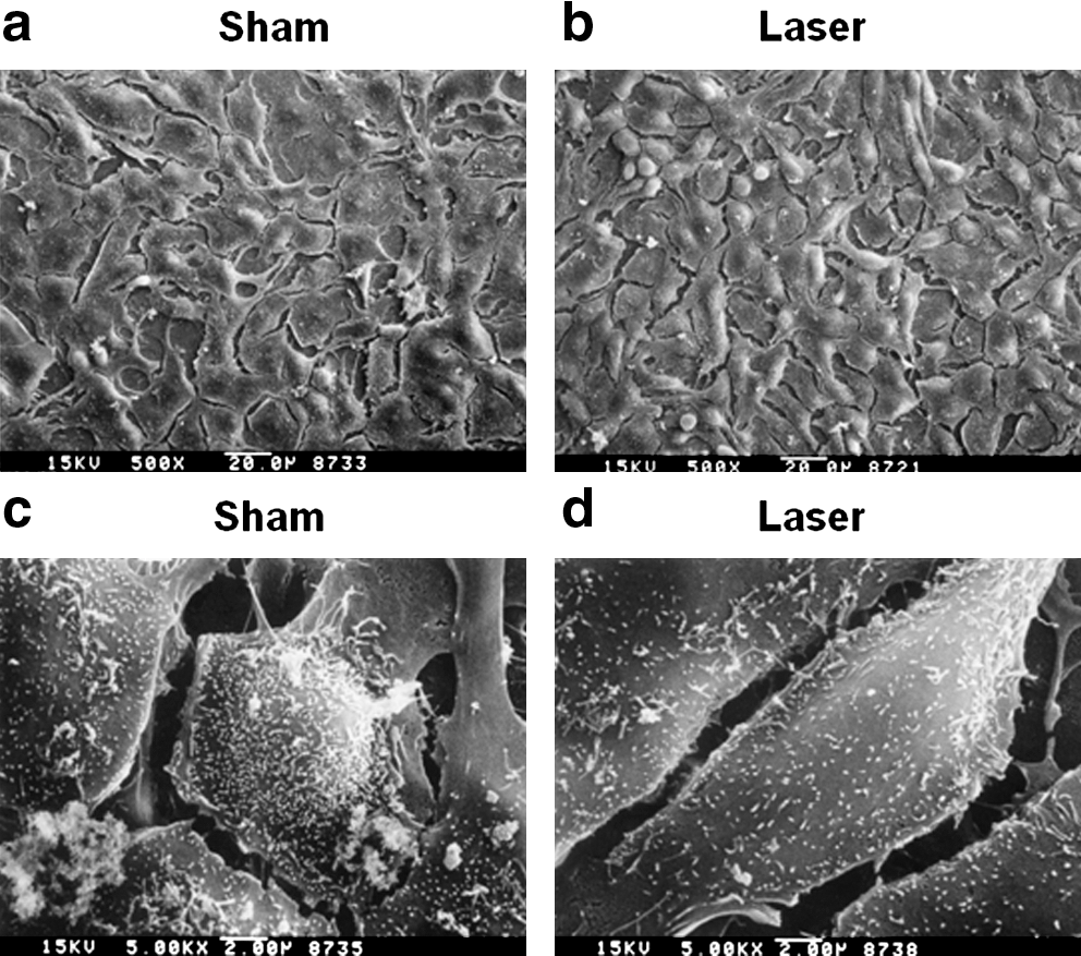
The morphology of ECs on tissue-culture polystyrene dish observed by SEM: (
The stimulative effects of laser on NO and Ca2+ concentrations were demonstrated on a per-cell basis (Fig. 2a, b). Laser induced higher levels of NO and Ca2+ concentrations.

The effect of laser on the NO (
Figure 3 shows the SEM images taken after 72 h of culture on PU graft surface. Preexposed cells were rather elongated, split apart, dragged, or translocated.
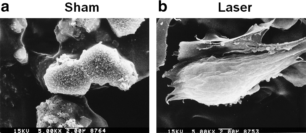
The morphology of ECs on biomaterial vascular grafts after 72 h of culture. (
Figure 4 shows that laser slightly increased the cellular secretion of sGAG after 72 h of culture on the PU graft surface. The increase was not remarkable but was statistically significant.
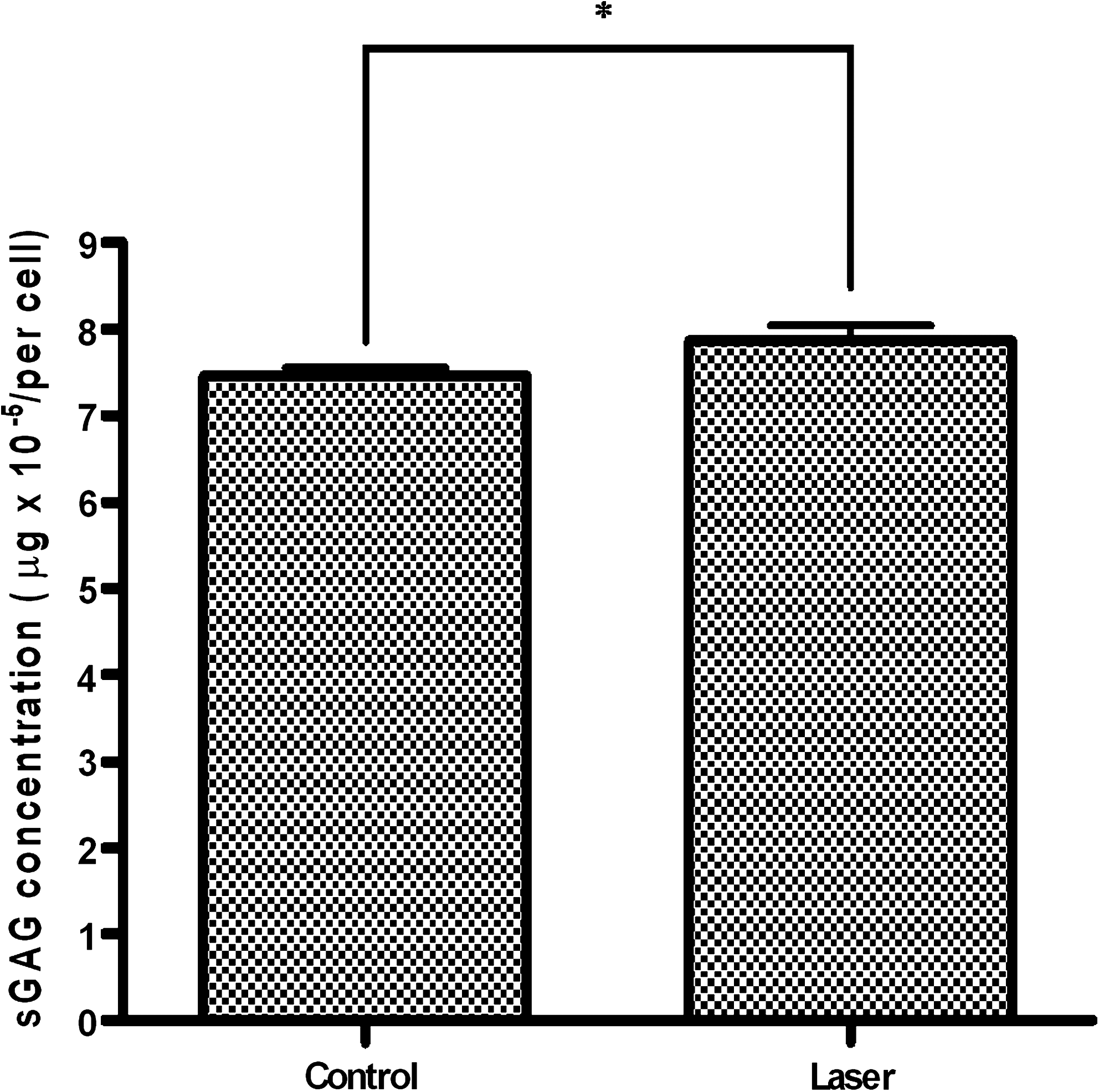
The matrix secretion of ECs after 72 h of culture when preexposed cells were seeded to PU grafts. All treatments were performed in triplicate (n = 3; *p < 0.05).
In Table 1, laser-preexposed ECs seeded on vascular grafts appeared to have a higher percentage of cell retention on the graft surface after perfusion in vitro. However, the difference was not very remarkable.
ECs were seeded by rolling at 0.16 rpm (12 h, 37°C).
In Fig. 5, we show the expressions for three genes (i.e., eNOS, laminin, and collagen type IV genes after treatments of sham, laser, or ultrasound for a period of 3 days. The eNOS gene expression for the treatments with laser and ultrasound showed a statistical difference from that of the sham treatment (p < 0.05). The laminin and collagen type IV mRNA exhibited no statistical difference after the treatments of control, laser, and ultrasound groups. However, the amount of total collagen was induced by ultrasound treatment, and the value was significantly higher (p < 0.05) than that under laser treatment (Fig. 6).
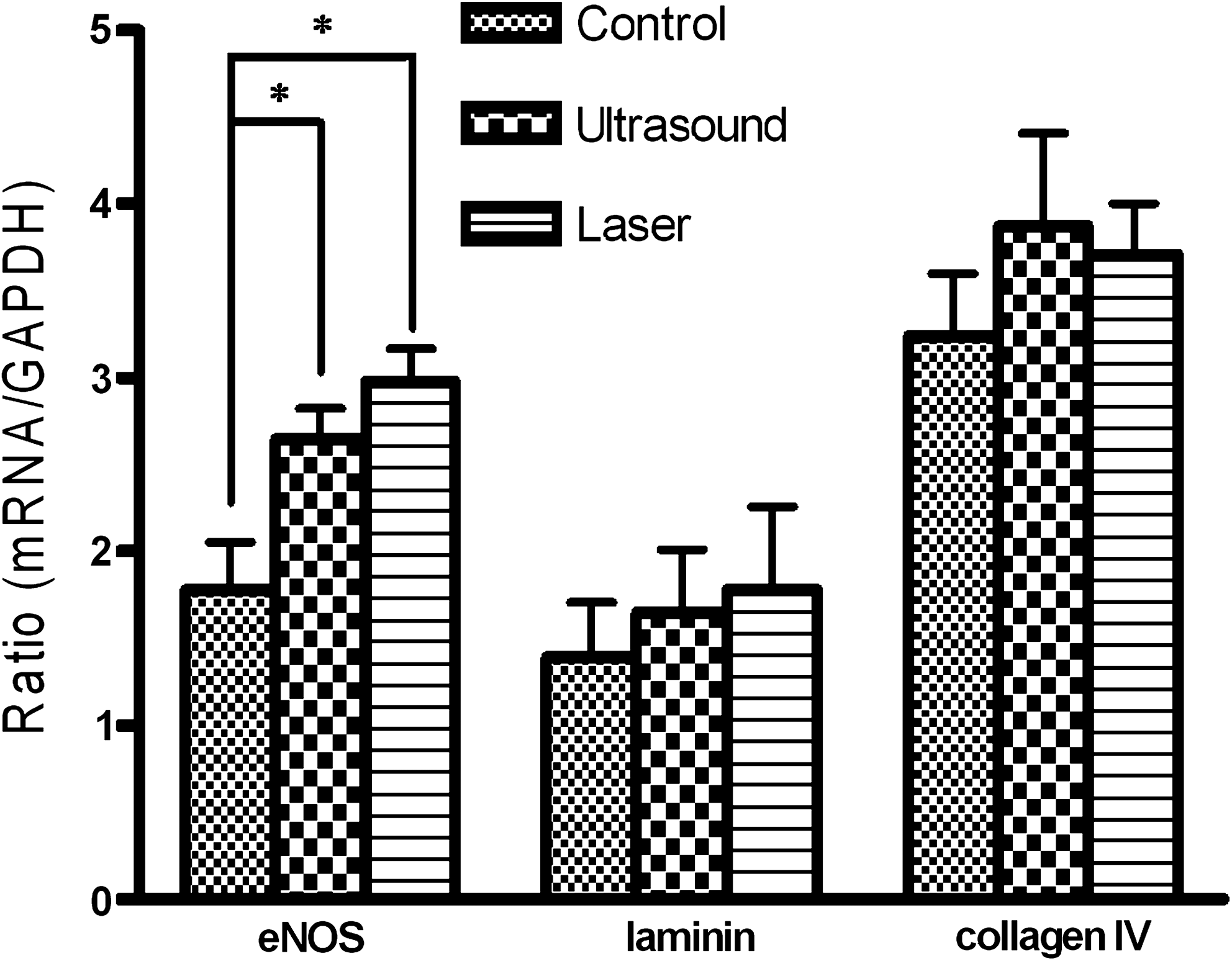
mRNA expression of eNOS, laminin, and collagen type IV for ECs treated with sham, laser, or ultrasound after 72 h of culture in a tissue-culture polystyrene dish. Ultrasound was used daily. Laser was used at days 1 and 3. The cells were harvested 12 h after the final exposure. All treatments were performed in triplicate (n = 3; *p < 0.05).
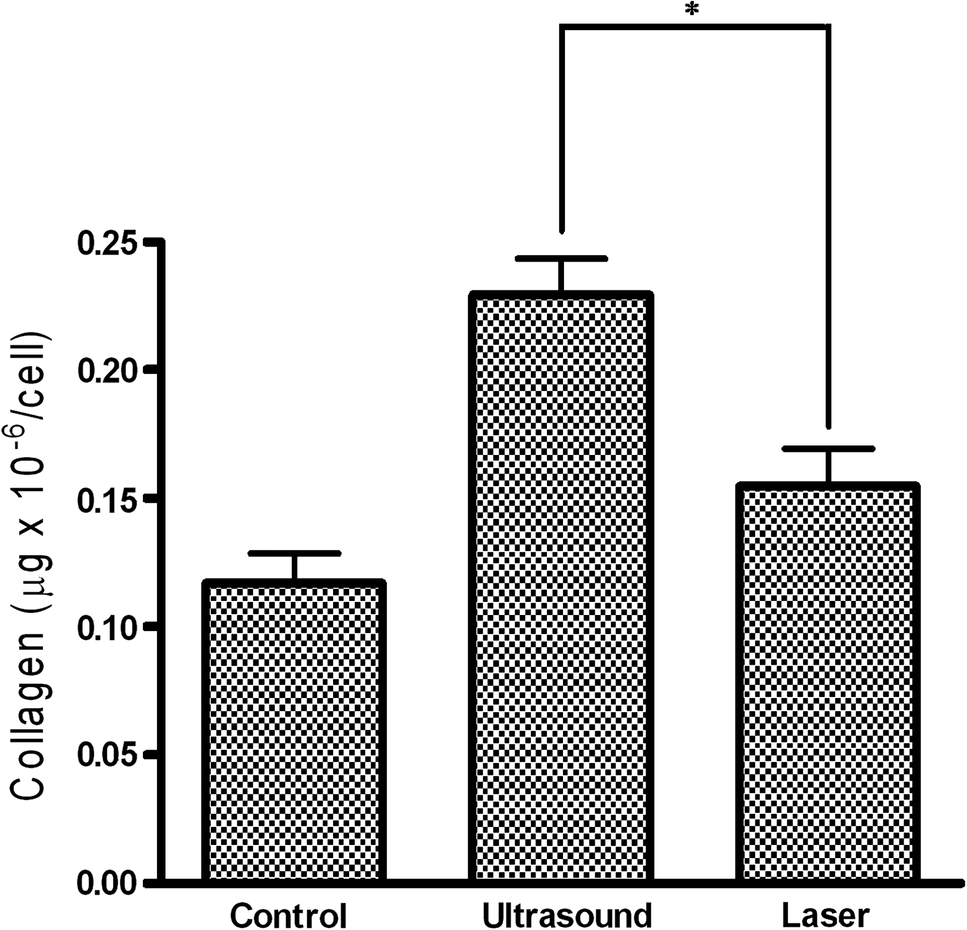
The amount of total collagen in ECs treated by sham, laser, or ultrasound after 72 h of culture in a tissue-culture polystyrene dish. Ultrasound was used daily. Laser was used at days 1 and 3. The cells were harvested 12 h after the final exposure. All treatments were performed in triplicate (n = 3; *p < 0.05)
Discussion
Many previous studies on LLL were directed to evaluate the effects of laser treatments on wound healing. The results showed positive effects of these treatment modalities on wound healing, mainly because of the accelerated biochemical reactions, fibroblast activity, and collagen metabolism. 4 –7 A wide variation in recommendations exists for the optimal energy of laser treatment for different conditions. The usual range is from 0.5 to 10 J/cm2. 27 Generally, a wavelength of 600- to 984-nm laser is used in physical medicine, and a wavelength of 632.8 nm He–Ne is most frequently used in wound healing. 28,29 It has been reported that LLL does not produce significant temperature changes 27 in tissues.
The present investigation focused on the effect of lasers after cells were exposed, trypsinized, and seeded onto another biomaterial substrate. Agreeing with our previous findings, the increase in extracellular calcium and nitric oxide concentrations was found for ECs exposed to laser. The Ca2+/calmodulin-dependent eNOS catalyzes the synthesis of NO in response to an increase in cytosolic Ca2+. Laser exposure increased the concentration of Ca2+, and such an increase may have led to NO synthesis by ECs. Ca2+ plays an important role in cell adhesion, 30 where it functions as a signal transducer and regulates the adhesive activity of integrins by altering either their matrix-binding site or their attachment to actin filaments. Based on our results, laser-preexposed cells secreted more sGAG after 72 h on the biomaterial substrate. LLL changed the morphology and matrix secretion of ECs, and such effects persisted when preexposed cells were seeded to biomaterial grafts for 72 h.
It is recognized that the increase in ECM production can have profound influences on binding between cells and a biomaterial substrate. However, in the case of laser treatment, such an effect depends on whether the cells on the biomaterial can efficiently receive the laser (i.e., the transmittance of laser). Many biomaterials, especially the porous ones, scatter the laser and diminish its efficacy. When cells are embedded or seeded in a porous biomaterial, laser can hardly penetrate the material and act on the cells. Therefore, in this study, we adapted the direct laser exposure (i.e., laser exposed to cells on biomaterials) to laser preexposure (i.e., cells exposed to laser before being seeded onto biomaterials) and examined the interaction between preexposed ECs and biomaterial interfaces.
The effects of low-intensity ultrasound and LLL on ECs appeared to be similar in many respects. To investigate the associated gene expression for cell adhesion induced by laser and ultrasound, three genes (eNOS, laminin, and collagen type IV) were selected and compared with RT-PCR. The results showed no statistical difference in the expression of laminin and collagen type IV genes, but eNOS revealed a significant upregulation from control by ultrasound and laser.
Bergh et al. 31 indicated that shear stress could influence a variety of genes for ECs, including eNOS upregulation. Therefore, we suspected that the ability of cell retention on the biomaterial surface after laser or ultrasound treatment may be through the eNOS-associated signaling pathway. However, the actual mechanism should be further explored. Although no obvious difference in the matrix secretion at the mRNA level was found between ultrasound and laser stimulation, cells stimulated by ultrasound showed higher production of total collagen than did those stimulated by laser. The larger amount of total collagen may lead to the greater adhesion force between cells and substrates and contribute to higher resistance to flushing. Therefore, ultrasound-treated ECs seeded on vascular grafts had a higher percentage of cell retention on the graft surface after perfusion in vitro. 17 In the literature, greater mRNA expression of type I collagen in fibroblasts after LLL treatment 32 and more type II collagen in chondrocytes after low-intensity ultrasound treatment 33 were reported. We suspected that the mechanical force (ultrasound) and light (laser), although both stimulated eNOS upregulation, may have provoked an effect on cells via different signaling pathways. It seems that laser tends to mobilize the cells, whereas ultrasound tends to halt the cells. The actual mechanisms for the change in transcriptional and translational levels of laminin and collagen in ECs by the treatment with LLL or low-intensity ultrasound is a further subject for study.
Conclusion
Endothelial cells were preexposed to low-level laser before being seeded onto polyurethane biomaterial vascular grafts. The retention of laser-preexposed endothelial cells on the graft surface was enhanced, but not as significantly as that of low-intensity ultrasound–preexposed endothelial cells.
Footnotes
Acknowledgments
The work was supported by National Science Council, Taiwan, R.O.C.
Author Disclosure Statement
No conflicting financial interests exist.