
Abstract
Introduction
Orthodontic treatment is based on the principle that if a prolonged force is applied to a tooth, the tooth will move itself as the alveolar bone is remodeling. 2
The search for rapid and safe orthodontic movement has led to several studies involving drugs or alternative or complementary therapeutic approaches or both. 3,4 Several studies using substances such as precursors of nitric oxide, prostacyclin, thromboxane A2, and tezosentan showed that tooth movement increases with their use. 5 –7 Conversely, interleukin-1, TNα; metalloproteinase, and γ-interferon may inhibit this movement. 8 –10
Previous reports found elsewhere on the literature have shown that the use of infrared (IR) laser light increases orthodontic movement. This effect has been attributed to the proliferation of periodontal cells, improved local circulation, as well as an increase of the activity of both osteoblasts and osteoclasts. 11 –14 The use of laser phototherapy (LPT) on bone has been well studied. 15 –17 Previous results found elsewhere on the literature are conflicting in regard to the amount of movement achieved because of the huge variation of the parameters used. 13,17,18
Several aspects are important for the outcome of treatments involving the use of light sources. Most of the proposed protocols used different parameters, and this resulted in conflicting results. The choice of appropriate parameters is essential for the results of the treatment. These parameters include wavelength, power density, energy, and time and frequency of application. 19
Laser phototherapy (LPT) may be used alone or with other techniques to stimulate tissues. It is known that the effects of LPT may not be the cause of healing. 20 –22 Some wavelengths possess therapeutic effects, and these are related to the use of adequate protocols. Laser light may affect cell metabolism in many ways and also may affect the secretion of β-endorphins, bradykinin, serotonin, and others. 23 In orthodontics, LPT has been used as analgesic and a stimulant of bone repair. 21,24
We aimed to study the effect of the use of IR LPT on orthodontic movement in rodents.
Methods
After approval by the Animal Experimentation Ethics Committee of the School of Dentistry of the Federal University of Bahia, we obtained 30 healthy Rattus norvegicus young adult-male Wistar rats with an average age of 3 months and weighing between 250 and 300 g. The animals were obtained from the Animal House of the School of Veterinary Medicine of the Federal University of Bahia and were kept at the Animal Experimentation Laboratory of the School of Dentistry of the Federal University of Bahia. The animals were kept in individual plastic cages lined with wood chips and maintained at 22°C in a day/night light cycle. The animals were fed a standard laboratory diet and had water available ad libidum. After a regular quarantine period, the animals were randomly distributed into two major groups with 15 animals in each. Group I acted as untreated control. The animals were subdivided into three subgroups according to the timing of the animal death (7, 13, and 19 days).
Orthodontic movement was carried out by using the apparatus devised by King et al. 25 The apparatus was installed on the upper arch of each animal. The force was applied by using a 0.010-inch wire (Morelli, Sorocaba, SP, Brazil) fixed to both extremities of one NiTi coil (Reflex Closed Coil Springs, light, 150 g, 3 mm; TP Orthodontics, La Porte, IN). The apparatus was installed under general anesthesia. To fix the wire anteriorly, a hole was drilled with a round bur (KG Sorensen, São Paulo, SP, Brazil) between the two central incisors. The teeth were then conditioned with 37% phosphoric acid (Alpha Acid; DFL, Rio de Janeiro, RJ, Brazil) solution. A force of 40 g/F was adjusted to the system with a dynamometer (25–250 dial type, HALDA, Halmstad, Sweden). The wire was marked on the lingual face and fixed with composite resin (Fill Magic Ortodôntico com Flúor; Vigodent, Rio de Janeiro, RJ, Brazil). Lower incisors were reduced in size to avoid damage to the apparatus during feeding. The first upper molar tooth was removed to eliminate mechanical interference (chewing) that could interfere with the desired mesial movement of the left upper first molar tooth. The apparatus used may be seen in Fig. 1.
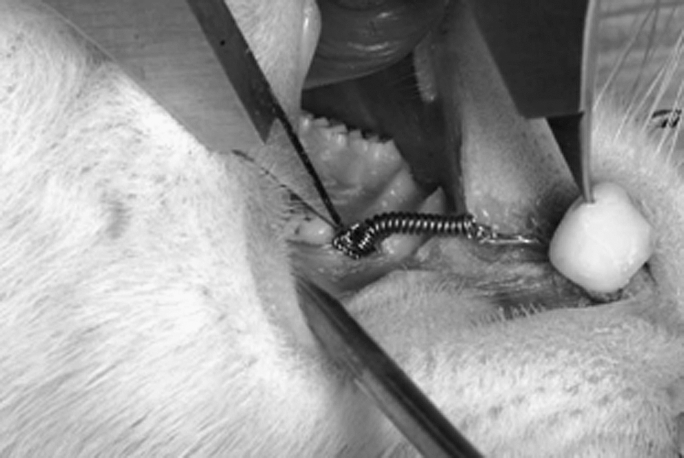
System used for the experiment.
LPT was carried out by using a diode laser (Laser Unit; Kondortech, São Carlos, SP, Brazil; λ790 nm, 40 mW φ ∼2 mm, SAEF of 20 J/cm2 per session). 21,22 The treatment was carried out every other day during the experimental time, and the session dose (SAEF) was divided into three parts: 4.5 J/cm2 applied both mesially and distally, and 11 J/cm2 on the buccal side, the last applied extraorally because of the anatomic impossibility of delivering the dose precisely intraorally. On this application point, the dose was increased in 20% to compensate for losses due to transcutaneous application of the light. 19 Animal death occurred after overdose of general anesthetics at days 7, 13, and 19 after installation of the apparatus. The animal weight did not change significantly during the experimental time in both groups.
The forces of the coil at the different times were calculated to verify whether they remained constant during the experiment. For this, Hooke's law was used, and this indicates that the deforming force is proportional to the elastic generation caused by the force.
Measurement of the distance between the most prominent point of the mesial surface of the first molar to the perforation on the resin bulk was performed in each animal immediately and at days 7, 13, and 19. This procedure was used to assess the dislocation of the tooth and was carried out with a precision digital caliper (Beerendonk Caliper 78532; Aesculap, Tuttlingen, Germany).
Statistical analysis used the Kolmogorov-Smirnov test. Intergroup testing was carried out with Student's t test. Significance level was set at 5%.
Results
The data were normally distributed (Student's t test). Table 1 shows the central tendency of measurements and dispersion of the data (in millimeters) of the distance between the reference points on the upper central incisor to the mesial surface of the upper first molar tooth on both groups during the experimental time. Table 2 shows the variation of the distance between these two points in relation to the time. The force of the coil at each time may be seen in Fig. 2. Although the force was slightly higher in the irradiated group than in the Controls, this was not significant at T7 (p = 0.079); T13 (p = 0.455), and T19 (p = 0.914).
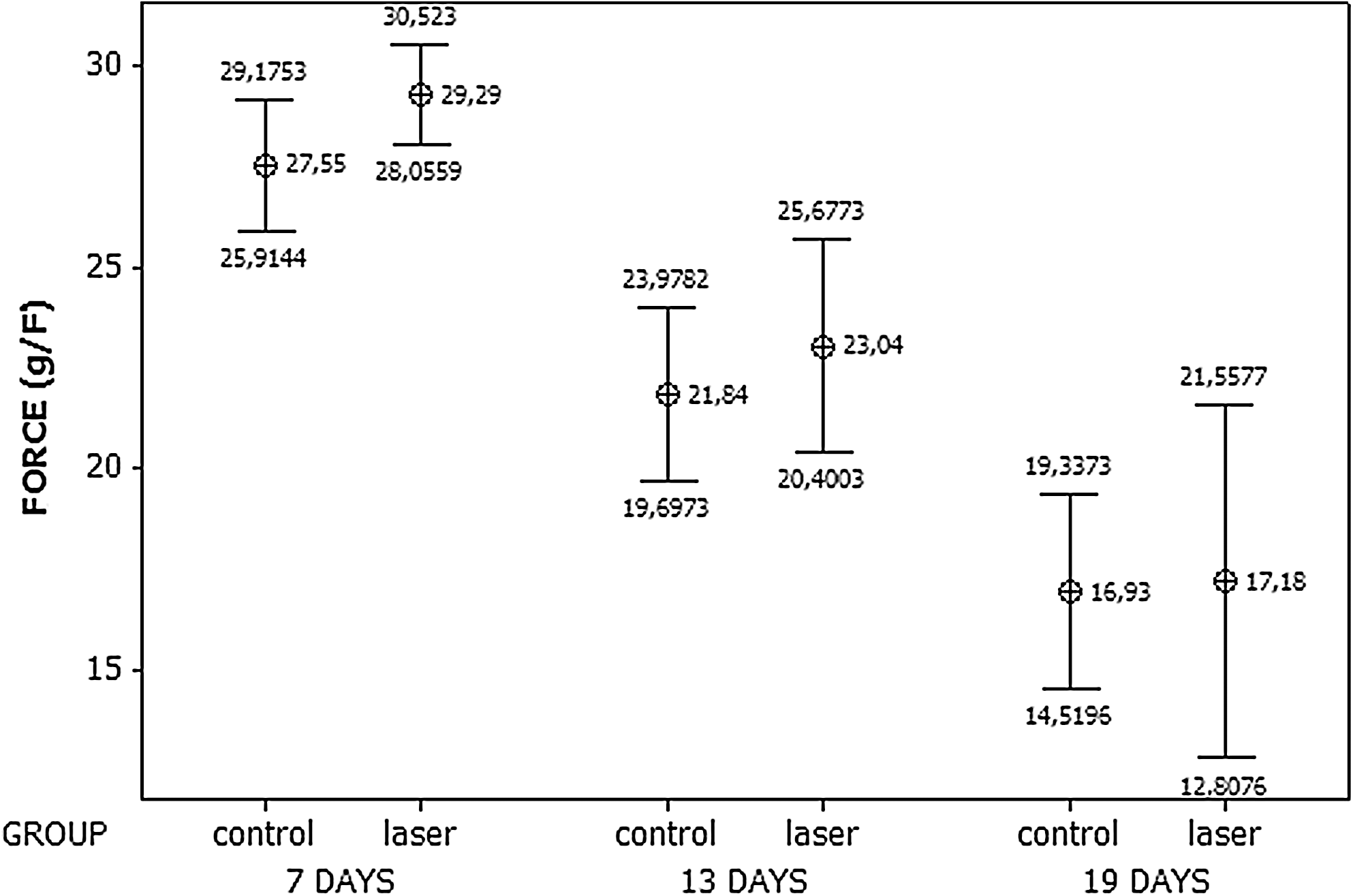
Mean and 95% confidence interval of the force (g/F) of the coil at the different experimental groups and times.
Student's t test.
SD, standard deviation.
Discussion
Rodents (Rattus norvegicus) have been widely used in different experimental models of orthodontic movement because of their accelerated metabolism. 2,5,7,8,10,13,26 –28 For the orthodontic movement to be efficient, some variables are essential, including the magnitude and duration of the force and the morphology and alveolar bone density. 1 In our study, the initial force (40 g/F) was applied continuously to induce more tooth movement. Similar forces have been used previously. 27
It is known that LPT stimulates cells of the periodontal ligament, increases numbers of osteoclasts, and improves the local blood supply. These factors may be indicative that this modality of treatment may accelerate tooth movement during orthodontic treatment. 29
The choice of the timing of the experiments was because most studies using the animal model of tooth movement use periods of observation ranging from 10 to 15 days. 2,9,30 –32 We used the treatment at 48-h intervals, and with this time, we could assess the effect of the light during different phases of the movement.
In the present study, we measured the distance between the mesial point of the first molar and a reference point on the incisors and did not measure directly the space between the first and second molars. The main point that influenced our decision was that, when we mesially move a tooth, the adjacent tooth may also move because the presence of the transseptum periodontal fibers 33 interferes with the measurement of the distance between the first and second molars.
Some previous reports on tooth movement used contralateral control 3,12,18 and did not consider the systemic effect of LPT, 25 perhaps making comparison with our results tricky. We opted for not using contralateral controls as did previous reports in the literature. 13,17 The protocol used on our study was also based on previous studies. 3,11,13,18
Another aspect to consider was the site of application of the LPT. We opted for both intra- and extraoral application because of anatomic constraints. This may also further complicate comparisons with studies that used only intraoral applications. 12,18
It is known that prolonged force application on one tooth will result in its displacement, 1,2,27,34 –38 as seen in the present study. We found a significant reduction in the displacement between the beginning and the end of the treatment in both control and irradiated subjects (p < 0.001 and p = 0.001), as seen in Table 1.
Our findings are indicative that the use of LPT did not significantly interfere with tooth displacement during orthodontic movement, as seen in Table 2, as mentioned previously. 18 It also must be considered that this late report was based on human data and a different protocol. Conversely, different experimental setups have shown increased tooth displacement associated with the use of LPT. 12,13 Our best result in this regard was found between T0 and T7 when LPT-treated subjects showed a smaller displacement than did the control.
The force used in the study seems not to have had a significant influence on the results, as no significant difference was found between the groups during the experimental time. We were not able to find any previous reports on the assessment of the degradation of the force during induced orthodontic movement in animals.
The fact that our results suggest that LPT does not significantly affect tooth displacement during orthodontic movement also considers previous reports that LPT possesses an analgesic effect. 24,39 It may be possible that postactivation pain may be ameliorated by using LPT, without compromising movement. Another aspect that may also be considered is that LPT has been shown effective in bone repair after maxillary expansion, postactivation pain, and the healing of traumatic soft-tissue lesions caused by the orthodontic apparatus. 40
A study from our group showed that LPT is capable of causing histologic changes related to the movement of teeth during orthodontic movement. Habib, 15 similar to us, used nonirradiated subjects as controls because of the systemic effect of the LPT, different from other studies that did not consider this aspect or did not use it. The parameters used in the present study were based on work described previously in the literature. Habib showed a significant increase in the number of osteoblasts from days 7 to 13 after activation in LPT-treated animals. It was suggested that newly formed bone after LPT shows better quality than does nonirradiated bone and may lead to a quickening of the movement and alveolar bone remodeling. The results of Habib's study showed that LPT was able to increase the number of osteoblasts on the tension side. This aspect may lead to an improved capacity of the alveolar bone to remodel. An increased amount of collagen matrix was reported on the pressure side in irradiated subjects at both 13 and 19 days, when compared with nonirradiated animals. The same was also seen on the tension side. It is known that LPT has the capacity to stimulate both the secretion and proliferation of fibroblasts and consequent increased collagen matrix deposition. The fibroblasts of the periodontal ligament are actively involved in alveolar bone remodeling during orthodontic movement. 41
The literature reports some previous works in which is suggested that laser treatment does interfere with tooth movement in rats, as this model features easy handling and standardization. 3,12,17,29,30,36,42 Although we were not able to find a significant difference between irradiated and nonirradiated groups, we found that tooth displacement was becoming more similar during the experimental time (T0–T19; p = 0.914). It is possible that the rapid metabolism in the animal model may also have influenced the outcome of the study. It is possible that earlier observation times may result in different findings. It is also possible that the amount of energy was too high, as other studies using less energy reported acceleration of the movement. 13
It may be concluded that LPT, using the parameters adopted in the present study, did not significantly increase the amount of teeth displacement during induced orthodontic movement in rodents
Author Disclosure Statement
No competing financial interests exist.