
Abstract
Introduction
A particular interest is focused on photodynamic therapy (PDT), in which cytotoxic lesions in cells are induced by a photosensitizer and light of an appropriate wavelength. 2,3 The photosensitizers used in these studies may be of both an exogenous and endogenous origin. One of latter type of sensitizers is protoporphyrin IX (PpIX), which can be accumulated in cells under the effect of 5-aminolevulinic acid (ALA). Depending on the cell type (normal or abnormal), differences in kinetics of PpIX accumulation and its final level in the cell were demonstrated. 2 –4 The studies on the application of PDT in endometriosis suggest that efficacy of PDT depends on several factors, including the patient's hormonal status. 2,3
Earlier studies have demonstrated that, in in vitro conditions, not all epithelial cells isolated from endometriotic foci synthesized PpIX in the presence of its natural precursor, 5-ALA. 3 Hypothetically, these cells may manifest the so-called multidrug resistance (MDR), frequently indicated as a cause of therapeutic failure in neoplastic diseases. The main cause of the removal of therapeutic molecules from the cells seems to involve an augmented activity of membranous transport proteins of ABC family, which contain the ATP-binding domain (ATP-binding cassette family). Several authors describe P-gp (MDR 1, 170–180 kDa) as the most important transport protein that takes part in this process. 5 –9 The mechanism of P-gp action has not yet been fully clarified. It is suggested to act as a flipase, removing hydrophobic substances from the cytoplasmic to the external leaflet of the cell membrane lipid bilayer, from which they may diffuse to the extracellular space. Another hypothesis implies that P-gp acts as a hydrophobic scavenger, which removes lipophylic compounds out of a cell from the lipid layer of cell membrane. Such mechanisms decrease the intracellular concentration of the drug and thus nullify its therapeutic effect. 9
Only a few studies report activity of P-gp in epithelial cells originating from endometriotic foci. The present research aimed to analyze MDR in endometriosis treated by PDT. The experiments conducted included the application of PDT, dependent on 5-ALA after blocking P-gp activity using verapamil. Verapamil has the ability to reverse MDR. This calcium channel blocker inhibits P-gp activity due to an increase in intracellular accumulation of tested drugs. 10 In parallel, the presence of MDR-1 protein in the analyzed material was detected by immunohistochemistry. Significance of the undertaken studies reflects the complete absence of reports related to modulation of P-gp activity and the efficacy of PDT in patients with endometriosis.
Materials and Methods
Chemicals
5-ALA, XTT salt, verapamil hydrochloride, and 3-3′-diaminobenzidine were obtained from Sigma (St. Louis, MO); anti-MDR-1, the polyclonal primary antibody, was purchased from Santa Cruz Biochemistry (Santa Cruz, CA); anti-cytokeratin 18, the primary monoclonal antibody, was obtained from Abcam Inc. (Cambridge, MA); and LSAB HRP Kit was purchased from Dako (Glostrup, Denmark). All employed chemicals were of analytical grade.
Sample collection
Tissue samples of normal endometria were obtained during hysterectomy from eight women diagnosed with cervical intra-epithelial neoplasia. Fragments of ovarian endometriosis were obtained at the time of laparoscopy or laparotomy from 15 women. The age of patients ranged from 28 to 42 years. Endometriosis was confirmed by a histopathologist. All women (with or without endometriosis) had regular menstrual cycles, and they had not been subjected to any hormonal treatment for at least 6 months before surgery. The specimens were obtained at the secretory phase of the menstrual cycle, which was confirmed by measurement of serum hormone levels. Every material was used for epithelial cell isolation, as well as for histological and immunohistochemical analysis. All biopsy specimens were collected at the Department of Mother's and Child's Health, Poznan University of Medical Sciences with the permission of the local ethics committee. Informed consent was obtained from every patient.
Cell isolation and culture
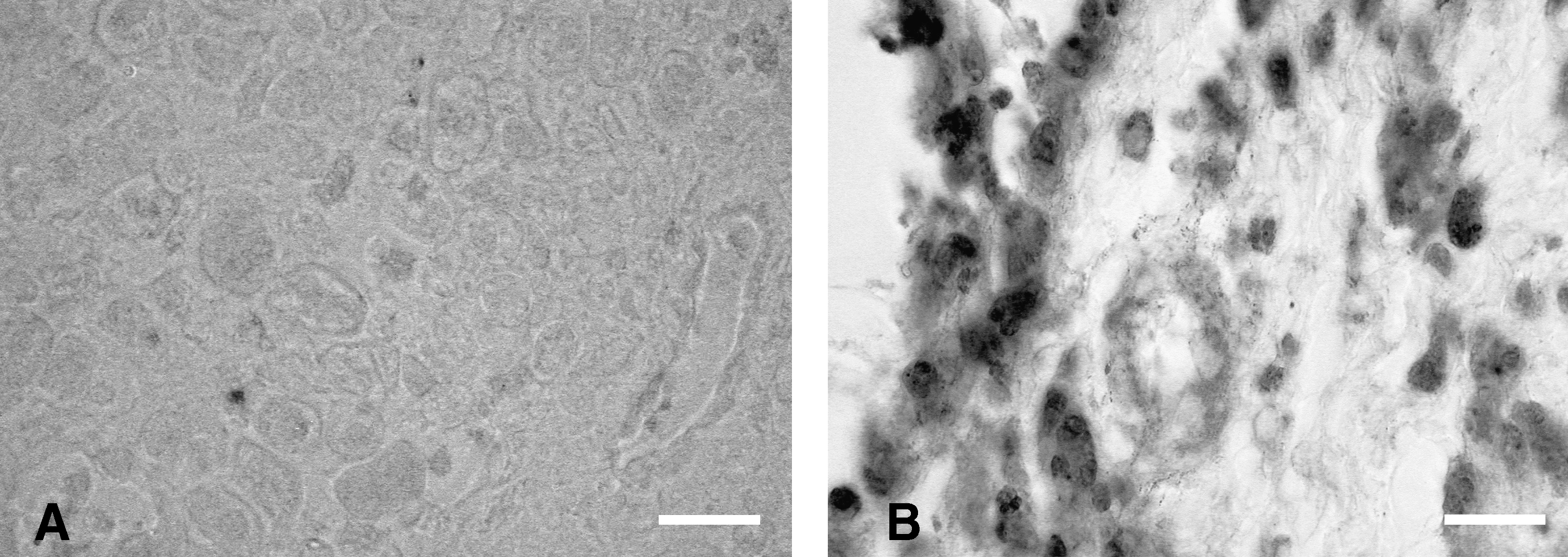
Epithelial cells of eutopic and ectopic endometria were isolated and cultured as described by Ryan et al. 11 All experiments on the cells were conducted following 4 days of culture (Fig. 1A and B), whenever purification of the epithelial cell population was confirmed by immunocytochemical staining with antibodies against cytokeratin-18 (Abcam). The cultures used in the experiments manifested purity of the epithelial cells greater than 92% (data not shown).

Representative gland freshly isolated from endometriosis used for isolation of epithelial cells
Blocking of P-gp activity using verapamil
In order to block P-gp activity, primary epithelial cells of normal endometrium and of endometriotic foci were seeded at 500 cells per well into 96-well microtiter plates in F-12 medium supplemented with 10% FBS (Fetal Bovine Serum; Sigma), glutamine (Sigma), and penicilin/streptomycin (Sigma). The cells were allowed to adhere, and after 4 days were preincubated for 8 h with 4 μmol/L verapamil (these conditions were selected experimentally). 11 Subsequently, the cells were suspended in a fresh medium containing 5-ALA (1, 2, 4, or 8 mmol/L) for 2 h. After incubation with ALA, the cultures were irradiated with the semiconductor laser (635 nm, exposure area 24 mm2; Uzor 2K, Kaluga, Russia) with a radiant exposure dose of 56 J/cm2 (laser head contacted lid of the plate). Twenty-four hours after irradiation, the viability of the cells was evaluated using the XTT test. The control consisted of cells exposed to ALA action but not irradiated, as well as irradiated cells incubated with verapamil alone. Every experiment conducted for eight eutopic endometrium and 15 ectopic endometrium was performed independently into two separate plates (16 measurements for eutopic endometrium and 30 for ectopic).
XTT viability test
The viability of cells was analyzed using the XTT colorimetric test, based on dynamics of reduction of XTT pigment (tetrazoline-2,3-bis[2-methoxy-4-nitro-5-sulphophenyl]-2H-5-carboxyanilide) by the living cells and production of a colourful product. The intensity of the fluorescence was measured at the wavelength of 450 nm.
12
The percentage of growth inhibition was calculated according to the following equation:
where OD is optical density, 12 and untreated cells are control cells without any drugs, cells treated with verapamil illuminated or not illuminated, and the cells incubated with verapamil and ALA without photoactivation. Statistical analysis of the results was done with the nonparametric Wilcoxon paired rank test and the Mann–Whitney U-test using Statistica software (V5; Statsoft, Krakow, Poland). A p value of <0.05 was considered to represent the threshold of significance.
Immunohistochemistry
For every formalin-fixed and paraffin-embedded sample, 4-μm serial sections were cut, dewaxed, and rehydrated. After quenching the endogenous peroxidase activity, achieving antigen retrieval and blocking of non-specific binding sites, incubation with anti-MDR1 (Santa Cruz) primary antibodies, diluted 1:50, was carried out overnight at room temperature. The binding of the primary antibody was detected by the LSAB-HRP (Dako) procedure, using DAB (Sigma) as a chromogen. Control experiments included reactions carried out under identical conditions except that primary antibodies were replaced by a non-immune serum.
Results
In order to evaluate the effect of P-gp on the phototoxic effect of 5-ALA exerted on epithelial cells isolated from normal endometrium, the activity of P-gp was blocked using verapamil.
The experiments showed that addition of 5-ALA to the incubation medium of analysed cells not subjected to the action of verapamil results in protoporhyrin IX accumulation. The subsequent photoactivation by the laser caused significant cells growth inhibition (Table 1, p < 0.05). Eight mmol/L ALA concentration was found to be most toxic for studied cells and resulted in 35.6% inhibition of cell growth. When the epithelial cells were preincubated with verapamil, the toxic effect of PDT-based on PpIX synthesis was still observed (Table 1). Statistical analysis, including comparison of results obtained in the absence of verapamil and upon using verapamil to block P-gp activity, demonstrated that verapamil did not affect phototoxicity of ALA at concentrations of 8 mmol/L (Table 1, p > 0.05). However, the compound clearly intensified the PDT effect when ALA concentrations of 2 or 4 mmol/L were used.
XTT assay of ALA-PDT-treated epithelial cells isolated from eutopic endometrium. The epithelial cells were preincubated for 8 h with 4 μmol/L verapamil (see Materials and Methods). Subsequently, the cells were incubated in a fresh medium containing an increasing concentration of 5-ALA for 2 h, and then they were irradiated. After 24 h, the viability of the cells was evaluated using the XTT test (columns: ALA + V%). Every experiment was performed independently into two separate plates (n = 8, 16 measurements for each epithelial cells isolated from eutopic endometrium). The statistical analysis of the results was done with a nonparametric Wilcoxon paired rank test and the means of growth inhibition of cells incubated with verapamil and treated ALA-PDT (ALA + V% data) were compared to the means of growth inhibition of cells exposed to ALA and light, without verapamil pretreatment (ALA% data).
ALA, 5-aminolevulinic acid; V, verapamil; SD, standard deviation; NS, not significant.
In the case of endometriotic epithelial cells, the toxic effect of PDT was also observed. The most pronounced inhibition of the cell growth was noted at 4 mmol/L ALA concentration (Table 2). The blocking of P-gp activity in endometriotic epithelial cells with verapamil caused a significant increase in the inhibition of cell proliferation (Table 2, p < 0.001). The effect was particularly strong in the case of ALA concentrations involving 1, 2, and 8 mmol/L. The addition of verapamil, together with these concentrations of ALA, resulted in an almost twofold higher inhibition of cell growth compared to cultures not treated with verapamil (Table 2).
XTT assay of ALA-PDT-treated epithelial cells isolated from ectopic endometrium. The cells were treated and the statistical analysis was done as described in Table 2. Every experiment was performed independently into two separate plates (n = 15, 30 measurements for each epithelial cells isolated from ectopic endometrium).
LA, 5-aminolevulinic acid; V, verapamil; SD, standard deviation.
Statistical analysis of the obtained results for cytotoxicity using the XTT test for eutopic end ectopic endometria (Table 1 vs. Table 2) indicates statistically significant differences between PDT-ALA alone and PDT-ALA verapamil-treated groups (for each concentration of ALA p < 0.05, Table 3). Epithelial cells isolated from ectopic endometria were more sensitive to PDT upon ALA alone, as well as after its preincubation with verapamil.
Statistical analysis of the obtained results for cytotoxicity using the XTT test (eutopic vs. ectopic endometria). The statistical analysis was done with the nonparametric Mann–Whitney U-test. p < 0.05 was considered to represent the threshold of significance.
Conc., concentrations of ALA; p, p-value for Mann–Whitney U-test; ALA, 5-aminolevulinic acid; V, verapamil; SD, standard deviation.
Subsequently in the study, the presence of P-gp was evaluated in the tissues, from which the above-described cell cultures were established. Using immunohistochemistry, the analysis demonstrated an absence of P-gp both in the epithelium and in the stroma of a normal endometrium (Fig. 2A). In turn, in cases of endometriosis, immunohistochemical staining provided proof for the presence of P-gp in both the epithelium and the stroma (Fig. 2B). The detailed analysis of material originating from patients with endometriosis showed a distinct localization of P-gp. The protein presence was cell-type dependent. While epithelial cells manifested a cytoplasmic localization of P-gp, in stromal cells both cytoplasmic and nuclear localization was detected.
P-Glycoprotein immunohistochemical staining in eutopic
Discussion
Previous studies on the application of 5-ALA in diagnosis and in PDT of endometriosis demonstrated that ALA allows the photodynamically localization and destruction of endometriotic foci (following exposure to light of appropriate wavelength). 2,3 The results of our earlier studies conducted on the primary cultures of endometriotic epithelial cells showed that ALA induces the accumulation of PpIX in these cells. The accumulation was distinct in endometriotic cells originating from different patients, as well as in cell colonies of epithelium isolated from the same focus. 3
It is suggested that in various types of cells, the low level of intracellular porphyrin depends on many factors, restricting efficiency of PDT. 8,13 –16 Several authors point to the phenomenon of MDR as one of the most important reasons for therapeutic failures in numerous diseases. It is believed that MDR represents primary or induced insensitivity of cells to therapeutic substances. 8,16 –18 The studies documenting significance of cancer cell resistance to chemotherapy imply the role of mdr-1 gene expression in this process. However, P-gp (MDR-1), encoded by this gene, was also detected in normal organs, including the liver, kidneys, small and large intestine, the brain, testes, and the placenta. 19 At present, the manifestation of MDR is used as a prognostic factor, which may define the quality of neoplastic lesions and related duration of a patient's survival. 18
Recently, significant attention has been focused on the potential of PDT and increased cell survival, indicating “resistance” to PDT. 13 Only a few reports have described the relationship between the therapeutic effects of PDT and activity of P-gp. 8,9,15
Up to now, the results on MDR phenomenon and on ALA-dependent PDT have been contradictory: they have pointed to the potential of inducing resistance to extracellular PpIX in some types of cancer cells of adenocarcinoma type. 13 In these studies, the PDT-resistant cells have been shown to carry changes in their cytoskeleton, involving the increased adhesion to the sublayer. In the in vivo conditions, the cells have manifested a decreased metastatic potential, reduced migration and infiltration, which are significant for metastatic development. 13,18 On the other hand, some data indicate the absence of any relationship between MDR and results of ALA-dependent PDT. 16
In the present study, we evaluated the variable effects of ALA-dependent PDT in cases where endometriotic epithelial cells may have reflected primary MDR. In previous research, related to PpIX accumulation under application of ALA and analysis of cell death, the effect of PpIX has been found to be accentuated by progesterone. 2,3 The hormone is thought to represent one of the substrates for P-gp. 5,7 It has been noted to augment accumulation of chemotherapeutic agents in cells manifesting MDR in the presence of progesterone. Results of our study have demonstrated that verapamil, a P-gp inhibitor, increases the phototoxic effect of PpIX on both normal epithelium and epithelium isolated from the foci of endometriosis. However, the mechanisms responsible for the increased light sensitivity of epithelial cells under the effect of verapamil may be different. This seems to be indicated by immunohistochemical analysis of the analysed material. Epithelial or stromal cells of normal endometrium contain no P-gp. Thus verapamil should not affect the phototoxic effects. However, at the ALA dose of 1, 2, and 4 mmol/L the compound clearly influenced ALA-dependent phototoxic effects. The reason for the observed phenomenon remains unclear and requires further studies. The inhibition of cell growth may reflect the effect of verapamil on the calcium ions homeostasis in the studied cells and photoactivation of PpIX accumulated in the cells. 20,21
In the case of epithelium isolated from endometriotic foci, the effect of verapamil has been shown to be significant. The increase in growth inhibition caused by verapamil has been proven, independent of the ALA dosage used in the experiments. Thus the application of verapamil allows the use of lower concentrations of ALA in therapy for endometriosis. Moreover, these cells were more sensitive to PDT upon ALA alone, as well as after preincubation with verapamil, than the epithelial cells isolated from eutopic endometrial.
Immunohistochemical analysis performed in this study has demonstrated the presence P-gp in both epithelial cells and stromal fibroblasts. Its different localization in cells has been proven: a cytoplasmic in epithelial and stromal cells and a nuclear in the case of stromal cells. Thus the results of the study have shown that P-gp may be responsible for phototherapeutic effects of PDT in endometriosis.
Conclusion and Summary
Toxicity of ALA-based PDT in endometriotic epithelium may be increased by appropriate treatment with verapamil. In view of the frequent failure of endometriosis treatment, it remains significant to test the cells for MDR, particularly before PDT. The potential to modulate the activity of P-gp may be a useful tool for the development of a new therapeutic approach to treat endometriosis.
Footnotes
Acknowledgments
The study was supported in the part by National Committee for Scientific Research, grant No NN 407 084 836.
Author Disclosure Statement
No competing financial interests exist.